Examination of Facial Sensation
Authors: Lewis, Steven L.
Title: Field Guide to the Neurologic Examination, 1st Edition
Copyright ©2004 Lippincott Williams & Wilkins
> Table of Contents > Section 2
– Neurologic Examination > Cranial Nerve Examination > Chapter 15
– Examination of Facial Sensation
– Neurologic Examination > Cranial Nerve Examination > Chapter 15
– Examination of Facial Sensation
Chapter 15
Examination of Facial Sensation
PURPOSE
The main purpose of the examination of facial sensation
is to assess for lesions involving the fifth (trigeminal) cranial
nerve. Another purpose is to assess for lesions of the sensory pathways
in the cerebral hemispheres and brainstem that may also cause facial
sensory loss.
is to assess for lesions involving the fifth (trigeminal) cranial
nerve. Another purpose is to assess for lesions of the sensory pathways
in the cerebral hemispheres and brainstem that may also cause facial
sensory loss.
WHEN TO EXAMINE FACIAL SENSATION
Facial sensation should be tested in any patient who has
a symptom of facial numbness or facial pain or in any patient suspected
of having a disorder affecting cranial nerves. In patients who have no
facial sensory symptoms or when there is no other particular clinical
concern for a fifth nerve lesion or a disorder affecting cranial
nerves, it is not imperative to test facial sensation routinely.
NEUROANATOMY OF FACIAL SENSATION
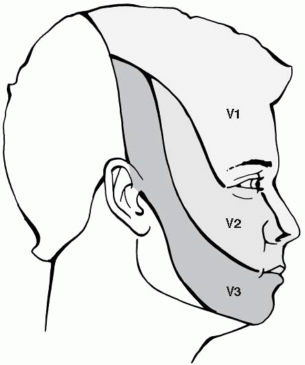
Sensation to the face is supplied by the three main
branches of the trigeminal nerve: the ophthalmic division (V1,
pronounced “vee one”), the maxillary division (V2), and the mandibular
division (V3). Figure 15-1 illustrates the cutaneous sensory distributions of the three divisions of the trigeminal nerve.
branches of the trigeminal nerve: the ophthalmic division (V1,
pronounced “vee one”), the maxillary division (V2), and the mandibular
division (V3). Figure 15-1 illustrates the cutaneous sensory distributions of the three divisions of the trigeminal nerve.
The trigeminal nerve enters the brainstem at the level
of the pons and then synapses with its sensory nucleus; from there,
sensory information crosses to the other side to travel to the
contralateral thalamus and parietal cortex.
of the pons and then synapses with its sensory nucleus; from there,
sensory information crosses to the other side to travel to the
contralateral thalamus and parietal cortex.
EQUIPMENT NEEDED TO TEST FACIAL SENSATION
-
A safety pin and a cotton swab.
-
See Chapter 29, Examination of Pinprick Sensation, for details on the use of a safety pin (or appropriate alternative) to test pin sensation.
HOW TO EXAMINE FACIAL SENSATION
-
Test light touch with your finger or with
a cotton swab. Simply lightly touch both sides of the patient’s
forehead (V1) simultaneously and ask if the sensation is “about the
same” on each side. Repeat the same question when touching both sides
of the patient’s cheeks (V2), and when touching both sides of the
patient’s chin (V3). -
To test pinprick sensation, inform the
patient that you’ll be lightly touching the patient’s face with the
point of the pin, and ask the patient to tell you if the pin sensation
is “about the same” on each side. Touch one side of the forehead (V1)
with the pin and then the other, and ask the patient if the pin
sensation is about the same on each side. Repeat the same process when
touching each cheek (V2) and each side of the chin (V3).
NORMAL FINDINGS
Normally, sensation to light touch and pin should be
approximately equal on each side of the face, in all three divisions of
the trigeminal nerve.
approximately equal on each side of the face, in all three divisions of
the trigeminal nerve.
P.51
 |
|
Figure 15-1 The approximate cutaneous sensory distributions of the three divisions of the trigeminal nerve.
|
ABNORMAL FINDINGS
-
Diminished sensation that corresponds to
the territory of one or more divisions of the trigeminal nerve on one
side of the face suggests a lesion of the trigeminal nerve. -
Diminished sensation of an entire side of
the face is suggestive of a central nervous system lesion affecting
somatosensory pathways, most likely involving the contralateral
thalamus or parietal cortex.
ADDITIONAL POINTS
-
The facial sensory examination, as with
most sensory tests, is of limited value in the absence of a sensory
complaint, such as numbness or tingling. Abnormalities found on facial
sensory testing should be interpreted within the clinical context. -
Asking the patient if the sensation to
pin or light touch is “about the same” on each side is preferable to
asking the patient if there is a “difference” in the sensation on each
side. When asked if there is a difference in sensation, the suggestible
patient may feel compelled to report (the unavoidable) minor
differences in the pressure exerted by the physician, instead of just
reporting areas of truly diminished sensation. -
It is a useful general rule that
trigeminal neuralgia (paroxysmal pain in the distribution of one or
more divisions of the trigeminal nerve) is usually not associated with
any significant loss of facial sensation. Actual loss of sensation that
maps out one or more divisions of the trigeminal nerve, however, is
suggestive of a trigeminal nerve lesion that may be due to significant
infiltrative or compressive etiologies.
