
Editors: Frassica, Frank J.; Sponseller, Paul D.; Wilckens, John H.
Title: 5-Minute Orthopaedic Consult, 2nd Edition
Copyright ©2007 Lippincott Williams & Wilkins
> Table of Contents > Syndactyly
Syndactyly
Dawn M. LaPorte MD
John J. Hwang MD
Description
-
Webbed fingers (or toes) (Fig. 1), usually detected at birth
-
Classification:
-
Internal involvement: Simple (skin only) versus complex (bony involvement/fusion)
-
Extent of syndactyly: Complete (entire length of digits involved) versus incomplete (some part of fingers joined)
-
-
Synonym: Webbed digits
Epidemiology
More common in Caucasian males
Incidence
-
Syndactyly occurs per ~1 per 2,000 births (1).
-
It is seen bilaterally in 50% of cases (1).
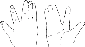 Fig. 1. Syndactyly, or webbing of the fingers, can be simple or complex.
Risk Factors
The presence of other congenital abnormalities constitutes a risk factor for this condition.
Genetics
-
10–40% of the cases are familial (2).
-
80% of cases are sporadic (3).
Etiology
-
Syndactyly occurs secondary to a failure of separation of the digits in the 6th–8th weeks of intrauterine life.
-
The specific cause is unknown.
-
Although syndactyly may be associated in
some cases with a positive family history or with a syndrome, in most
cases it is an isolated finding.
Associated Conditions
-
Apert syndrome: Acrocephalosyndactyly
-
Poland syndrome: Associated with chest wall anomalies and cardiac anomalies
-
Congenital constriction band syndrome
-
Fenestrated syndactyly (joined at the tips)
-
Proteus syndrome
-
NF: A slight increase in incidence
Signs and Symptoms
No pain is associated with this condition.
Physical Exam
-
Observe joined fingers, which can be associated with many anomalies.
-
Examine the joints for active and passive ROM.
-
Test the 2 joined digits at each level for independent movement.
-
The ability to move separately indicates no bony or complex syndactyly.
-
-
The amount of excess skin between the digits is a sign of the difficulty of reconstruction.
-
Inspect the nails.
-
If they are joined, it is likely that the underlying bones are joined also.
-
Tests
Imaging
-
Plain radiography is indicated to differentiate simple from complex syndactyly.
-
Angiography or MRA may be needed in
difficult cases of syndactyly to assess the structure of the underlying
vascular supply of the 2 digits.-
Vasculature branching distally instead of proximally may limit the extent of possible separation.
-
Pathological Findings
-
Insufficient amount of skin present
-
Abnormal fascial interconnections
-
Abnormal interconnection between flexor and extensor tendons
-
Various anomalies of bones and joints
P.435
General Measures
-
Release of webbing:
-
Can improve cosmesis and function
-
Webs less than a few millimeters
distally, or those causing minimum inhibition to spread of the fingers,
do not need surgical intervention.
-
-
Complex syndactyly, especially with only 1 branching neurovascular bundle, can be difficult to correct surgically.
-
The timing of surgery is controversial, but usual recommendations are:
-
>6 months of age for border digits (thumb/index finger and ring/small finger)
-
>12 months for central digits
-
Special Therapy
Physical Therapy
Physical therapy is not needed unless for postoperative scar management, web space splinting, and/or motion.
Surgery
-
The degree of syndactyly dictates which of the many available release techniques is used.
-
A large dorsal flap with a wider proximal base than the distal end is a good technique for simple and incomplete syndactyly.
-
Skin grafting usually is necessary, depending on the amount of skin defect after release.
-
Division of bones is needed in complex syndactyly.
-
Postoperative dressing is an important part of treatment.
-
The dressing is extended above the elbow, and an above-the-elbow plaster cast can be beneficial.
-
The same dressing is continued until postoperative day 14.
-
Prognosis
The prognosis is good, although minor differences in width and appearance of the reconstructed digit are common.
Complications
-
Stiffness
-
Wound dehiscence
-
Scar contracture
-
Partial web recurrence
-
Circulatory deficit, resulting in loss of digit:
-
Rare
-
Can be minimized by operating on only 1 side of the digit at a time, so that a collateral vessel is preserved.
-
Patient Monitoring
As children grow, they should be monitored for partial recurrence of the web or scar contracture.
References
1. Kay
SP, McCombe D, Kozin SH. Deformities of the hand and fingers. In: Green
DP, Hotchkiss RN, Pederson WC, et al., eds. Green’s Operative Hand
Surgery, 5th ed. Philadelphia: Elsevier Churchill Livingstone,
2005:1381–1444.
SP, McCombe D, Kozin SH. Deformities of the hand and fingers. In: Green
DP, Hotchkiss RN, Pederson WC, et al., eds. Green’s Operative Hand
Surgery, 5th ed. Philadelphia: Elsevier Churchill Livingstone,
2005:1381–1444.
2. Ger
E. Syndactyly. In: Buck-Gramcko D, ed. Congenital Malformations of the
Hand and Forearm. London: Churchill Livingstone,1998:131–140.
E. Syndactyly. In: Buck-Gramcko D, ed. Congenital Malformations of the
Hand and Forearm. London: Churchill Livingstone,1998:131–140.
3. Trumble
TE. Congenital hand deformities. In: Trumble TE, ed. Principles of Hand
Surgery and Therapy. Philadelphia: WB Saunders,2000:579–601.
TE. Congenital hand deformities. In: Trumble TE, ed. Principles of Hand
Surgery and Therapy. Philadelphia: WB Saunders,2000:579–601.
Additional Reading
Fearon JA. Treatment of the hands and feet in Apert syndrome: an evolution in management. Plast Reconstr Surg 2003;112:1–12.
Codes
ICD9-CM
755.1 Syndactyly
Patient Teaching
-
The patient or parents should be educated about the complexity of procedure, which they often underestimate.
-
In particular, the need for obtaining
soft-tissue coverage and the difficulty of increasing ROM of an
abnormal joint should be explained.
FAQ
Q: Is syndactyly typically bilateral?
A: It occurs bilaterally 50% of the time.
Q: What is the classification system for syndactyly?
A:
Syndactyly is “complete” if the web space extends to include the
fingertip and “incomplete” when the web space occurs anywhere between
the normal web and the fingertips. “Simple” syndactyly has only
skin/soft tissue connections, and “complex” syndactyly is marked by
skeletal anomalies.
Syndactyly is “complete” if the web space extends to include the
fingertip and “incomplete” when the web space occurs anywhere between
the normal web and the fingertips. “Simple” syndactyly has only
skin/soft tissue connections, and “complex” syndactyly is marked by
skeletal anomalies.
Q: At what age should surgery be performed for syndactyly?
A:
Syndactyly release usually is performed in patients ~12 months old. It
is performed earlier for the border digits or with increasing angular
deformity.
Syndactyly release usually is performed in patients ~12 months old. It
is performed earlier for the border digits or with increasing angular
deformity.