
Editors: Chelly, Jacques E.
Title: Peripheral Nerve Blocks: A Color Atlas, 3rd Edition
Copyright ©2009 Lippincott Williams & Wilkins
> Table of Contents > Section V – Pediatric Peripheral Blocks > 55 – Peripheral Nerve Blockade of the Head and Neck
55
Peripheral Nerve Blockade of the Head and Neck
Kristine Henderson
Pediatric patients undergoing many types of head and
neck procedures can benefit from peripheral nerve blockade for
postoperative analgesia. Common procedures include cleft lip and palate
repair, otoplasty, rhinoplasty and septoplasty, mastoidectomy,
craniotomy, and ventricular shunt placement.
neck procedures can benefit from peripheral nerve blockade for
postoperative analgesia. Common procedures include cleft lip and palate
repair, otoplasty, rhinoplasty and septoplasty, mastoidectomy,
craniotomy, and ventricular shunt placement.
The scalp is innervated by two groups of nerves: the first division of the trigeminal nerve, which divides into the supraorbital and supratrochlear nerves, and cervical root C2, which gives rise to the occipital nerves.
The supraorbital and supratrochlear nerves supply the anterior part of
the scalp, and the occipital nerves supply the posterior part of the
scalp. These two blocks are often performed together for analgesia of
the frontal scalp.
The supraorbital and supratrochlear nerves supply the anterior part of
the scalp, and the occipital nerves supply the posterior part of the
scalp. These two blocks are often performed together for analgesia of
the frontal scalp.
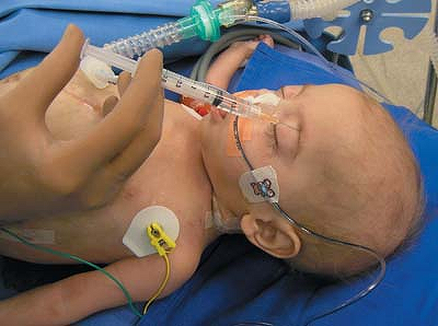
For the first block, the supraorbital
notch is palpated. After antiseptic preparation of the skin, a 27-gauge
needle is inserted perpendicularly into the notch and 1 mL of
bupivacaine (0.25% with 1:200,000 epinephrine) is injected after
aspiration to prevent intravascular injection (Fig. 55-1).
notch is palpated. After antiseptic preparation of the skin, a 27-gauge
needle is inserted perpendicularly into the notch and 1 mL of
bupivacaine (0.25% with 1:200,000 epinephrine) is injected after
aspiration to prevent intravascular injection (Fig. 55-1).
To then block the supratrochlear nerve,
the needle is withdrawn to the skin level and directed medially toward
the tip of the nose; 1 mL of bupivacaine is injected. Gentle pressure
should then be applied to the supraorbital area to prevent the
dissection of local anesthetic and the formation of ecchymosis (Fig. 55-2).
the needle is withdrawn to the skin level and directed medially toward
the tip of the nose; 1 mL of bupivacaine is injected. Gentle pressure
should then be applied to the supraorbital area to prevent the
dissection of local anesthetic and the formation of ecchymosis (Fig. 55-2).
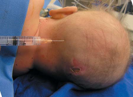
The greater occipital nerve
provides cutaneous innervation to the major portion of the posterior
scalp. Blocking this nerve provides relief of occipital pain following
posterior fossa surgery and posterior shunt revisions. For this block,
the patient’s head is turned to one side, or with the patient prone,
the occipital artery is palpated at the level of the superior nuchal
line. The occipital nerve is located medial to the occipital artery; 1
to 2 mL of bupivacaine (0.25% with 1:200,000 epinephrine) is injected
to form a skin wheal. Blockade of these three nerves together can
provide effective analgesia for most craniotomies (Fig. 55-3).
The infraorbital nerve is
the termination of the second division of the trigeminal nerve. It is
entirely sensory. The nerve emerges in front of the maxilla through the
infraorbital foramen and divides into four branches, innervating the
lower eyelid, lateral inferior portion of the nose and its vestibule,
the upper lip, and the vermilion. Blocking this nerve helps provide
postoperative pain relief in cleft lip repair, septoplasty,
rhinoplasty, and in patients undergoing endoscopic sinus surgery. An
intraoral approach or an extraoral approach can be used. The intraoral
approach may be more aesthetic, in that any small hematoma that is
formed will be less obvious (Fig. 55-4).
P.365
 |
|
Figure 55-1. Supraorbital nerve block.
|
The sphenopalatine ganglion
(SPG) appears to be a pain pathway, especially when trigeminal nerve
divisions 1 (ophthalmic, sensory) and 2 (maxillary, sensory) are
involved. This ganglion is located in the pterygopalatine fossa, behind
the middle nasal turbinate under 1 mm of mucous membrane, and anterior
to the pterygoid canal. Due to this superficial location, the block can
be performed by topical application of local anesthetic or by injection.
The sphenopalatine ganglion is classified as a parasympathetic
ganglion because only preganglionic parasympathetic axons appear to
synapse within the ganglion. It contains the cell bodies of the
postganglionic parasympathetic neurons. However, postganglionic
sympathetic neurons as well as somatic sensory afferent branches of the
trigeminal nerve also pass through the ganglion (without terminating),
all of which may be inhibited by this block.
 |
|
Figure 55-2. Supratrochlear nerve block.
|
P.366
 |
|
Figure 55-3. Greater occipital nerve block.
|
Postganglionic parasympathetic neurons then distribute
to the lacrimal glands, paranasal sinuses, palate, and upper pharynx.
They also innervate the major cerebral arteries, along with
postganglionic sympathetic fibers. A portion of the pain relief from
this block is probably secondary to blocking the parasympathetic
contribution to intracranial vasodilation.
The somatic sensory afferent axons traveling through the
ganglion arise from the maxillary division of the trigeminal nerve by
way of five branches that extend from the nasopharynx, nasal cavity,
palate, and orbit.
ganglion arise from the maxillary division of the trigeminal nerve by
way of five branches that extend from the nasopharynx, nasal cavity,
palate, and orbit.
The simplest, least invasive approach to block this ganglion is via the intranasal
approach. The most common technique utilizes 2 cotton applicators
soaked with 4% lidocaine placed intranasally until the posterior
pharyngeal wall is contacted. This technique can be modified by using
an intratracheal cannula to deliver the local anesthetic instead of
approach. The most common technique utilizes 2 cotton applicators
soaked with 4% lidocaine placed intranasally until the posterior
pharyngeal wall is contacted. This technique can be modified by using
an intratracheal cannula to deliver the local anesthetic instead of
P.367
cotton
applicators, in order to deliver a more perfectly weight-appropriate
dose, if this is a consideration. The patient is placed supine and a
small-gauge intratracheal cannula preloaded with the dose of 4%
lidocaine may be inserted into the nose, passing alongside the inferior
turbinate and directed posteriorly until the upper posterior wall of
the nasopharynx is reached. As you can see in this example, sometimes
the intranasal approach is not as feasible.
 |
|
Figure 55-4. Infraorbital nerve block.
|
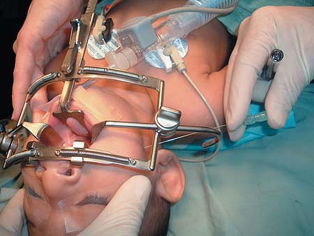 |
|
Figure 55-5. Transfacial approach.
|
The transfacial approach may
also be used in the anesthetized child. A 22-gauge B-bevel needle is
inserted anterior to the mandible and under the zygoma. The needle is
advanced until it contacts the pterygoid plate, and is then withdrawn
approximately 1 mm. In Figure 55-5,
a spinal needle was used because the oral retractor was in the way.
Careful aspiration must confirm that the needle is not intravascular
before 1 mL of local anesthetic is injected.
 |
|
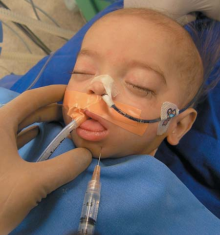
Figure 55-6. Mental nerve block using a transfacial or intraoral approach.
|
P.368
 |
|
Figure 55-7. Greater auricular nerve block.
|
The mental nerve is easily blocked using a transfacial or intraoral
approach. A 25-gauge needle and 0.5 to 2 mL local anesthetic are used.
Typically, a slightly greater volume of anesthetic is used with the
intraoral approach as there is greater local anesthetic spread in the
loose mucosal tissues. This block is useful for plastic surgical
procedures or in the emergency room setting.
The intraoral approach is favored by dentists and oral
surgeons, but also has the advantage of aesthetics; as with the SPG
block discussed previously, any small hematoma formed is less visible.
Another advantage is that a small cotton ball soaked with 2% viscous
lidocaine can be used to provide topical anesthetic prior to the block.
With a helpful parent and the patient’s eyes closed, the child may
never realize that a needle was used (Fig. 55-6).
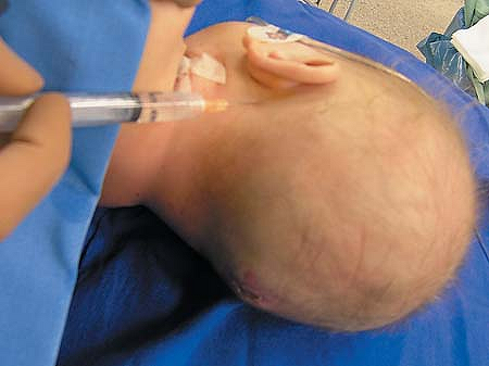
The greater auricular nerve,
arising from fibers of the 2nd and 3rd cervical nerves, innervates the
ear. Blockade of this nerve supplies good postoperative pain relief for
otoplasty. It can also be used for pain relief in patients suffering
from Ramsay Hunt syndrome, herpetic involvement of the geniculate
ganglion.
arising from fibers of the 2nd and 3rd cervical nerves, innervates the
ear. Blockade of this nerve supplies good postoperative pain relief for
otoplasty. It can also be used for pain relief in patients suffering
from Ramsay Hunt syndrome, herpetic involvement of the geniculate
ganglion.
Palpate the mastoid process. A 25-gauge B-bevel needle
is inserted at the level of the mastoid process, then when the
periosteum is reached, is redirected toward the earlobe.
is inserted at the level of the mastoid process, then when the
periosteum is reached, is redirected toward the earlobe.
Inject a total of 3 mL of local anesthetic in a fan-like pattern, moving medially during injection (Fig. 55-7).