
Authors: Koval, Kenneth J.; Zuckerman, Joseph D.
Title: Handbook of Fractures, 3rd Edition
Copyright ©2006 Lippincott Williams & Wilkins
> Table of Contents > I – General Considerations > 7 – Orthopaedic Analgesia
7
Orthopaedic Analgesia
PHARMACOLOGY: CLASSES OF DRUGS
-
Local anesthetics
-
Vasoconstrictors
-
Opioids
-
Sedatives (benzodiazepines)
-
Others
LOCAL ANESTHETICS
-
Basic function
-
These drugs act by blocking voltage-gated sodium channels in axons, preventing action potential.
-
-
Local effect
-
Block is most effective in smaller, myelinated fibers that fire at high frequency.
-
Pain and temperature fibers are much more
sensitive than pressure fibers, which are more sensitive than motor and
proprioceptive fibers.
-
-
Toxicity
-
Central nervous system (CNS)
-
They block inhibitory pathways, leading to unopposed excitatory components.
-
Symptoms include dizziness, nystagmus, and seizures (tonic-clonic).
-
-
Cardiovascular-depressive effects
-
Weaker contraction and arteriolar dilatation occur.
-
-
Neurotoxicity
-
In high concentrations, they can directly damage peripheral nerve fibers.
-
-
-
Lidocaine: rapid, potent, high penetration
-
Short acting
-
Most widely used local anesthetic: local anesthesia, regional, spinal, epidural
-
-
Bupivacaine: slower, potent
-
Longer lasting than lidocaine
-
Can separate motor and sensory block by altering concentration
-
Increased cardiac toxicity (?)
-
-
Maximal dose of commonly used local anesthetics
-
Lidocaine: 5 mg/kg (7 mg/kg if combined with epinephrine)
-
Calculation example:
-
Percent concentration × 10 = mg/mL of drug
-
1% lidocaine = 10 mg/mL of lidocaine
-
-
30-kg child, 1% lidocaine without epinephrine
-
10 mg/mL of lidocaine
-
5 mg/kg × 30 kg = 150 mg allowed
-
150 mg/10 mg/mL = 15 mL of 1% lidocaine
-
-
-
Bupivacaine: 1.5 mg/kg (3 mg/kg with epinephrine)
-
P.53
VASOCONSTRICTORS
-
Allow for longer-lasting blockade (decreased blood flow, less drug leaves area).
-
They may also decrease local blood loss.
-
Epinephrine
-
Most widely used, diluted to 1/200,000
-
Should not be used for a digital block
-
Mnemonic for areas not to use epinephrine: nose, hose (penis), fingers, toes
-
-
Phenylephrine is occasionally used in spinal anesthesia.
OPIOIDS
-
They are derived from the seed of the opium poppy, Papaver somniferum.
-
Morphine and codeine are directly from the plant; others are synthesized.
-
They act by binding to specific opioid receptors in the CNS (µ, δ, κ).
-
The µ receptor is the one most responsible for the analgesic effect.
-
The action is both presynaptic and postsynaptic.
-
-
Central action/pain modulation
-
When activated, the µ receptor inhibits
γ-aminobutyric acid (GABA)-ergic neurons that would otherwise inhibit
pain inhibitory neurons. -
It may also effect neurons in the thalamus and midbrain, to modulate pain stimuli.
-
-
CNS effects
-
Analgesia, euphoria, sedation, respiratory depression, cough suppression, miosis, nausea
-
-
Peripheral effects
-
Cardiovascular: bradycardia
-
Gastrointestinal: decreased motility, constipation, constriction of biliary tree
-
Genitourinary: decreased renal function and increased sphincter tone
-
-
Morphine
-
Naturally occurring, oldest member of this drug class
-
Dosing for adults
-
Loading dose of 0.05 to 0.10 mg/kg intravenously (IV) followed by 0.8 to 10.0 mg/hour IV titrated to pain
-
-
Onset: 5 minutes
-
Relatively long lasting: 3 to 4 hours
-
Better for continuous dull pain rather than sharp/severe pain
-
-
Meperidine (Demerol)
-
Most common emergency department narcotic
-
One-tenth as potent as morphine
-
Dosing for adults
-
15 to 35 mg/hour slow IV infusion or 50 to 150 mg subcutaneously/intramuscularly every 3 to 4 hours as needed
-
-
Poorly titrated: 5- to 10-minute onset and 2- to 3-hour duration
-
Potential for CNS stimulation
-
-
FentanylP.54
-
100× more potent and 7,000× more lipophilic than morphine
-
Rapid uptake: 30 to 60 seconds with peak analgesia in 2 to 3 minutes
-
Duration: 20 to 30 minutes
-
Dose: 1 µg/kg slowly, with sedation often at 3 to 4 µg/kg
-
Risks: “tight chest syndrome,” bradycardia, respiratory depression
-
-
Naloxone, naltrexone (Narcan)
-
Opioid antagonist
-
Strong affinity for µ receptor
-
Binds to receptor, but does not activate it, rapidly reversing the opioid effect within 1 to 3 minutes
-
Usual dose: 0.1 to 0.4 mg IV (0.01 mg/kg in children)
-
Shorter half-life than most agonists, so multiple doses may be necessary
-
SEDATIVES
-
Benzodiazepines
-
In general, they produce anxiolysis and sedation and encourage sleep.
-
They are metabolized in the liver and excreted in the urine.
-
Mechanism
-
They act centrally, bind to, and activate the GABA-A receptor.
-
GABA is major inhibitory neurotransmitter in the CNS.
-
The GABA receptor is the chloride channel.
-
When activated, they hyperpolarize the membrane, making it less excitable.
-
-
Effects
-
Sedation, hypnosis, anesthesia, amnesia
(anterograde), anticonvulsant effects, muscle relaxation, respiratory
depression (especially in pulmonary patients) -
Often increased when combined with opioids
-
-
-
Midazolam
-
Peak effect: 2 to 3 minutes
-
Water soluble, hepatic metabolization
-
Easily titrated with doses every 5 to 7 minutes
-
1 to 2 mg per dose (0.1 mg/kg/dose in children)
-
-
Flumazenil
-
Blocks the effect of benzodiazepines at the GABA receptor level.
-
It has a much shorter half-life than most benzodiazepines that are used clinically.
-
The dose is 0.1 to 0.2 mg IV (0.02 mg/kg in children).
-
Use with caution because it may precipitate seizures.
-
-
Ketamine
-
Dissociative anesthetic
-
Catatonic, amnestic, without loss of consciousness or loss of protective reflexes
-
Blockade of glutamic acid at the N-methyl-D-aspartate receptor subtype
-
May stimulate cardiovascular system and increase blood flow
-
Dose: 1 mg/kg IV
-
Rapid onset: 1 to 3 minutes
-
Duration: 15 to 20 minutes
-
Occasional hallucinations on emergence: can be avoided with a small dose of midazolam
-
May increase salivation: atropine, 0.01 mg/kg, given before ketamine
P.55 -
-
Propofol
-
Isopropylphenol compound
-
Rapid onset, short duration (half-life 30 minutes but lipid soluble so clinical duration is less)
-
Minimal gastrointestinal side effects or nausea
-
Provides general anesthesia: sedation, hypnosis, without analgesia or amnesia
-
Complications: respiratory depression, hypotension, pain at injection site
-
Need for anesthesia/emergency department assistance with airway
-
Dose: 0.5 to 1.0 mg/kg for induction of sedation
-
Highly titratable: 25 to 100 ug/kg/minute infusion after initial bolus
-
NITROUS OXIDE
-
Inhaled agent
-
Given in 50/50 mixture with oxygen
-
Flow controlled by patient holding the mask
-
Provides analgesia and anxiolysis, some sedation
-
Rapid onset: 5 minutes
-
Short duration: resolves within 5 minutes of removing mask
-
Often used as an adjunct with other forms of anesthesia, or for short procedures
-
Very safe for brief procedures
REGIONAL BLOCKS AND CONSCIOUS SEDATION
Hematoma block, regional blocks, Bier block (if proper
equipment and training available), and conscious sedation can all be
effectively used by orthopaedists for fracture reduction and select
procedures.
equipment and training available), and conscious sedation can all be
effectively used by orthopaedists for fracture reduction and select
procedures.
Hematoma Block
-
This replaces the fracture hematoma with local anesthetic.
-
It provides analgesia for closed reductions.
-
It provides postreduction analgesia.
-
Technique
-
Sterile preparation of the fracture site is indicated.
-
Enter the fracture hematoma with a large-bore needle, aspirating hematoma fluid.
-
Replace the hematoma with 10 to 15 mL of 1% lidocaine without epinephrine.
-
Bupivacaine may be added to help with postreduction pain.
-
-
Wait 5 to 7 minutes, then perform the reduction maneuver.
-
-
Risks
-
Systemic toxicity
-
Potential risk of the local anesthetic’s entering the bloodstream directly via the bone’s blood supply.
-
Infection
-
Theoretically converting a closed fracture to an open one.
-
Single case report in orthopaedic literature.
-
-
-
P.56
Regional Blocks
 |
|
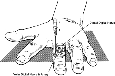
Figure 7.1. Surgical anatomy for the Digital Block Technique
(Reproduced with permission from Consins MJ, Bridenbaugh PO, eds. Neural Blockade ed. Philadelphia: Lippincott-Raven, 1998.)
|
-
They provide anesthesia to a certain area of the body, without general whole-body effects.
-
They are useful in fracture-dislocation reduction, as well as minor and major surgical procedures on the extremities.
-
They are also beneficial for postprocedure analgesia.
-
Local anesthetic is injected around the peripheral nerves.
-
Length of block depends on the choice of anesthetic, as well as the use of epinephrine.
Digital Block
-
Indications include finger fracture, laceration, nailbed injury, and finger/nailbed infection.
-
Do not use epinephrine.
-
Technique (Fig. 7.1)
-
Pronate the hand (skin on the dorsum is less sensitive).
-
Use two injection sites, at each side of the metacarpophalangeal.
-
Use about 2 mL per nerve (8 mL total).
-
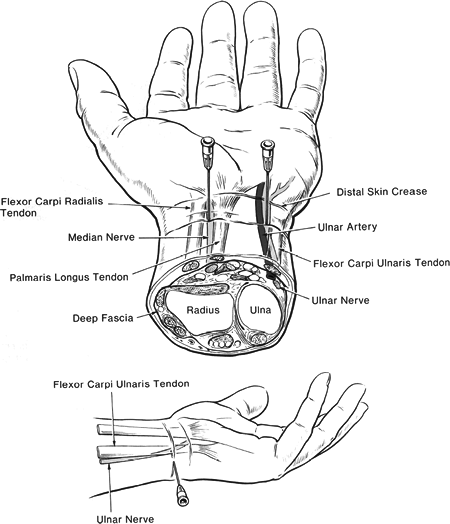
Wrist Block (Fig. 7.2)
-
Median nerve
-
Indications include multiple finger fractures and finger/ nailbed lacerations.
-
Technique
-
Supinate the forearm.
-
The needle is placed between the palmaris longus and the flexor carpi radialis, 2 cm proximal to the wrist flexion crease.
-
If paresthesia is elicited, inject 3 to 5 mL at this site.
![]() Figure 7.2. Surgical anatomy for the Wrist Block Technique(Reproduced with permission from Consins MJ, Bridenbaugh PO, eds. Neural Blockade ed. Philadelphia: Lippincott-Raven, 1998).
Figure 7.2. Surgical anatomy for the Wrist Block Technique(Reproduced with permission from Consins MJ, Bridenbaugh PO, eds. Neural Blockade ed. Philadelphia: Lippincott-Raven, 1998). -
If no paresthesia occurs, then inject 5 mL in fan-shaped fashion.
P.57 -
-
-
Ulnar nerve
-
Indications: ulnar-sided lacerations, reductions of boxer’s fracture (if anesthesia is required)
-
Technique: supinated hand, 6 cm proximal
to wrist crease, just radial to flexor carpi ulnaris, 8 to 10 mL (more
distal block will miss the dorsal branch, which can be blocked by a
wheal ulnar to the flexor carpi ulnaris.
-
-
Radial nerve
-
Indications include thumb and dorsum of hand lacerations.
-
Technique
-
Elbow Block
-
Indications include procedures of the hand and wrist.
-
Four nerves are involved: median, ulnar, radial, and lateral antebrachial cutaneous.
-
Median nerve
-
Draw a line between the medial and lateral condyles of the humerus.
-
The skin wheal is just medial to the brachial artery.
-
Advance the needle until paresthesia is obtained.
-
Inject 3 to 5 mL of lidocaine.
-
-
Ulnar nerve
-
The elbow is flexed.
-
Inject 1 cm proximal to the line that connects the medial epicondyle and the olecranon.
-
Use 3–5 mL of lidocaine.
-
Inject very superficially.
-
-
Radial/musculocutaneous (lateral antebrachial cutaneous nerve)
-
At the intercondylar line, inject 2 cm lateral to the biceps tendon.
-
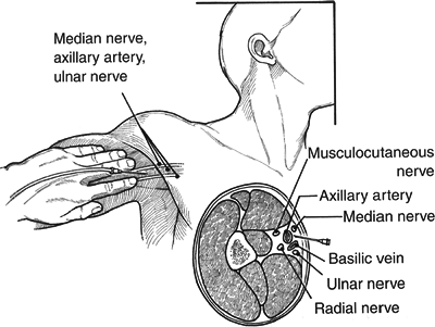
Axillary Block
-
Indications
-
These include hand and forearm procedures and some elbow procedures.
-
-
Technique (Fig. 7.3)
-
The patient is supine with the shoulder abducted and externally rotated.
-
Palpate the axillary artery in the distal axilla.
-
Some advocate going through the artery,
depositing two-thirds of the total anesthetic (20 to 30 mL) behind the
artery and one-third superficial to it. -
Others suggest going on either side of the palpable artery.
-
Think of the four nerves in four quadrants:
-
Musculocutaneous: 9 to 12 o’clock
-
Median: 12 to 3 o’clock
-
Ulnar: 3 to 6 o’clock
-
Radial: 6 to 9 o’clock
-
-
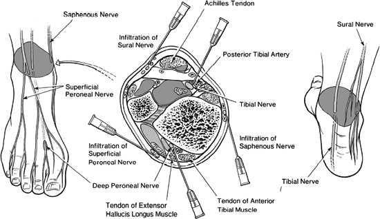
Ankle Block
-
Indications include any foot and ankle procedure.
-
The block must include all five nerves: tibial, superficial and deep peroneal, saphenous and sural nerves (Fig. 7.4).
-
Tibial
-
Posterior to the posttibial artery, halfway between the medial malleolus and the calcaneus.
-
-
Deep peroneal
-
Just lateral to the anterior tibial artery and the extensor hallucis longus.
-
-
Superficial peroneal and saphenous
-
Field block medially and laterally from a deep peroneal site
-
-
Sural
-
Lateral border of the Achilles tendon, halfway between the lateral malleolus and the calcaneus
-
P.59 -
 |
|
Figure 7.3. Surgical anatomy for the Axillary Block Technique
(Reproduced with permission from Doyle JR, Hand and Wrist. Philadelphia: Lippincott Williams & Wilkins, 2006.)
|
Popliteal Block
-
Indications include foot and ankle surgery.
-
Technique
-
The patient is prone, with the knee flexed.
-
Identify the popliteal fossa.
![]() Figure 7.4. Surgical anatomy for the Ankle Block Technique(Reproduced with permission from Consins MJ, Bridenbaugh PO, eds. Neural Blockade ed. Philadelphia: Lippincott-Raven, 1998.)
Figure 7.4. Surgical anatomy for the Ankle Block Technique(Reproduced with permission from Consins MJ, Bridenbaugh PO, eds. Neural Blockade ed. Philadelphia: Lippincott-Raven, 1998.) -
Inject 5 cm superior to the skin crease, 1 cm lateral to the midline, lateral to the artery.
-
Advance in an anterosuperior direction.
P.60 -
-
Add a field block of the saphenous distal to the medial tibial plateau for a more complete block.
Bier Block (Fig. 7.5)
-
It is also known as regional IV anesthesia.
-
This was developed by August Bier in 1908.
-
Indications include hand/wrist procedures and fracture reductions.
-
Technique
-
Start the IV infusion in the hand.
-
Place double tourniquets around the upper arm.
-
Exsanguinate the upper extremity.
-
Inflate the tourniquet.
-
Inject lidocaine without epinephrine (1.5 mg/kg dilute solution or 3 mg/kg, ~50 mL 0.5%).
-
The tourniquet must stay inflated for 25 to 30 minutes.
-
-
Risks
-
Tourniquet pain
-
Length of block most often limited by the ability to tolerate the tourniquet
-
Systemic toxicity
-
Theoretic risks: severe cardiovascular
and CNS side effects with early release of the tourniquet and a large
intravascular bolus of lidocaine
-
CONSCIOUS SEDATION
-
Alteration in consciousness
-
Decreased anxiety
-
Pain relief
-
-
Patient able to maintain patent airway and have intact protective airway reflexes
-
Patient able to respond to verbal or physical stimuli
-
Sedation a continuum
-
Awake/light sedation
-
Anxiolysis, patient essentially responding normally
-
Conscious sedation
-
Response requiring verbal or physical stimuli; airway maintained
-
-
Deep sedation
-
Repeated or painful stimuli necessary for response, airway patency questionable
-
-
General anesthesia
-
Unarousable, airway not protected
-
-
-
When to use it?
-
Any time a potentially painful procedure needs to be performed in the outpatient setting
-
For procedures not requiring general anesthesia and that are reasonably short in duration
-
When appropriate monitoring equipment is available
-
-
Contraindications
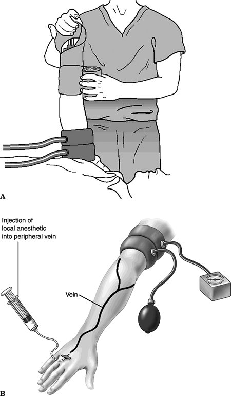 Figure 7.5. (A and B): Intravenous regional blockade (Bier block).(From Bucholz RW, Heckman JD, eds. Rockwood and Green’s Fractures in Adults, 5th ed. Baltimore: Lippincott Williams & Wilkins, 2002:102.)
-
Clinically unstable patient requiring other more urgent procedures
-
Refusal by a competent patient
-
Relative contraindication: long-lasting procedures, likely to require general anesthetic for success
-
-
Appropriate equipment
-
IV access
-
Pulse oximetry
-
Electrocardiographic monitor
-
Blood pressure cuff
-
Airway management equipment
-
Supplemental oxygen
-
Reversal medications (naloxone, flumazenil)
-
-
Technique
-
This typically involves combining an
opioid (morphine or fentanyl) for analgesia and a benzodiazepine
(midazolam) for sedation, relaxation, and amnesia. -
Titrate dosing to achieve appropriate level of sedation while minimizing the risk of adverse outcome.
-
The patient should at all times be
responsive to physical or verbal stimuli (therefore should have
protective airway reflexes intact). -
Remember that these patients have likely already had large doses of opioids for pain control.
-
Be aware of “dose stacking,” giving additional doses of narcotics before waiting to see the effects of the prior doses.
-
-
Risks
-
Respiratory depression/hypoventilation
-
Risk of respiratory depression potentiated by a combination of opioids and benzodiazepines
-
Moderated by appropriate dosing, monitoring, and presence of reversal agents
-
Aspiration
-
Theoretic risk in nonfasted, sedated patients
-
No reported incidences of aspiration during emergency department conscious sedation in the current literature
-
-
Disposition
-
Vital signs, mental status, motor function returning to baseline
-
Pain control with oral analgesics
-
Adequate oral intake
-
Responsible adult present to monitor for continued effects of sedatives
-
P.61
P.62