
Authors: Doyle, James R.
Title: Hand and Wrist, 1st Edition
Copyright ©2006 Lippincott Williams & Wilkins
> Table of Contents > Section I – Basic Anatomy > 1 – Anatomy > 1.1 – Surface Anatomy
1.1
Surface Anatomy
The most appropriate starting point is the hand’s
surface anatomy. Much can be learned about the deeper structures in the
hand by correlating external landmarks such as skin creases and
eminences to underlying anatomic structures.
surface anatomy. Much can be learned about the deeper structures in the
hand by correlating external landmarks such as skin creases and
eminences to underlying anatomic structures.
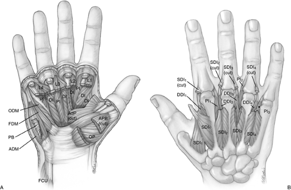
Palmar Hand
External Landmarks
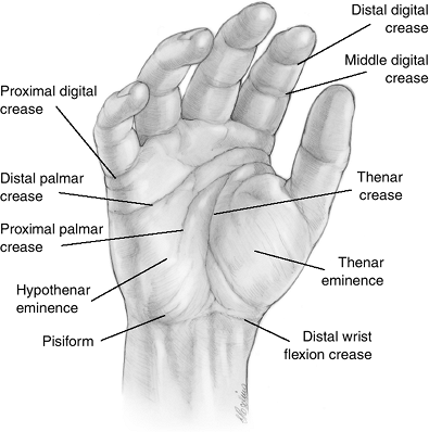
The landmarks of the palmar side of the hand are depicted in Figure 1.1-1.
These landmarks are identified by inspection of the skin creases and
eminences, and by palpation for the bony landmarks of the pisiform and
the hook process of the hamate bone.
These landmarks are identified by inspection of the skin creases and
eminences, and by palpation for the bony landmarks of the pisiform and
the hook process of the hamate bone.
Flexion Creases
The wrist, thenar, palmar, and digital flexion creases
are skin flexion lines seen near synovial joints. These creases provide
“folding points” in the skin, similar to the creases in a road map. Two
creases are present over the proximal interphalangeal (PIP) joints,
which account for the increased angles of flexion at these joints. By
comparison, only one crease is found adjacent to the
metacarpophalangeal (MCP) and distal interphalangeal (DIP) joints.
Flexion creases are usually at right angles to the long axis of the
metacarpals and phalanges, and parallel to the flexion-extension joint
axis of motion. The pronounced obliquity of the thenar crease reflects
the opposing movement of the thumb. It must be noted, however, that
only one of the 17 creases (the thumb MCP joint) lies directly over the
joint. Look at your own hand and note that the MCP flexion crease lies
at the midpoint between the MCP and PIP joints.
Figure 1.1-2 depicts the
relationship between these various skin creases and the underlying
joints, and will allow you to locate the underlying joint structures
with a high degree of confidence.
relationship between these various skin creases and the underlying
joints, and will allow you to locate the underlying joint structures
with a high degree of confidence.
Thenar and Hypothenar Eminences
The thenar eminence is formed by the abductor pollicis
brevis (APB), the most superficial of the thenar group, and the flexor
pollicis brevis (FPB). Both overlie the deeper opponens pollicis (OP).
The ulnar-sided counterpart of the thenar eminence is the hypothenar
eminence, which is formed by the abductor and flexor digiti minimi
(ADM, FDM) and the opponens digiti minimi (ODM).
Bony Landmarks
Pisiform Bone
This relatively superficial and easily palpated carpal
bone is located on the ulnar side of the base of the hand, and it aids
in the identification of the flexor carpi ulnaris (FCU) tendon, the
underlying ulnar neurovascular bundle, and the more distal and radial
hook process of the hamate bone.
bone is located on the ulnar side of the base of the hand, and it aids
in the identification of the flexor carpi ulnaris (FCU) tendon, the
underlying ulnar neurovascular bundle, and the more distal and radial
hook process of the hamate bone.
Hook Process of the Hamate Bone
This process of the hamate may be palpated approximately
2 cm distal and 1 cm radial to the more prominent pisiform. It marks
the beginning of the oblique course of the motor branch of the ulnar
nerve.
2 cm distal and 1 cm radial to the more prominent pisiform. It marks
the beginning of the oblique course of the motor branch of the ulnar
nerve.
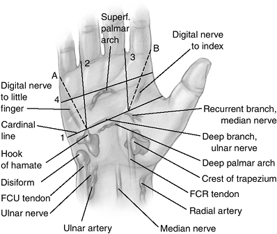
Relationship of the Superficial Landmarks and the Deeper Structures
A unique system of lines may be drawn on the hand that
will permit the examiner to accurately locate the underlying deeper
structures. These lines and the underlying structures are depicted in Figure 1.1-3.
will permit the examiner to accurately locate the underlying deeper
structures. These lines and the underlying structures are depicted in Figure 1.1-3.
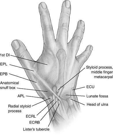
Dorsal Hand
External Landmarks
The external landmarks on the dorsum of the hand are illustrated in Figure 1.1-4.
P.2
 |
|
Figure 1.1-1 Landmarks of the palmar hand.
|
 |
|
Figure 1.1-2 Wrist, thenar, palmar, and digital skin flexion creases and their relationship to the underlying joints and bones.
|
 |
|
Figure 1.1-3
Kaplan described a unique system of lines that may be drawn on the palmar side of the hand and that coincide with the underlying structures. |
 |
|
Figure 1.1-4 Landmarks on the dorsal hand.
|
P.3
Bony Landmarks
Lister’s Tubercle
This bony landmark is located about 0.5 cm proximal to
the dorsal articular margin of the distal radius, in line with the
cleft between the index and middle finger metacarpals. It is easily
palpated and marks the fulcrum, or turning point, for the extensor
pollicis longus (EPL) tendon on its way to the terminal phalanx of the
thumb. It lies in a groove just ulnar to Lister’s tubercle. The
extensor carpi radial brevis (ECRB) tendon is just radial to Lister’s
tubercle in a similar groove on the distal aspect of the radius.
 |
|
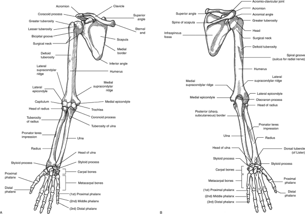
Figure 1.2-1 Bones of the upper limb: anterior (A) and posterior view (B).
|
P.4
Styloid Process of the Middle Finger Metacarpal
The styloid process of the middle finger metacarpal is
located on the metacarpal’s dorsal and radial base. It points to the
articular interface between the capitate and the trapezoid, and is just
proximal to the point of insertion of the ECRB tendon.
located on the metacarpal’s dorsal and radial base. It points to the
articular interface between the capitate and the trapezoid, and is just
proximal to the point of insertion of the ECRB tendon.
Radial Styloid
This distal projection of the radial side of the radius
forms a visible and easily examined landmark that is palpable both
palmar and dorsal to the abductor pollicis longus (APL) and extensor
pollicis brevis (EPB) tendons that run across its apex.
forms a visible and easily examined landmark that is palpable both
palmar and dorsal to the abductor pollicis longus (APL) and extensor
pollicis brevis (EPB) tendons that run across its apex.
P.5
 |
|
Figure 1.2-2 (A) Flexor forearm muscles. (B) Extensor forearm muscles.
|
 |
|
Figure 1.2-3 (A) Palmar hand muscles. (B) Dorsal hand muscles.
|
P.6
 |
|
Figure 1.2-4 Arteries of the upper extremity.
|
Distal Head of the Ulna
The distal aspect of the ulna is slightly expanded and
contains a head and a comparatively small styloid process. The head is
most noticeable and prominent when the forearm is pronated. The short
styloid process is a rounded dorsoulnar projection from the ulnar head
that is most palpable in supination, and is about 1 cm proximal to the
plane of the radial styloid. The apex of the triangular fibrocartilage
attaches to the palmar-radial base of the styloid process. The extensor
carpi ulnaris (ECU) tendon runs in a groove along the dorsal aspect of
the ulnar head.
Other Dorsal Landmarks
Anatomic Snuff Box
This depression on the radial side of the wrist is a
narrow triangle with its apex distal that is bordered dorsoulnarly by
the EPL, radially by the abductor pollicis longus (APL),
narrow triangle with its apex distal that is bordered dorsoulnarly by
the EPL, radially by the abductor pollicis longus (APL),
P.7
and
extensor pollicis brevis (EPB) tendons, and proximally by the distal
margin of the extensor retinaculum. It contains the dorsal branch of
the radial artery; in its dorsoulnar corner, the tendon of the extensor
carpi radialis longus (ECRL); and superficially, one or more branches
of the superficial branch of the radial nerve. The carpal scaphoid bone
lies beneath this fossa and tenderness in this area following trauma
may indicate an injury of this bone.
Lunate Fossa
The lunate fossa is a palpable central depression
located on the dorsum of the wrist, in line with the longitudinal axis
of the third metacarpal just ulnar and distal to Lister’s tubercle, and
beginning immediately distal to the dorsal margin of the radius. It is,
on average, approximately the size of the pulp of your thumb, and it
marks the location of the carpal lunate in the proximal carpal row.