
Editors: Frassica, Frank J.; Sponseller, Paul D.; Wilckens, John H.
Title: 5-Minute Orthopaedic Consult, 2nd Edition
Copyright ©2007 Lippincott Williams & Wilkins
> Table of Contents > Genu Varum (Bowed legs)
Genu Varum (Bowed legs)
Paul D. Sponseller MD
Description
-
The knee goes through normal phases of
changing alignment in childhood: Genu varum (bowed legs) is physiologic
in infants and young children up to 2 years of age, and its appearance
is maximal at 12–18 months of age (1–3). -
Bowing is most obvious when children start walking.
-
It may be combined with internal tibial torsion, which makes it appear more pronounced.
-
Bowing may seem greater with weightbearing.
-
This condition usually resolves by 2 years of age and changes to physiologic genu valgum (knock-knee) (2).
-
Tibia vara (Blount disease) (see “Blount Disease”
chapter), rickets, fibrocartilaginous dysplasia of the proximal tibia,
and other genetic disorders can cause pathologic genu varum (Fig. 1-1).
Epidemiology
-
Physiologic (normal) bowing is ~1,000 times more common than pathologic bowing (e.g., Blount disease) (1,3).
-
It occurs equally in boys and girls.
Risk Factors
Family history
Genetics
-
Some causes of bowed legs are familial:
-
Blount disease
-
Renal rickets
-
Skeletal dysplasia
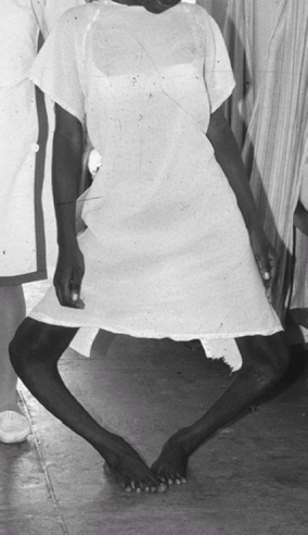 Fig. 1. Patient with severe untreated infantile varus now in adolescence.
-
Etiology
-
Bowing is an imbalance between the load and growth plate development.
-
It may be caused by:
-
Overweight
-
Rickets (4)
-
Skeletal dysplasia
-
-
Physiologic causes: Normal growth
patterns of the femoral and tibial growth plates include a period of
normal varus in early infancy. -
Pathologic causes:
-
Tibia vara (Blount disease)
-
Rickets (nutritional or renal)
-
Achondroplasia
-
Epiphyseal and metaphyseal dysplasias
-
Focal fibrocartilaginous dysplasia
-
-
In most of these conditions, the varus results from inability of the growth plate to respond normally to load (3).
Associated Conditions
-
Early walker
-
Heavy weight
Signs and Symptoms
-
Parental concern about the appearance of the legs is the most common reason for the presentation of children.
-
The patient should be pain free; if pain exists, another cause should be sought.
-
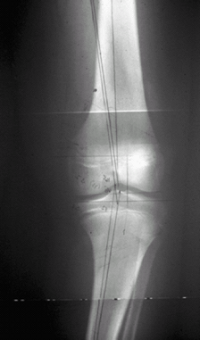
Genu varum may develop spontaneously in
the overweight adolescent who previously had straight legs (adolescent
Blount disease), and it usually requires treatment (Fig. 2).
History
If a patient has physiologic bowing, the parents should start to notice improvement after the 2nd birthday (3).
 |
|
Fig. 2. 13-year-old boy with adolescent genu varum. Note the widened medial physis.
|
Physical Exam
-
Obtain a medical, family, and developmental history.
-
Determine the patient’s height and weight percentiles.
-
Estimate the angulation of the knee.
-
Check the rotation of the tibia and femur.
-
To monitor the patient’s progress, document the distance between the medial surfaces of the knees (intercondylar distance) (5,6).
Tests
Lab
-
In routine cases, tests are not indicated if varus appears mild and physiologic.
-
If metabolic causes are suspected, serum
calcium, phosphate, alkaline phosphatase, 1,25-vitamin D, and
creatinine levels may be measured (3,4).
Imaging
-
Radiographic evaluation of bowed legs in
children <18 months old should be reserved for asymmetric bowing or
for patients suspected of having a pathologic condition other than
benign physiologic varus. -
A single AP radiograph of the lower
extremity from hip to ankle on a standing film is the most appropriate
1st imaging study; care should be taken that the knee is pointing
straight ahead. -
Widening of physis suggests rickets;
delayed ossification of the distal femoral and proximal tibial
epiphyses may be a result of excessive pressure on 1 side of the knee. -
The femorotibial angle and the metaphyseal–diaphyseal angle of the tibia should be measured (5).
-
If the metaphyseal–diaphyseal angle is <11°, physiologic bowing is assured.
-
If the metaphyseal-diaphyseal angle is >16°, the child has Blount disease.
-
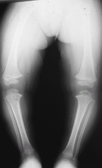
If the metaphyseal bowing in the femur is
equal to or greater than that in the tibia, the bowing is more likely
to be physiologic (1) (Fig. 3).![]() Fig.
Fig.
3. This 2-year-old patient had bowed legs. The metaphyseal–diaphyseal
angle is 10° on each side. The bowing is more pronounced on the femoral
than the tibial metaphysis. It resolved without treatment.
P.155
Differential Diagnosis
-
Achondroplasia
-
Rickets
-
Infantile or adolescent Blount disease
-
Metaphyseal or epiphyseal dysplasia
General Measures
-
Physiologic conditions:
-
Physiologic bowing always resolves without treatment; bracing is not needed
-
At ≥18 months of age, follow-up
examination and imaging are needed to differentiate physiologic bowing
from tibia vara (may be difficult) (6).
-
-
Pathologic conditions:
-
Rickets or other metabolic bone disease:
-
The underlying disease is treated, with osteotomy reserved for those patients with persisting varus after treatment.
-
-
Achondroplasia and epiphyseal or metaphyseal dysplasia:
-
The patient may need surgical treatment, depending on the degree of deformity.
-
-
Tibia vara (Blount disease):
-
Brace treatment is appropriate for children <3 years old; a knee-ankle-foot brace may be used for walking.
-
If the patient is >4 years of age, osteotomy is recommended.
-
-
Special Therapy
Physical Therapy
-
Not necessary for physiologic bowing
-
Not an effective treatment for pathologic varus
Surgery
-
Many different types of osteotomy are
available for correcting varus deformity, including dome, oblique,
closing wedge, or opening wedge osteotomy. -
The tibia or the femur may require surgery, depending on the site of the deformity.
-
Physeal bar resection or hemiepiphysiodesis may be indicated for some cases.
Prognosis
-
Physiologic genu varum has an excellent prognosis for spontaneous improvement.
-
The prognosis of pathologic genu varum varies.
-
Knee pain and worsening of the bow are likely in adulthood if the deformity is >10–15° (5).
Complications
-
Untreated genu varum may cause pain on the medial part of the knee and eventual arthritis during adulthood.
-
Adolescent genu varum may be painful.
-
Complications occasionally seen from surgery may include:
-
Infection
-
Compartment syndrome
-
Recurrence of deformity
-
Growth disturbance
-
Patient Monitoring
-
The frequency of follow-up varies, depending on the individual surgeon or pediatrician.
-
Physiologic bowing does not need frequent
follow-up unless the condition is not improving; resolution is a slow
process and may take a year. -
Pathologic bowing needs more prolonged follow-up.
References
1. Bowen
RE, Dorey FJ, Moseley CF. Relative tibial and femoral varus AS a
predictor of progression of varus deformities of the lower limbs in
young children. J Pediatr Orthop 2002;22:105–111.
RE, Dorey FJ, Moseley CF. Relative tibial and femoral varus AS a
predictor of progression of varus deformities of the lower limbs in
young children. J Pediatr Orthop 2002;22:105–111.
2. Salenius P, Vankka E. The development of the tibiofemoral angle in children. J Bone Joint Surg 1975;57A:259–261.
3. Schoenecker PL, Rich MM. The lower extremity. In: Morrissy RT, Weinstein SL, eds. Lovell and Winter’s Pediatric Orthopaedics, 6th ed. Philadelphia: Lippincott Williams & Wilkins, 2006:1157–1211.
4. Biser-Rohrbaugh A, Hadley-Miller N. Vitamin D deficiency in breast-fed toddlers. J Pediatr Orthop 2001;21:508–511.
5. Gordon JE, King DJ, Luhmann SJ, et al. Femoral deformity in tibia vara. J Bone Joint Surg 2006;88A:380–386.
6. Langenskiold A. Tibia vara. A critical review. Clin Orthop Relat Res 1989;246:195–207.
Codes
ICD9-CM
736.42 Genu varum
Patient Teaching
-
Parents should be told that physiologic
genu varum will resolve spontaneously and slowly; if it is not starting
to improve at least by 2–3 years of age, additional evaluation is
needed. -
No restriction on activity is recommended.
-
Exercises do not help genu varum resolve.
FAQ
Q: Do infant “jumper” devices contribute to bowed legs?
A: No evidence suggests that they do. The children are supported in these devices, so the load on the legs is controlled.
Q: When should a child with bowing be referred to a specialist?
A: If the bowing gets worse after 18 months or persists after the 2nd birthday.