
Editors: Chelly, Jacques E.
Title: Peripheral Nerve Blocks: A Color Atlas, 3rd Edition
Copyright ©2009 Lippincott Williams & Wilkins
> Table of Contents > Section
II – Single-Injection Peripheral Blocks > B – Lower Extremity >
14 – Lower Extremity Multiple-Stimulation Techniques
II – Single-Injection Peripheral Blocks > B – Lower Extremity >
14 – Lower Extremity Multiple-Stimulation Techniques
14
Lower Extremity Multiple-Stimulation Techniques
Andrea Casati
The lower limb is innervated by two plexus: the lumbar
and the sacral. Among the nerves that emerge from these plexus, the
sciatic nerve, which divides into the tibial and common peroneal
nerves; the femoral nerve, which divides into seven terminal branches
including the saphenous nerve (Fig. 14-1; [1]); and the nerve to the vastus intermedius muscle (Fig. 14-1; [2]) and the nerve to the vastus lateralis (Fig. 14-1; [3]) may be blocked using a multiple-stimulation technique. As discussed in Chapter 8,
the use of a multiple-stimulation technique allows one to minimize the
volume of local anesthetic required to produce surgical anesthesia.
This is especially important for lower limb blocks because both a block
of the femoral and sciatic nerve are required, which results in an
increased risk of local anesthetic toxicity due to overdosing when the
single-stimulation technique is used. Another important advantage of
the multiple-injection technique is the reduced toxicity associated
with intravascular injections of local anesthetics. Thus, it is well
known that a negative blood aspiration before the injection of local
anesthetics—although necessary—does not prevent the risk for
unintentional intravascular injection.
It has been reported that withdrawing and redirecting
the stimulating needle to elicit multiple motor responses might result
in slightly more discomfort to the patient. For this reason an
appropriate sedation with small doses of benzodiazepine combined with
appropriate analgesia with opioids is indicated with the use of
multiple-stimulation techniques. To minimize the risk of intraneural
injection, it is important that patients remain able to report any pain
during the local anesthetic injection. For this same reason, lower
extremity blocks should also be performed only after the complete
resolution of neuroaxial blocks.
P.150
A. Multiple-Stimulation Technique to Block the Femoral Nerve
To perform a femoral nerve block using a
multiple-stimulation technique, three different muscular stimulations
should be elicited: (a) the contractions of the vastus medialis,
induced by stimulation of the vastus medialis/saphenous nerve; (b) the
contractions of the vastus intermedius muscle with movements of the
patella, induced by the stimulation of the vastus intermedius nerve;
and (c) the contractions of the vastus lateralis muscle, induced by
stimulation of the vastus lateralis nerve.
The insulated needle connected to a nerve stimulator (1.5 mA, 2 Hz, 0.1
ms) is inserted immediately lateral to the femoral artery above the
inguinal crease and advanced perpendicularly to the skin. The
contraction of the vastus lateralis is usually the first to be
elicited. After the appropriate motor response is obtained, the needle
position is then adjusted to maintain the same motor response with a
stimulating current intensity ≤0.5 mA. After negative aspiration, 5 to
6 mL of local anesthetic is injected slowly. The stimulating needle is
then withdrawn from the skin, and the current intensity is increased to
1.5 mA. The needle is reoriented 2° to 5° laterally and introduced
slightly deeper in search of a contraction of the vastus intermedius
nerve and movement of the patella. After the appropriate motor response
is obtained, the needle position is then adjusted to maintain the motor
response with a stimulating current intensity ≤0.5 mA. After negative
aspiration, another 5 to 6 mL of the same anesthetic solution is
injected. Finally, the insulated needle is withdrawn to the level of
the skin, and the current intensity is increased to 1.5 mA. The needle
is redirected more laterally by increasing the angle by another 5° and
reintroduced in search of the contraction of the vastus lateralis
muscle. After the appropriate motor response is obtained, the needle
position is then adjusted to maintain the motor response with an
intensity ≤0.5 mA. After negative aspiration, a final 5 to 6 mL of
local anesthetic solution is injected.
-
To minimize the risk for arterial
puncture, the initial insertion of the needle should be the closest to
the femoral artery. Thereafter the needle is redirected laterally. -
The contraction of the vastus lateralis
muscle is often associated with residual movements of the patella and
sometimes can be only seen distally at the level of the knee. -
Very often, the contraction of the vastus
lateralis is preceded by a contraction of the sartorius muscle. To
obtain the desired motor response, the needle needs to be introduced
slightly deeper.P.151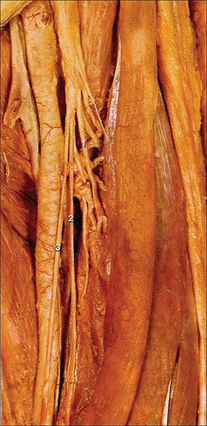 Figure 14-1. Saphenous nerve. (1) Vastus lateralis nerve; (2) Vastus medialis nerver; (3) Saphenous nerve.
 -
In contrast to a three-in-one
perivascular block, the multiple-stimulation technique to block the
femoral nerve does not allow the possibility of also blocking the
lateral femoral cutaneous nerve and the obturator nerve. If these
blocks are required, they need to be performed individually. -
With this technique, a complete sensory and motor femoral block occurs within 20 minutes.
P.152
B. Multiple-Stimulation Technique to Block the Sciatic Nerve
The sciatic nerve block produces motor responses that
vary according to the level at which the nerve is being stimulated. At
a high level (parasacral, posterior, and sub-gluteal), three types of
responses are elicited: (a) the dorsiflexion or eversion of the foot,
induced by the stimulation of the common peroneal branch of the sciatic
nerve; (b) the plantar flexion of the foot and toes produced by the
stimulation of the tibial branch of the sciatic nerve; and (c) the
contraction of the biceps femoris induced by the stimulation of the
branches innervating the hamstring muscles. Above the knee (lateral and
posterior popliteal nerve block), only contraction of the foot and toe
muscles are observed. Although the sciatic nerve divides into the
common peroneal and tibial nerves above or at the popliteal fossa, the
two branches can be clearly identified from its origin; and in about
10% of cases they are two separate nerves already at the ischiatic
foramen. Nevertheless, and except in amputated limb cases, the distal
motor responses (dorsiflexion or eversion) and flexion of the foot and
toes are searched to block the common peroneal and tibial nerves,
respectively.
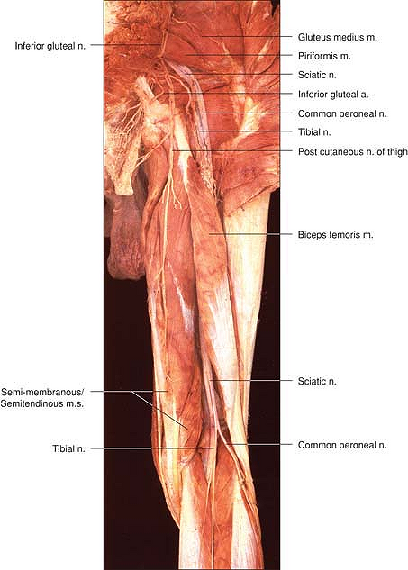
High Sciatic Approaches
Classic Posterior (Labat) Approach
The greater trochanter, the sacral hiatus, and the posterosuperior
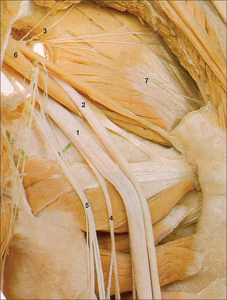
iliac spine. The sciatic nerve is often divided into a common peroneal (Fig. 14-2; [1]) and tibial branch (Fig. 14-2; [2]), close to the origin of the sciatic nerve. At this level it is also possible to identify the superior gluteal nerve (Fig. 14-2; [3]), the nerve to the hamstring muscle (Fig. 14-2; [4]), the posterior cutaneous nerve of the thigh (Fig. 14-2; [5]), the piriformis muscle (Fig. 14-2; [6]), and the medius muscles (Fig. 14-2; [7]).
Subgluteal Approach
The needle is introduced perpendicularly to the skin and advanced
slowly until a stimulation of either the common peroneal or the tibial
nerve is elicited. After the appropriate motor response is obtained,
the needle position is then adjusted to maintain the same motor
response with a stimulating current intensity ≤0.5 mA. After negative
aspiration, 8 to 10 mL of the local anesthetic solution is injected.
The stimulating needle is withdrawn to the level of the skin, the
current intensity is set back to 1.5 mA, and the needle is then
redirected 2° to 5° (if the tibial nerve was stimulated first) or
medially (if the common peroneal nerve was stimulated first) and
P.153
advanced
until the appropriate motor response is elicited. The position of the
needle is then adjusted to maintain the same motor response with a
stimulating current ≤0.5 mA. After negative blood aspiration, 8 to 10
mL of local anesthetic solution is injected.
 |
|
Figure 14-2.
Vastus intermedius muscle. (1) Tibial nerve; (2) Common Peroneal nerve; (3) Inferior gluteal nerve; (4) Posterior cutaneous nerve of thigh; (5) Nerve to quadrus femoris; (6) Inferior gluteal nerve; (7) Gluteus minimus. |
-
Considering the length of the stimulating
needle and the depth at which the sciatic nerve is located, changes in
the angle of the stimulating needle should be minimal (no more than 2°
to 5°). Consideration should also be given not to flex the stimulating
needle. -
The depth at which the first motor
response is obtained allows the clinician to control the limit at which
the needle should be introduced next (the tibial nerve is slightly more
posterior and medial to the common peroneal nerve). -
The subgluteal approach is as successful as the Labat classic posterior approach and has the advantage of being less painful.
-
If bone contact occurs, the needle is
withdrawn from the skin. For either the Labat or the subgluteal
approach, bone contact usually indicates the sciatic nerve should be
found more superficially.
P.154
Low Sciatic Approaches
Popliteal Approaches
At the level of the popliteal fossa, the two branches of
the sciatic nerve clearly divide. The more caudal the sciatic approach,
the more separated are the two branches of the sciatic nerve (common
peroneal and tibial nerves) (Fig. 14-3).
There is also a spatial distribution of common peroneal and tibial
nerves that needs to be considered. The tibial nerve lies closer to the
bone and more medial. Thus it is slightly more anterior and deeper than
the common peroneal nerve when approached from the lateral side of the
thigh. In contrast, the common peroneal nerve is located more laterally
and superficially, and it exits the popliteal fossa just above the head
of the fibula.
To perform a multiple-stimulation sciatic block at the popliteal fossa, a proximal approach is usually recommended.
Posterior Popliteal Sciatic Nerve Block
The popliteal crease, the medial border of the femoris biceps
laterally, and the lateral border of the semi-tendinous tendon medially.
The needle is introduced perpendicular to the skin in search of a motor
response related to either the stimulation of the common peroneal or
tibial nerve (usually the common peroneal nerve). The needle position
is adjusted to maintain the same motor response with a stimulating
current ≤0.5 mA. After negative aspiration for blood, 10 mL of local
anesthetic solution is injected. As with all approaches to the sciatic
nerve, the first motor response determines how the needle is oriented
when searching for the next motor response. If the first motor response
elicited is the dorsiflexion of the foot (common peroneal nerve), the
needle will be directed more medially and slightly deeper in search of
a stimulation of the tibial nerve (plantar flexion with flexion of the
toes). If the first motor response is the plantar flexion of the foot
(tibial nerve), the stimulating needle is redirected more laterally.
Lateral Popliteal Sciatic Nerve Block
P.155
 |
|
Figure 14-3. Sciatic nerve at the level of the popliteal fossa.
|
A vertical line is drawn at the level of the superior border of the
patella. The site of needle introduction is demarcated by the
intersection of this line and the line drawn at the level of the groove
between the femoralis biceps and the vastus lateralis muscles. The
needle is inserted at an angle 20° to 30° posterior to the horizontal
plane with a slight caudal direction. It is advanced slowly, usually in
search of the common peroneal nerve, which produces either a
dorsiflexion and toes extension or an eversion of the foot. The needle
position is then adjusted to maintain the same motor response with a
stimulating current intensity ≤0.5 mA. After negative aspiration, 8 to
10 mL of local anesthetic solution is injected. The needle is then
withdrawn from the skin and oriented at 45° in search of the tibial
nerve, which is located more medially and
P.156
slightly
deeper than the common peroneal nerve. Its stimulation produces a
plantar and toes flexion. The needle position is then adjusted to
maintain the same motor response with a stimulating current intensity
≤0.5 mA. After negative aspiration, 8 to 10 mL of local anesthetic
solution is injected.
Suggested Readings
Bailey SL, Parkinson SK, Little WL, et al. Sciatic nerve block: a comparison of single versus double injection technique. Reg Anesth 1994;19:9–13.
Casati
A, Fanelli G, Beccaria P, et al. Effects of the single or multiple
injection technique on the onset time of peripheral nerve blocks with
0.75% ropivacaine. Anesth Analg 2000;91:181–184.
A, Fanelli G, Beccaria P, et al. Effects of the single or multiple
injection technique on the onset time of peripheral nerve blocks with
0.75% ropivacaine. Anesth Analg 2000;91:181–184.
Casati
A, Fanelli G, Beccaria P, et al. Effects of single or multiple
injections on the volume of 0.5% ropivacaine required for femoral nerve
blockade. Anesth Analg 2001;93:183–186.
A, Fanelli G, Beccaria P, et al. Effects of single or multiple
injections on the volume of 0.5% ropivacaine required for femoral nerve
blockade. Anesth Analg 2001;93:183–186.
Davies MJ, McGlade DP. One hundred sciatic nerve blocks: a comparison of localization techniques. Anesth Intensive Care 1993;21:76–78.
di
Benedetto P, Bertini L, Casati A, et al. A new posterior approach to
the sciatic nerve block: a prospective, randomized comparison with the
classic posterior approach. Anesth Analg 2001;93:1040–1044.
Benedetto P, Bertini L, Casati A, et al. A new posterior approach to
the sciatic nerve block: a prospective, randomized comparison with the
classic posterior approach. Anesth Analg 2001;93:1040–1044.
Fanelli
G, Casati A, Garancini P, et al. Nerve stimulator and multiple
injection technique for upper and lower limb blockade: failure rate,
patient acceptance and neurologic complications. Anesth Analg 1999;88:847–852.
G, Casati A, Garancini P, et al. Nerve stimulator and multiple
injection technique for upper and lower limb blockade: failure rate,
patient acceptance and neurologic complications. Anesth Analg 1999;88:847–852.
Kinirons
BP, Bouaziz H, Paqueron X, et al. Sedation with sufentanil and
midazolam decreases pain in patients undergoing upper limb surgery
under multiple nerve block. Anesth Analg 2000;90:1118–1121.
BP, Bouaziz H, Paqueron X, et al. Sedation with sufentanil and
midazolam decreases pain in patients undergoing upper limb surgery
under multiple nerve block. Anesth Analg 2000;90:1118–1121.
Paqueron
X, Bouaziz H, Macalou D, et al. The lateral approach to the sciatic
nerve at the popliteal fossa: one or two injections? Anesth Analg 1999;89:1221–1225.
X, Bouaziz H, Macalou D, et al. The lateral approach to the sciatic
nerve at the popliteal fossa: one or two injections? Anesth Analg 1999;89:1221–1225.