
Editors: Frassica, Frank J.; Sponseller, Paul D.; Wilckens, John H.
Title: 5-Minute Orthopaedic Consult, 2nd Edition
Copyright ©2007 Lippincott Williams & Wilkins
> Table of Contents > Friedreich Ataxia
Friedreich Ataxia
Paul D. Sponseller MD
Description
-
Friedreich ataxia is an uncommon, heritable disorder causing progressive spinocerebellar degeneration (1,2).
-
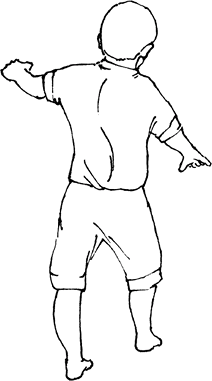
Scoliosis, ataxia, and foot deformities are the most common findings (Fig. 1).
-
Systems affected: Central nervous system; Heart; Skeleton
-
Classification:
-
No subclassifications are used in Friedreich ataxia.
-
It is classified under the category spinocerebellar degeneration, and it is the most common example of this class.
-
Another disorder in this class is spinocerebellar ataxia.
-
-
Synonym: Spinocerebellar degeneration
Epidemiology
Incidence
Prevalence is ~1 per 50,000 persons (1).
Prevalence
-
It may become apparent anytime from 5–25 years of age.
-
Males and females are affected equally.
Risk Factors
The condition is more common in people of French Canadian descent (1).
Genetics
Transmission is autosomal recessive.
 |
|
Fig. 1. Scoliosis, cavovarus feet, and ataxia as seen in Friedreich ataxia.
|
Etiology
-
The cause is a defect in a gene for a protein, frataxin, found on chromosome 9 (1).
-
The pathogenesis of the findings is not well known.
-
Variations in characteristics of the
disease (e.g., age at onset and rate of progression) may be caused by
different mutations at one of the loci.
Associated Conditions
-
Diabetes mellitus
-
Cardiomyopathy
-
Scoliosis
-
Foot deformity
The diagnosis is made on a clinical basis, usually confirmed by a neurologist.
Signs and Symptoms
-
Common signs:
-
Ataxia
-
Wide-based gait
-
Weakness and loss of position sense in the lower extremities
-
Frequently, loss of upper extremity reflexes
-
-
Less common signs:
-
Pes cavus
-
Optic atrophy
-
Nystagmus is possible.
-
-
Symptoms:
-
Partial deafness, depression, loss of coordination, painful muscle spasms, and weakness
-
Symptoms of diabetes mellitus also may be related because of the increased coexistence of these disorders.
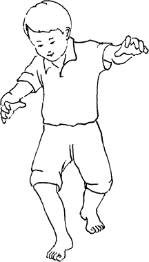 Fig. 2. Ataxia and cavovarus feet in Friedreich ataxia.
-
Physical Exam
-
Examine heel-to-toe walking and finger-to-nose positioning (Fig. 2).
-
Look for the presence of increased kyphosis on routine standing.
-
Perform the forward-bend test for scoliosis.
-
Test upper and lower extremity reflexes.
-
Note the flexibility of the feet and the correctability of any deformity.
-
Test muscle strength (proximal muscles are affected earlier are than distal muscles).
-
The gluteus maximus is the 1st muscle to be affected clinically.
Tests
Lab
-
Creatine phosphokinase levels are normal.
-
Fasting serum glucose for diabetes mellitus should be obtained.
-
Other tests:
-
Electrocardiography should be performed
before surgery, with echocardiography as indicated, because of the
increased incidence of hypertrophic cardiomyopathy. -
Electromyography shows polyphasic potentials and mild slowing of nerve conduction velocity.
-
Imaging
-
Standing posteroanterior and lateral radiographs of the spine should be ordered when scoliosis and kyphosis are found.
-
Periodic monitoring then is indicated, even after maturity.
-
-
Foot radiographs may be indicated.
Differential Diagnosis
-
Cerebellar tumors
-
Chiari malformation
-
Muscular dystrophy
-
Spinal dysraphism (as a cause of scoliosis and foot deformity)
P.151
General Measures
-
Foot and spine deformities should be followed by an orthopaedic surgeon, even if surgery is not contemplated.
-
Brace treatment of scoliosis is appropriate for curves of 25–45° and may slow the rate of progression.
-
Stretching and night bracing may be helpful in preventing worsening of the foot deformities.
-
Walking should be maintained as long as possible.
Special Therapy
Physical Therapy
-
Therapy is essential, to keep up strength and skills after surgery.
-
Stretching of plantar fascia and ankle muscles help to prevent deformity.
Medication
-
No medical treatment currently is available.
-
If painful muscle spasms occur, baclofen or diazepam may be helpful.
Surgery
-
Scoliosis:
-
A cardiopulmonary evaluation is indicated preoperatively.
-
To prevent progressive decompensation of
the spine, posterior fusion and instrumentation with 2 contoured rods
is indicated for curves of >50°. -
If the curve is large, rigid, and unbalanced, an anterior release also may be indicated to increase the correctability (2–4).
-
Spinal cord monitoring may be more challenging and should include sensory and motor modalities.
-
Postoperative immobilization usually is unnecessary.
-
-
Cavovarus feet: Correction may include:
-
Plantar fasciotomy
-
Achilles tendon lengthening and transfer or lengthening of the PTT
-
Possible osteotomy
-
Disposition
Issues for Referral
A neurologist is the best specialist for coordinating the overall care of these patients.
Prognosis
-
If scoliosis develops before the patient is 15 years old, it most likely will become severe and require surgery.
-
Most affected individuals stop walking by 20–30 years of age and are wheelchair dependent.
-
Death usually occurs by the 4th or 5th decade; causes most often include pneumonia, aspiration, and cardiomyopathy.
Complications
-
Cardiomyopathy
-
Calluses or skin breakdown on the foot
-
Pneumonia
Patient Monitoring
-
This disease is progressive.
-
Walking distance and status of all physical findings should be monitored every 3–6 months.
-
Scoliosis, if present, should be checked every 6 months.
References
1. Cady RB, Bobechko WP. Incidence, natural history, and treatment of scoliosis in Friedreich’s ataxia. J Pediatr Orthop 1984;4:673–676.
2. Delatycki MB, Paris DBBP, Gardner RJM, et al. Clinical and genetic study of Friedreich ataxia in an Australian population. Am J Med Genet 1999;87: 168–174.
3. Labelle H, Tohme S, Duhaime M, et al. Natural history of scoliosis in Friedreich’s ataxia. J Bone Joint Surg 1986;68A:564–572.
4. Shapiro
F, Bresnan MJ. Orthopaedic management of childhood neuromuscular
disease. Part II: Peripheral neuropathies, Friedreich’s ataxia, and
arthrogryposis multiplex congenita. J Bone Joint Surg 1982;64A:949–953.
F, Bresnan MJ. Orthopaedic management of childhood neuromuscular
disease. Part II: Peripheral neuropathies, Friedreich’s ataxia, and
arthrogryposis multiplex congenita. J Bone Joint Surg 1982;64A:949–953.
Codes
ICD9-CM
334.0 Friedreich ataxia
Patient Teaching
-
Patients should be counseled about the
natural history of the disease, which consists of slow degeneration, so
they can plan appropriately. -
Patient support groups often are helpful.
-
Genetic counseling should be offered.
FAQ
Q: Will physical therapy help to restore coordination?
A: No. Coordination depends most on the progression of the disease.