
Editors: Frassica, Frank J.; Sponseller, Paul D.; Wilckens, John H.
Title: 5-Minute Orthopaedic Consult, 2nd Edition
Copyright ©2007 Lippincott Williams & Wilkins
> Table of Contents > Peroneal Tendon Subluxation
Peroneal Tendon Subluxation
Brett M. Cascio MD
Description
-
The 2 main peroneal tendons include the peroneal longus and brevis.
-
The accessory peroneus quartus muscle is variably present in up to 21% of people (1).
-
-
The peroneal longus and brevis share a common tenosynovial sheath as they course posterior to the lateral malleolus.
-
The tendons are held in their groove by the superior and inferior peroneal retinacula.
-
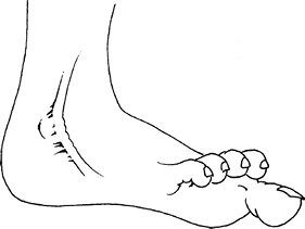
The peroneal tendons can sublux or
dislocate out of the groove with ankle motion, which can be associated
with pain and a palpable or audible snap (Fig. 1). -
Subluxation can be secondary to acute trauma or chronic in nature.
Epidemiology
Most common in adults
Incidence
Rare
Risk Factors
Athletic patients are at risk.
Genetics
No known Mendelian predisposition exists.
 |
|
Fig. 1. The peroneal tendons may subluxate anteriorly over the fibular head if the retinaculum is torn or stretched.
|
Etiology
-
Classically occurs in persons
participating in sports, such as skiing, in which the ankle is
dorsiflexed forcefully with the foot in eversion, tearing the superior
peroneal retinaculum -
May develop spontaneously in predisposed
persons, resulting from a shallow groove behind the fibula or
generalized ligamentous laxity
Associated Conditions
-
Ligamentous laxity
-
Shallow peroneal groove
-
Ankle instability
-
Varus hindfoot alignment
Signs and Symptoms
-
Lateral ankle pain with activity that does not resolve
-
Snapping of the peroneal tendons over the fibula
-
Tenderness behind the lateral malleolus
along the peroneus brevis muscle; subluxation elicited with the patient
attempting to dorsiflex the affected foot from a plantarflexed, everted
position
History
-
Running athlete or skier with lateral, painful snapping of the ankle
-
Recurrent ankle instability
Physical Exam
-
Acutely, ecchymosis and swelling are present posterior to the fibula.
-
Assess the neurovascular status of the ankle and foot.
-
Assess lateral ligament stability, including anterior drawer and inversion tilt tests.
-
Have the patient dorsiflex the ankle from a plantarflexed and everted position to reproduce posterolateral symptoms.
-
Have the patient invert and evert the foot while the examiner palpates behind the lateral malleolus muscle.
-
The peroneal tendons may palpably subluxate out of their groove and over the fibula.
-
-
Peroneal tendons normally snap or click
while in place within their sheath; only subluxation with reproduction
of symptoms or pain is diagnostic.
Tests
Lab
No laboratory tests for this condition
Imaging
-
Ankle radiographs are used to evaluate for fracture: An avulsion fracture of lateral malleolus occurs in ~10% of cases (2).
-
CT shows the shape of the peroneal groove.
-
MRI may show attenuation or tearing of retinaculum, fluid in the peroneal sheath, or a longitudinal tear of the peroneus brevis.
Diagnostic Procedures/Surgery
Diagnostic injection of the peroneal sheath with local anesthetic may help confirm tendon abnormality.
Pathological Findings
-
Possible shallow groove for the peroneal tendons behind the fibula
-
Attenuated superior peroneal retinaculum
-
Patients with chronic subluxation may have longitudinal tearing of the peroneus brevis tendon.
Differential Diagnosis
-
Ankle sprain
-
Chronic ankle instability
-
Lateral malleolar fracture
-
Posterolateral ankle impingement
-
Osteochondral talar dome fracture
General Measures
-
Attempt nonoperative treatment initially.
-
After an acute injury, cast or boot immobilization may help relieve the patient’s symptoms and reduce inflammation.
-
Cast treatment is unlikely to be successful in chronic cases.
-
NSAIDs, ice, and rest
-
An ankle brace may limit the excursion of the foot and may decrease the episodes of painful subluxation.
-
Taping or lateral crescent- or J-shaped pads also can be used to stabilize tendons in athletes.
-
-
Surgical intervention:
-
Competitive athletes usually require operative treatment early to expedite return to play.
-
If nonoperative treatment fails, surgery commonly is necessary to resolve symptoms in patients with recurrent subluxation.
-
P.321
Activity
Activity modification may reduce the occurrence of subluxation in certain patients if the subluxation is activity-specific.
Special Therapy
Physical Therapy
-
Physical therapy alone is not an effective treatment.
-
Usually used postoperatively for functional rehabilitation of the ankle
Medication
NSAIDs can be used acutely and during rehabilitation to relieve pain and to facilitate physical therapy.
Surgery
-
Surgical treatment (3,4) addresses all pathologic elements present; options include:
-
Tendon repair
-
Retinacular repair
-
Peroneal groove deepening or bone block procedures
-
Rerouting procedures deep to the CFL
-
Prognosis
-
Few cases are successfully managed nonsurgically (5).
-
Athletic individuals who require surgery
usually can return to their sporting activity, but possibly not to
their previous performance levels. -
Patients can return to activity gradually
after a 2–3-month postoperative rehabilitation regimen, but return to
full sports activity often requires 5–6 months.
Complications
Complications of surgery include recurrence, sural nerve entrapment, and sural neuroma.
Patient Monitoring
Re-education in ankle strengthening exercises is sometimes necessary.
References
1. Sobel M, Levy ME, Bohne WHO. Congenital variations of the peroneus quartus muscle: an anatomic study. Foot Ankle 1990;11:81–89.
2. Alm A, Lamke LO, Liljedahl SO. Surgical treatment of dislocation of the peroneal tendons. Injury 1975;7:14–19.
3. Kollias SL, Ferkel RD. Fibular grooving for recurrent peroneal tendon subluxation. Am J Sports Med 1997;25:329–335.
4. Zoellner G, Clancy W, Jr. Recurrent dislocation of the peroneal tendon. J Bone Joint Surg 1979; 61A:292–294.
5. Eckert WR, Davis EA, Jr. Acute rupture of the peroneal retinaculum. J Bone Joint Surg 1976; 58A:670–672.
Additional Reading
Brage ME, Hansen ST, Jr. Traumatic subluxation/ dislocation of the peroneal tendons. Foot Ankle 1992;13:423–431.
Keene JS. Foot and ankle. Section G: Tendon injuries of the foot and ankle. In: DeLee JC, Drez D, Jr, Miller MD, eds. DeLee & Drez’s Orthopaedic Sports Medicine: Principles and Practice, 2nd ed. Philadelphia: WB Saunders, 2003:2409–2446.
Codes
ICD9-CM
718.3 Peroneal tendon subluxation
Patient Teaching
-
Educate the patient about the anatomy of
the lower limb and which positions of eversion and dorsiflexion of the
ankle are most likely to reproduce the subluxation. -
The condition does not lead to degenerative joint disease, but it may:
-
Cause the ankle to give way unexpectedly
-
Lead to peroneal tendinopathy or partial tearing of the peroneus brevis
-
Activity
-
For some patients, activity modification can provide symptomatic relief.
-
Protected weightbearing after surgery in a cast or boot brace for 6 weeks, followed by a stirrup brace for 6 additional weeks
FAQ
Q: What is the typical mechanism of injury of the superior peroneal retinaculum?
A: Dorsiflexion and eversion.
Q: What examination finding is pathognomonic for peroneal subluxation?
A: Painful snapping and subluxation of tendons with dorsiflexion and eversion.
Q: How are most athletes treated for symptomatic peroneal subluxation?
A: Surgically.