
Editors: Chelly, Jacques E.
Title: Peripheral Nerve Blocks: A Color Atlas, 3rd Edition
Copyright ©2009 Lippincott Williams & Wilkins
> Table of Contents > Section IV – Ultrasound > 39 – Ultrasound Guided Femoral Nerve Block
39
Ultrasound Guided Femoral Nerve Block
Anahi Perlas
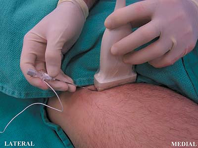
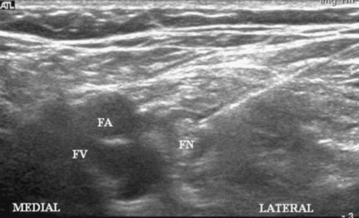
Sterile prep of the skin. The probe is placed just below the inguinal
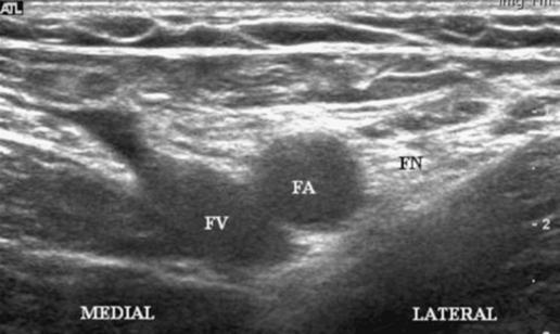
crease with the probe oriented in the axial plane. The femoral artery
is identified and the hyperechoic nerve is identified lateral to the
artery (Fig. 39-3).
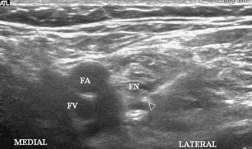
The needle is inserted at the lateral edge of the probe and advanced
until a contraction is seen in the quadriceps femoris muscle. A
stimulating current of 0.5 mA is usually appropriate to elicit a
contraction (Fig. 39-4). Injection of local anesthetic proceeds until the nerve is surrounded by a hypoechoic ring (Fig. 39-5).
P.295
 |
|
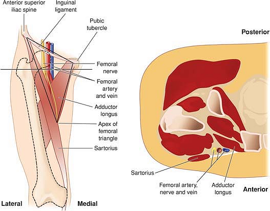
Figure 39-1.
Gross anatomy of the femoral triangle together with a drawing or section of the femoral vessels and nerve in the transverse plane. |
 |
|
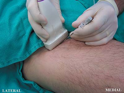
Figure 39-2. Transducer position.
|
 |
|
Figure 39-3. Femoral artery and hyperechoic nerve.
|
P.296
 |
|
Figure 39-4. A stimulating current is appropriate to elicit a contraction.
|
 |
|
Figure 39-5. Injection of local anesthetic until the nerve is surrounded by a hypoechoic ring.
|
 |
|
Figure 39-6. Catheter technique.
|
-
If a catheter technique is appropriate,
the practitioner may wish to insert the needle perpendicular to the
long axis of the probe (Fig. 39-6).
This allows advancement of the catheter into the femoral sheath without
an acute angulation. The needle is advanced toward the nerve and its
tip is inferred by tissue distortion. Once a contraction has been
elicited, local anesthetic may be injected and seen spreading around
the nerve. The catheter is then inserted into the nerve sheath.
Suggested Reading
Casati
A, Baciarello M, DiCianni S, et al. Effects of ultrasound guidance on
the minimum effective anaesthetic volume required to block the femoral
nerve. Br J Anaesth 2007;98:823–827.
A, Baciarello M, DiCianni S, et al. Effects of ultrasound guidance on
the minimum effective anaesthetic volume required to block the femoral
nerve. Br J Anaesth 2007;98:823–827.