
Editors: Chelly, Jacques E.
Title: Peripheral Nerve Blocks: A Color Atlas, 3rd Edition
Copyright ©2009 Lippincott Williams & Wilkins
> Table of Contents > Section IV – Ultrasound > 42 – Ultrasound Guided Posterior Tibial Nerve Block
42
Ultrasound Guided Posterior Tibial Nerve Block
Luiz Guilherme L. Soares
Colin McCartney
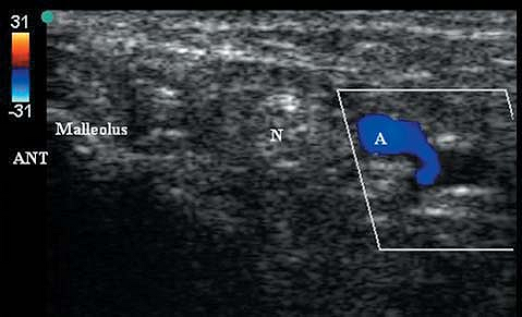
The medial malleolus is an hyperechoic curvilinear structure. The
posterior tibial artery and hyperechoic tibial nerve are found
posterior and superficial to the medial malleolar bony shadow (Figs. 42-2, 42-3).
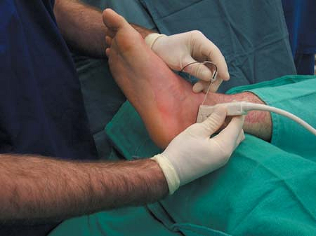
Sterile prep of the skin. A 13-MHz linear transducer is placed
posterior to the medial malleolus. The needle is placed anterior to the
probe in the longitudinal plane at an angle which is nearly tangential
to the skin. The needle is advanced with a current of 0.5 mA until
plantar flexion of the toes is elicited or until a paresthesia is
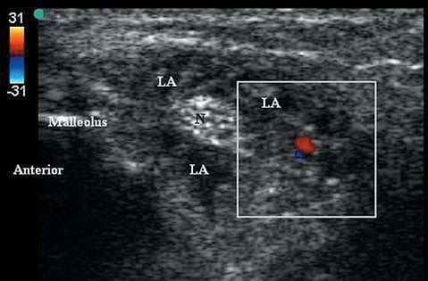
obtained. Injection of 3 to 5 mL of local anesthetic should surround
the nerve with a block hypoechoic ring (Fig. 42-4).
P.306
 |
|
Figure 42-1. Illustration of needle and probe position for ultrasound guided posterior tibial nerve block.
|
 |
|
Figure 42-2.
Sonogram (with color Doppler) of anatomy posterior to the medial malleolus. Ant, anterior; N, posterior tibial nerve; A, posterior tibial artery. |
 |
|
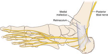
Figure 42-3. Anatomy of the nerves at the ankle.
|
 |
|
Figure 42-4.
Sonogram (with color Doppler indicating arterial pulse) demonstrating local anesthetic spread surrounding the posterior tibial nerve. N, posterior tibial nerve; LA, local anesthetic. |
P.307
-
Some practitioners prefer to perform the block without a stimulating needle.
-
For anesthesia of the forefoot, the deep
peroneal, saphenous, sural, and superficial peroneal nerves must also
be blocked. The later three are best blocked with simple infiltration
as described elsewhere in the text.