
Editors: Frassica, Frank J.; Sponseller, Paul D.; Wilckens, John H.
Title: 5-Minute Orthopaedic Consult, 2nd Edition
Copyright ©2007 Lippincott Williams & Wilkins
> Table of Contents > Clinodactyly
Clinodactyly
John J. Hwang MD
Dawn M. LaPorte MD
P.78
Description
-
Clinodactyly presents as a painless bent finger with angulation in a radial or ulnar direction (Fig. 1).
-
It is most commonly the little finger bent in a radial direction.
-
Most often, the finger has a short, delta-shaped middle phalanx.
-
This condition may be associated with mental retardation, especially when clinodactyly is severe.
-
Synonym: Bent finger
General Prevention
No evidence suggests that the deformity may be prevented or its natural history changed by intervention.
Epidemiology
-
Detected at birth
-
More common in males, in whom it is usually bilateral
Incidence
1–19.5% in otherwise normal children; least common in Caucasians (1)
Risk Factors
-
In children with Down syndrome, the incidence of clinodactyly is 35–70% (1).
-
It also is seen in children with many other syndromes, especially Kline-Felter and trisomy 18.
Genetics
-
The condition is autosomal dominant, with variable expressivity.
-
Some cases are sporadic.
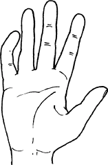 Fig. 1. Clinodactyly refers to bony angulation of a finger, usually the 5th.
Etiology
Abnormal shape of the underlying phalanx develops as a result of asymmetrical longitudinal growth.
Associated Conditions
-
Symphalangism
-
Brachydactyly (short fingers)
-
Trisomies
-
Treacher Collins syndrome
-
Silver syndrome
-
Holt-Oram syndrome
-
Prader-Willi syndrome
Signs and Symptoms
-
The finger (usually the little finger) is curved in a radial or ulnar direction.
-
Deviation can occur at the PIP joint, middle phalanx, or DIP joint (Fig. 1).
-
It is most common in the DIP joint.
-
This condition is painless.
Physical Exam
-
The angle of deviation of a finger at the PIP joint, the middle phalanx, or the DIP joint should be measured.
-
Active and passive motion at each joint should be recorded.
-
The remainder of the skeleton also should be inspected.
Tests
Lab
Chromosome analysis should be undertaken if an underlying syndrome is suspected.
Imaging
-
Conventional plain radiography of the affected finger is recommended, especially when considering surgical correction.
-
<10° of angulation is within normal limits (2).
Pathological Findings
Maldevelopment of 1 of the phalanges causes an angulation of the joint surface.
Differential Diagnosis
-
Delta phalanx (a wedge-shaped phalanx with a sloped joint surface)
-
Malunion after fractures
General Measures
-
Most cases are cosmetic problems.
-
Slight deformity does not need surgical correction.
-
Because nonoperative treatment, including
manipulation and casting, usually is futile, and patients find such
modalities difficult to tolerate, treatment choices are no intervention
or surgery. -
Surgical correction can be considered for substantial deformity persisting after the age of 6 years.
-
Surgical procedures are elective because the problem is mainly cosmetic.
Activity
No restrictions on activity
P.79
Special Therapy
Physical Therapy
Therapy may be helpful for regaining motion after surgery.
Surgery
-
Surgical procedures include osteotomy and growth plate reconstruction with a free-fat graft.
-
For a child <6 years old, a fat-graft
placement should be performed after resection of the midportion of the
continuous epiphysis and underlying physis (growth plate). -
After age 6, a simple closing osteotomy can be done easily and with few complications.
Prognosis
The prognosis is good, with no evidence of degenerative joint disease.
Patient Monitoring
Patients may monitor the angulation of the finger and return for surgical treatment if it becomes unacceptable.
References
1. Flatt AE. Crooked fingers. In: The Care of Congenital Hand Anomalies. St. Louis: Quality Medical Publishing, Inc., 1994:196–227.
2. Dudding
BA, Gorlin RJ, Langer LO. The oto-palato-digital syndrome. A new
symptom-complex consisting of deafness, dwarfism, cleft palate,
characteristic facies, and a generalized bone dysplasia. Am J Dis Child 1967;113:214–221.
BA, Gorlin RJ, Langer LO. The oto-palato-digital syndrome. A new
symptom-complex consisting of deafness, dwarfism, cleft palate,
characteristic facies, and a generalized bone dysplasia. Am J Dis Child 1967;113:214–221.
Additional Reading
McCombe D, Kay SP. Deformities of the hand and fingers. Clinodactyly. In: Green DP, Hotchkiss RN, Pederson WC, et al., eds. Green’s Operative Hand Surgery, 5th ed. Philadelphia: Elsevier Churchill Livingstone, 2005;1431–1434.
Codes
ICD9-CM
759.59 Clinodactyly
Patient Teaching
Educating patients about the excellent prognosis and benign nature of the condition is helpful.
FAQ
Q: Is splinting helpful?
A: No. The deformity can be corrected only by surgical intervention.
Q: Is surgery recommended for all patients?
A:
No. Most patients do not have a functional deficit. Surgery for
cosmetic improvement alone should be avoided because of the risks of
scarring and stiffness.
No. Most patients do not have a functional deficit. Surgery for
cosmetic improvement alone should be avoided because of the risks of
scarring and stiffness.
Q: How is clinodactyly different from camptodactyly?
A:
Clinodactyly is angulation of the digit in a radioulnar plane distal to
the MCP joint, versus camptodactyly, which is angulation in an AP plane.
Clinodactyly is angulation of the digit in a radioulnar plane distal to
the MCP joint, versus camptodactyly, which is angulation in an AP plane.
Q: What is the preferred treatment for clinodactyly?
A:
No treatment is required for mild to moderate clinodactyly. More severe
clinodactyly requires realignment of the digit through osteotomy.
No treatment is required for mild to moderate clinodactyly. More severe
clinodactyly requires realignment of the digit through osteotomy.