
Editors: Frassica, Frank J.; Sponseller, Paul D.; Wilckens, John H.
Title: 5-Minute Orthopaedic Consult, 2nd Edition
Copyright ©2007 Lippincott Williams & Wilkins
> Table of Contents > Scoliosis
Scoliosis
Paul D. Sponseller MD
Description
-
Scoliosis is a 3D curvature of the spine, best appreciated on an AP radiograph and physical examination.
-
Both thoracic and lumbar segments of the spine may be affected.
-
It is defined as a curve >10°.
-
Classification (1–3):
-
Etiology:
-
Idiopathic
-
Congenital
-
Neuromuscular
-
Connective tissue
-
Degenerative
-
-
Location (of the apex or middle of the curve):
-
Thoracic
-
Thoracolumbar
-
Lumbar
-
-
Subclassification of idiopathic scoliosis by age:
-
Infantile (<3 years)
-
Juvenile (3–10 years)
-
Adolescent (≥11 years)
-
-
Epidemiology
-
The most common type is idiopathic scoliosis.
-
Scoliosis may occur at any age.
-
The most common age at diagnosis of idiopathic scoliosis is 11–13 years (3).
Prevalence
-
Small curves of idiopathic scoliosis are almost equally prevalent in males and females.
-
Females, however, are 3–4 times more likely to develop progression of the curve.
-
In scoliosis other than the idiopathic type, less difference in gender-related prevalence is noted.
-
Prevalence of curves >10° is ~2–3% (3).
-
Prevalence of curves requiring bracing (>25°) is ~0.3% (3).
-
Prevalence of curves requiring surgery is ~1 in 1,000 (3).
Risk Factors
-
Progressive idiopathic scoliosis
-
Positive family history
-
Female gender
-
Premenarchal status
-
Paralytic scoliosis
-
Severe spinal cord injury before adolescence
-
Scoliosis in cerebral palsy, including total involvement
Genetics
Idiopathic scoliosis is transmitted as autosomal dominant, with incomplete penetrance and variable expressivity.
Etiology
-
Idiopathic scoliosis:
-
Theories about the cause of idiopathic scoliosis include a subtle connective-tissue abnormality or neurohormonal defect.
-
Causes of congenital scoliosis include hemivertebrae and fusions between vertebrae.
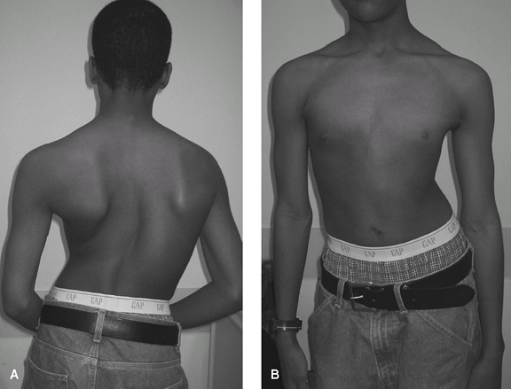 Fig. 1. Clinical appearance of severe scoliosis. A: Posterior view. B: Anterior view.
-
-
Neuromuscular scoliosis:
-
Cerebral palsy
-
Traumatic paralysis
-
Spina bifida
-
Poliomyelitis
-
Friedreich ataxia
-
Virtually any systemic neurologic condition that affects the trunk
-
-
Connective tissue-associated scoliosis:
-
Marfan syndrome
-
Ehlers-Danlos syndrome
-
NF
-
Down syndrome
-
Associated Conditions
-
Almost any systemic neurologic disorder that affects the trunk
-
Most connective-tissue disorders
Signs and Symptoms
-
Varied, depending on the location of the spine affected (Fig. 1)
-
For thoracic curves, the ribs are rotated
on the convex side, producing a “rib hump” and a more prominent scapula
on the same side. -
With thoracolumbar and lumbar curves, 1 side of the pelvis becomes more prominent, giving the appearance of a “high hip.”
-
Many, but not all, teens develop increased pain in the area of the curve.
-
Symptoms are few until adulthood, when back pain and nerve root pain may develop.
P.373
Physical Exam
-
Examine the patient, while he or she is standing, to see shoulder, rib, and hip asymmetry.
-
Measure leg lengths.
-
Perform the forward bend test with the
patient’s legs straight and observe the entire spine for asymmetry
between the right and left sides (Fig. 2).-
This test is most useful and highly sensitive, and it is used in school screening programs.
-
If asymmetry is present, measure the slope between the right and left sides of the rib cage.
-
Some patients with a positive forward bend test do not have severe scoliosis.
-
Follow-up a positive test with a radiograph if the rib slope is >6°.
-
Repeat the test if an abnormality is found.
-
-
Quantify rib prominence or a hump by a scoliometer.
-
Observe any kyphosis and lordosis.
-
Inspect the skin over the entire spine
for dimples, hair, or vascular markings, which may signal an underlying
congenital anomaly. -
Rule out ligamentous laxity.
-
Examine for café-au-lait spots or neurofibromas.
-
Perform a careful neurologic examination,
which can be practically done by observing gait and 1-legged hop and by
testing reflexes. -
Assess the patient’s physical maturity by
checking for secondary sexual characteristics such as axillary and
facial hair, skin changes, breast development. -
Measure the height for serial comparison.
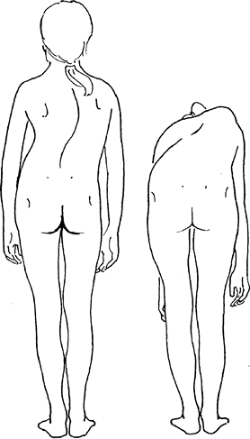 Fig. 2. The forward bend test exaggerates the rib deformity in scoliosis and allows sensitive diagnosis.
Tests
Imaging
-
Radiography:
-
Standing posteroanterior radiography of the entire spine is indicated.
-
Lateral films should be obtained if associated abnormal kyphosis is present.
-
Spine films usually show the iliac crests and allow determination of the Risser stage (3) for skeletal maturity.
-
The Risser stage is the amount of ossification of the iliac growth cartilage.
-
Risser 0, unossified, skeletally immature; Risser V, fully ossified, mature.
-
-
Presence of an open triradiate cartilage of the hip indicates that the growth spurt has not been completed.
-
-
MRI is indicated only if spinal cord disease is possible.
Pathological Findings
-
The vertebrae are rotated toward the convexity of the curve.
-
In addition, individual vertebrae are misshapen because of growth while curved.
Differential Diagnosis
-
Isolated rib rotation may occur without scoliosis.
-
Kyphosis (a curvature in the sagittal
plane only, which may be confused with scoliosis), clavicle fracture,
or Sprengel deformity may give the appearance of a “high shoulder.” -
Leg-length inequality may cause the appearance of a “high hip.”
General Measures
-
The spine in patients with scoliosis is not unstable.
-
Encourage patients to be as active as possible.
-
Physical therapy and exercise if pain or stiffness is present
-
Patients with minor curves (<25°)
should be observed if they are still growing, but they can be
discharged if skeletal maturity has been reached. -
Patients with moderate curves (25–40°) should be braced if substantial growth remains.
-
Patients with large curves (>45°):
-
See an orthopaedic surgeon to determine whether correction is indicated.
-
Surgery offered.
-
Special Therapy
Physical Therapy
-
Strengthening and stretching of abdominal and extensor muscles if pain exists
-
Not indicated for routine cases of scoliosis
-
Does not help correct the curves
Complementary and Alternative Therapies
Yoga may be helpful for back discomfort.
P.374
Surgery
-
If a curve is to be fused, 1 or 2 rods (Fig. 3) are used to correct the curve, and a bone graft is placed along the rod to cause the vertebrae to fuse.
-
Only the curved region is fused.
-
The neurologic risk is currently <1% (2).
-
Patients should be followed at least until maturity.
Prognosis
-
Pulmonary compromise, including cor
pulmonale in congenital or neuromuscular curves, occurs mainly in
patients with curves >100° (4). -
Most untreated curves >40–50° in adulthood slowly become worse (3).
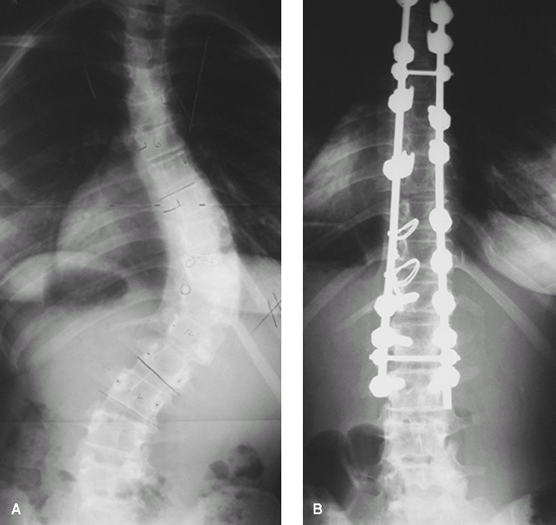 Fig. 3. Patient with severe scoliosis before (A) and after (B) posterior instrumentation and fusion.
Complications
-
Severe curves (>70°) occasionally may progress to the point where they compromise pulmonary function.
-
Curves >40° pose an increased risk of back pain in adulthood (3).
-
Surgical complications include neurologic injury (<1%), infection, and failure of the vertebrae to fuse (3).
Patient Monitoring
-
Growing children should be seen every 4–6 months, usually with radiographs.
-
Adults should be seen every 1–5 years.
-
Patients with congenital scoliosis should be monitored for associated anomalies.
References
1. Hedequist D, Emans J. Congenital scoliosis. J Am Acad Orthop Surg 2004;12:266–275.
2. Lenke
LG, Edwards CC, II, Bridwell KH.The Lenke classification of adolescent
idiopathic scoliosis: How it organizes curve patterns AS a template to
perform selective fusions of the spine. Spine 2003;28:S199–S207.
LG, Edwards CC, II, Bridwell KH.The Lenke classification of adolescent
idiopathic scoliosis: How it organizes curve patterns AS a template to
perform selective fusions of the spine. Spine 2003;28:S199–S207.
3. Newton PO, Wenger DR.Idiopathic scoliosis. In: Morrissy RT, Weinstein SL, eds. Lovell and Winter’s Pediatric Orthopaedics, 6th ed. Philadelphia: Lippincott Williams & Wilkins, 2006:693–762.
4. Weinstein
SL, Dolan LA, Spratt KF, et al. Health and function of patients with
untreated idiopathic scoliosis: A 50-year natural history study. JAMA 2003;289:559–567.
SL, Dolan LA, Spratt KF, et al. Health and function of patients with
untreated idiopathic scoliosis: A 50-year natural history study. JAMA 2003;289:559–567.
P.375
Codes
ICD9-CM
-
737.30 Idiopathic scoliosis
-
754.2 Congenital scoliosis
Patient Teaching
-
Instruct patients in the general guidelines and treatment options.
-
Remind parents of the genetic nature of
the condition so that relatives and young siblings with scoliosis may
be detected while bracing is still an option.
Prevention
-
Curve worsening may be effectively prevented in growing children by use of a brace.
-
It must be worn 18–23 hours per day.
-
This intervention is effective in ~75% of patients (2).
-
Bracing does not correct curves.
-
FAQ
Q: What is the cause of scoliosis?
A: It is not known. It often is passed along in families. It may be a subtle disorder of balance or spinal growth.
Q: Can physical therapy or exercises slow or halt the worsening of the curve?
A: No evidence suggests that it can.
Q: Can bracing correct scoliosis?
A: It rarely produces any permanent correction.
Q: Does scoliosis affect internal organs?
A:
It is associated with decreased pulmonary function in curves ≥70°.
There is little documentation of effects on cardiac, gastrointestinal,
or genitourinary function.
It is associated with decreased pulmonary function in curves ≥70°.
There is little documentation of effects on cardiac, gastrointestinal,
or genitourinary function.