
Editors: Frassica, Frank J.; Sponseller, Paul D.; Wilckens, John H.
Title: 5-Minute Orthopaedic Consult, 2nd Edition
Copyright ©2007 Lippincott Williams & Wilkins
> Table of Contents > Olecranon Fracture
Olecranon Fracture
Andrew M. Richards MD
Jinsong Wang MD
Description
-
The olecranon:
-
Is the proximal bony projection of the ulna at the elbow
-
Articulates with the trochlea of the distal humerus to form the ulnohumeral portion of the elbow joint:
-
This articulation is responsible for flexion and extension of the elbow joint.
-
-
Is the insertion site of the triceps tendon
-
-
Contraction of the triceps pulling on the olecranon produces extension of the elbow.
-
Fracture of the olecranon disrupts the extensor mechanism of the elbow, which is critical to arm function (Fig. 1).
-
If these intra-articular fractures are not repaired anatomically, posttraumatic arthritis will develop.
-
Classification:
-
By characteristic
-
Nondisplaced or displaced
-
Transverse or oblique
-
Simple or comminuted
-
-
By the amount of involvement of the articular surface in the olecranon notch (1)
-
Type 1: Proximal 1/3 of the notch
-
Type 2: Middle 1/3
-
Type 3: Distal 1/3
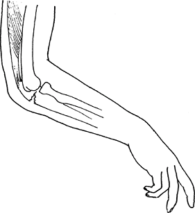 Fig. 1. Olecranon fractures involve the proximal ulna and enter the elbow joint.
-
-
General Prevention
Elbow pads should be used for contact sports and skating (2).
Epidemiology
-
Olecranon fracture may occur after a fall, sports injury, or motor vehicle accident.
-
Elderly or osteoporotic patients may sustain an olecranon fracture after a low-energy fall.
-
Olecranon fractures occur less frequently
in children than in adults because in children, this type of force is
more likely to produce a fracture of the distal humerus (supracondylar)
(3).
Risk Factors
Activities with high fall rates, such as inline skating, have a high risk of elbow fractures.
Etiology
-
The most common mechanisms of injury are:
-
Fall on the outstretched hand with the elbow in flexion
-
Direct blow to the tip of the elbow
-
Associated Conditions
-
Elbow dislocation
-
Radial head fracture
-
Triceps avulsion
-
Elbow instability
-
Neurologic damage (ulnar, median, and radial nerves)
Signs and Symptoms
-
Pain, swelling, ecchymosis, and deformity of the elbow
-
Inability to extend the elbow
-
These injuries often are associated with radial head fractures and elbow dislocations.
History
History should be obtained to determine the mechanism of injury.
Physical Exam
-
Initial evaluation should include
particular attention to the function of the triceps muscle, the
function of the radial and ulnar nerves, and the vascular status of the
upper extremity. -
Palpable defect often detected on posterior elbow
-
Test for triceps mechanism integrity by asking the patient to actively extend the elbow against gravity.
-
Examine the degree of soft-tissue injury and determine whether the fracture is open or closed.
Tests
Lab
Before surgery, routine preoperative laboratory tests are performed, depending on the age and medical condition of the patient.
Imaging
-
Radiography:
-
Obtain AP and lateral radiographs of the elbow.
-
A radiocapitellar view may be helpful if there appears to be an associated radial head injury.
-
-
Obtain a CT scan of the elbow to evaluate complex fracture dislocations of the elbow.
Differential Diagnosis
-
Distal humerus fracture
-
Elbow dislocation
-
Radial head fracture
P.281
General Measures
-
Nondisplaced fractures are treated with
immobilization in an above-the-elbow splint or cast with the elbow in
90° of flexion for 4 weeks. -
Follow-up radiography is necessary 7–10 days after injury to ensure that the fracture has not displaced.
-
Displaced fractures usually require surgical fixation to restore extensor mechanism function.
Activity
-
After fracture, the arm should be splinted comfortably at 90° of flexion, and a sling should be offered.
-
Patients should be encouraged to move the hand and shoulder to prevent stiffness.
Nursing
-
Patients in a sling should be helped with personal hygiene in the armpit area.
-
Care should be taken that the splint is comfortable and does not rub.
Special Therapy
Physical Therapy
-
Initially, strengthening and gentle
passive ROM exercises to address the common sequelae (decreased ROM and
muscle strength) of elbow immobilization after olecranon fractures -
Gradually progress to active ROM exercises when radiographic evidence shows callus formation and fracture healing.
-
Motion of the ipsilateral shoulder and hand should be encouraged.
Medication
First Line
Narcotic medicines may be necessary for pain relief after fracture.
Surgery
-
Most olecranon fractures are treated surgically because they disrupt the extensor mechanism.
-
Fractures must be repaired anatomically to restore the joint surface.
-
Choice of repair technique depends on the
size of the fragment, the direction of the fracture line, and the
amount of fracture comminution.-
Stable, nondisplaced fractures may be treated nonoperatively.
-
Displaced fractures require open reduction and fixation.
-
Open fractures should be treated with surgical débridement and fixation.
-
Small avulsion fractures are treated with excision and repair.
-
The triceps tendon is sutured back to the olecranon.
-
Bone removal does increase joint pressures and, if possible, bony fixation should be attempted (4).
-
-
Transverse fractures are repaired using 2 Kirschner wires and a tension band wire to resist the pull of the triceps muscle.
-
Oblique fractures may be repaired using interfragmentary screw fixation and an accompanying tension band wire.
-
Fixation with screws has been shown to be stronger than that with tension band wires (5).
-
-
Severely comminuted fractures are not amenable to tension wiring and require fixation with plating.
-
A 3.5-mm reconstruction plate can be bent to fit the olecranon, or a precontoured plate can be used.
-
-
Fractures with bone loss may require bone grafting to repair defects (6).
-
Disposition
After surgery, the arm is splinted until skin healing occurs, and then early ROM exercises are started.
Prognosis
-
The prognosis is good for >90% of patients (7).
-
Fractures with more articular involvement and more severity have been shown to have worse outcomes (8).
Complications
-
Painful hardware requiring removal occurs in 20–80% of patients (9,10).
-
Radial neuropathy
-
Ulnar neuropathy
-
Flexion contracture
-
Elbow arthritis
-
Malunion
-
Nonunion
Patient Monitoring
-
Patients should be monitored until fracture healing is observed radiographically.
-
ROM of the elbow and strength of the arm should be monitored.
References
1. Crenshaw
AH, Jr. Fractures of the shoulder, arm, and forearm. In: Canale ST, ed.
Campbell’s Operative Orthopaedics, 10th ed. St. Louis: Mosby,
2003:2985–3069.
AH, Jr. Fractures of the shoulder, arm, and forearm. In: Canale ST, ed.
Campbell’s Operative Orthopaedics, 10th ed. St. Louis: Mosby,
2003:2985–3069.
2. Jerosch
J, Heidjann J, Thorwesten L, et al. Injury pattern and acceptance of
passive and active injury prophylaxis for inline skating. Knee Surg Sports Traumatol Arthrosc 1998;6:44–49.
J, Heidjann J, Thorwesten L, et al. Injury pattern and acceptance of
passive and active injury prophylaxis for inline skating. Knee Surg Sports Traumatol Arthrosc 1998;6:44–49.
3. Carson S, Woolridge DP, Colletti J, et al. Pediatric upper extremity injuries. Pediatr Clin North Am 2006;53:41–67.
4. Moed
BR, Ede DE, Brown TD. Fractures of the olecranon: an in vitro study of
elbow joint stresses after tension-band wire fixation versus proximal
fracture fragment excision. J Trauma 2002;53:1088–1093.
BR, Ede DE, Brown TD. Fractures of the olecranon: an in vitro study of
elbow joint stresses after tension-band wire fixation versus proximal
fracture fragment excision. J Trauma 2002;53:1088–1093.
5. Hutchinson DT, Horwitz DS, Ha G, et al. Cyclic loading of olecranon fracture fixation constructs. J Bone Joint Surg 2003;85A:831–837.
6. Ikeda
M, Fukushima Y, Kobayashi Y, et al. Comminuted fractures of the
olecranon. Management by bone graft from the iliac crest and multiple
tension-band wiring. J Bone Joint Surg 2001;83B:805–808.
M, Fukushima Y, Kobayashi Y, et al. Comminuted fractures of the
olecranon. Management by bone graft from the iliac crest and multiple
tension-band wiring. J Bone Joint Surg 2001;83B:805–808.
7. Karlsson MK, Hasserius R, Karlsson C, et al. Fractures of the olecranon: a 15- to 25-year followup of 73 patients. Clin Orthop Relat Res 2002;403:205–212.
8. Rommens PM, Kuchle R, Schneider RU, et al. Olecranon fractures in adults: factors influencing outcome. Injury 2004;35:1149–1157.
9. Bailey CS, MacDermid J, Patterson SD, et al. Outcome of plate fixation of olecranon fractures. J Orthop Trauma 2001;15:542–548.
10. Karlsson
MK, Hasserius R, Besjakov J, et al. Comparison of tension-band and
figure-of-eight wiring techniques for treatment of olecranon fractures.
J Shoulder Elbow Surg 2002;11: 377–382.
MK, Hasserius R, Besjakov J, et al. Comparison of tension-band and
figure-of-eight wiring techniques for treatment of olecranon fractures.
J Shoulder Elbow Surg 2002;11: 377–382.
Additional Reading
Hak DJ, Golladay GJ. Olecranon fractures: treatment options. J Am Acad Orthop Surg 2000;8:266–275.
Nork SE, Jones CB, Henley MB. Surgical treatment of olecranon fractures. Am J Orthop 2001;30:577–586.
Codes
ICD9-CM
-
813.01 Closed olecranon fracture
-
813.11 Open olecranon fracture
Patient Teaching
-
Even with a perfect reduction, patients may still have decreased ROM.
-
Patients often lose 5–10° of extension.
Activity
Until fracture healing, patients should limit lifting with the arm.
Prevention
individuals involved in sports with high risks of falls, such as skating or rollerblading, should use elbow pads (2).
FAQ
Q: How long do olecranon fractures take to heal?
A: Fracture healing usually occurs in 2–3 months. Additional therapy is required to strengthen the arm over the next 3 months.