
VI – Continuous Nerve Blocks in Infants and Children > 60 –
Continuous Sciatic Nerve Blocks
Anesthesia and postoperative analgesia for surgery below the knee;
postoperative physiotherapy and complex regional pain syndrome.
The depth of the sacral plexus at this level has not yet been
investigated in children. The plexus can be found at 15- to 50-mm depth.
A line is drawn between the posterior superior iliac spine and the
ischial tuberosity; this line is divided in three. In an appropriately
anesthetized/sedated child, the needle is introduced perpendicular to
the skin, at the junction of the cranial third and the caudal
two-thirds of that line. A sciatic nerve stimulation is elicited. With
an appropriate muscle response still present at a current of 0.5 mA and
after negative aspiration for blood the appropriate amount of local
anesthetic solution is slowly injected. Maintaining the introducer
needle in the same position, the catheter is threaded 2 cm beyond the
needle tip. The introducer needle is removed and the catheter is
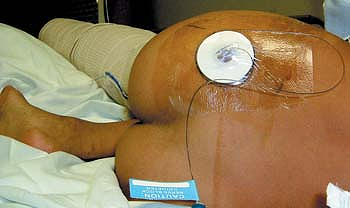
secured in place with benzoin and a transparent adhesive dressing (Fig. 60-1).
|
Table 60-1. Maximum Initial Bolus Volume of Ropivacaine 0.2%—Parasacral Approach
|
||||||||||||||||||||
|---|---|---|---|---|---|---|---|---|---|---|---|---|---|---|---|---|---|---|---|---|
|
 |
|
Figure 60-1. Parasacral catheter placement.
|
-
The needle is introduced perpendicular to the skin or 30° in the cranial direction.
-
At this level the sciatic nerve is close to the internal iliac vessels (sciatic vascular trunk).
-
If there is bone contact, the needle
needs to be inserted more caudally on the line between the posterior
superior iliac spine and the ischial tuberosity. -
A stimulating catheter can be used in older children.
C, Pirat Ph, Raux O, et al. Perioperative continuous peripheral nerve
blocks with disposable infusion pumps in children: a prospective
descriptive study. Anesth Analg 2003;97:687–690.
Anesthesia and postoperative analgesia for surgery below the knee;
postoperative physiotherapy and complex regional pain syndrome.
A line is drawn between the anterior superior iliac spine and the pubic
tubercle (inguinal ligament line). Next, a parallel line is drawn
passing
through
the greater trochanter. A perpendicular line is drawn at the junction
of the medial third and the lateral two-thirds of these lines. The
intersection of the perpendicular line and the line passing through the
greater trochanter presents the site of needle insertion. In an
appropriately anesthetized/sedated child, the insulated needle,
connected to a nerve stimulator (1.5 mA, 2 Hz, 0.1 ms) is introduced
perpendicular to the skin. Within 3 to 11 cm, a sciatic nerve
stimulation is elicited. With an appropriate muscle response still
present at a current of 0.5 mA and after negative aspiration for blood
the appropriate amount of local anesthetic solution is slowly injected.
Maintaining the introducer needle in the same position, the catheter is
threaded 2 cm beyond the needle tip. The introducer needle is removed
and the catheter is secured in place with benzoin and a transparent
adhesive dressing.
 |
|
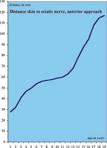
Figure 60-2. Distance skin to sciatic nerve, anterior approach distance.
|
|
Table 60-2. Maximum Initial Bolus Volume of Ropivacaine 0.2%—Anterior Approach
|
||||||||||||||||||||
|---|---|---|---|---|---|---|---|---|---|---|---|---|---|---|---|---|---|---|---|---|
|
-
A femoral nerve stimulation may be elicited within a depth of 1 to 4 cm.
-
The needle has to be withdrawn very carefully in order not to displace the catheter.
-
If the femur is contacted the needle has to be introduced more medially.
-
To the authors’ knowledge this approach
was not yet used for continuous infusion in smaller children. The
youngest patient with continuous infusion was 15 years old (43 kg). -
A stimulating catheter can be used in older children.
C, Pirat Ph, Raux O, et al. Perioperative continuous peripheral nerve
blocks with disposable infusion pumps in children: a prospective
descriptive study. Anesth Analg 2003;97:687–690.
Anesthesia and postoperative analgesia for surgery below the knee;
postoperative physiotherapy and complex regional pain syndrome.
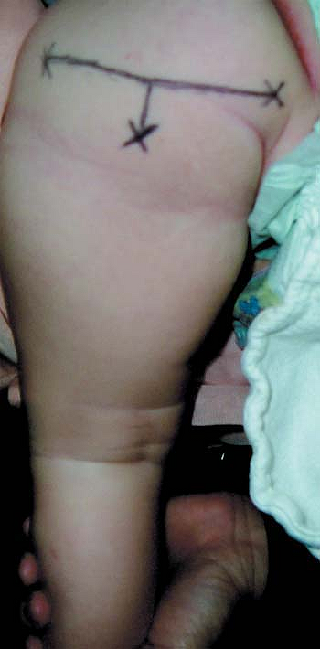
A line is drawn between the greater trochanter of the femur and the
ischial tuberosity. At its midpoint a 2- to 5-cm long, perpendicular,
subgluteal line is drawn. The end point of this perpendicular line
presents the site of needle insertion. In an appropriately
anesthetized/sedated child, the insulated needle, connected to a nerve
stimulator (1.5 mA, 2 Hz, 0.1 ms) is introduced perpendicular to the
skin. Within
2
to 6 cm, a sciatic nerve stimulation is elicited. With an appropriate
muscle response still present at a current of 0.5 mA and after negative
aspiration for blood the appropriate amount of local anesthetic
solution is slowly injected. Maintaining the introducer needle in the
same position, the catheter is threaded 2 cm beyond the needle tip. The
introducer needle is removed and the catheter is secured in place with
benzoin and a transparent adhesive dressing (Fig. 60-4).
 |
|
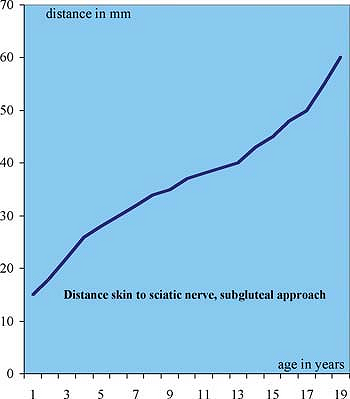
Figure 60-3. Distance skin to sciatic nerve, subgluteal approach.
|
|
Table 60-3. Maximum Initial Bolus Volume of Ropivacaine 0.2%—Subgluteal Approach
|
||||||||||||||||||||
|---|---|---|---|---|---|---|---|---|---|---|---|---|---|---|---|---|---|---|---|---|
|
-
A groove can be felt between the
semitendinous muscle and the biceps femoris muscle; the sciatic nerve
is located deep in that groove. -
A local stimulation of the biceps femoris
muscle can be elicited, but is not sufficient; only a plantar flexion
of the foot with flexion of the toes and/or a dorsiflexion and eversion
of the foot indicate correct needle placement. -
The ultrasound can be used to localize
the sciatic nerve, to position the needle, and to verify that the local
anesthetic is injected via the catheter around the nerve. -
A stimulating catheter can be used in older children.
 |
|
Figure 60-4. Placement subgluteal catheter.
|
C, Pirat Ph, Raux O, et al. Perioperative continuous peripheral nerve
blocks with disposable infusion pumps in children: a prospective
descriptive study. Anesth Analg 2003;97:687–690.
Geffen G, Gielen M. Ultrasound-guided subgluteal sciatic nerve blocks
with stimulating catheters in children: a descriptive study. Anesth Analg 2006;103(2):328–333.
Anesthesia and postoperative analgesia for surgery below the knee;
postoperative physiotherapy and complex regional pain syndrome.
 |
|
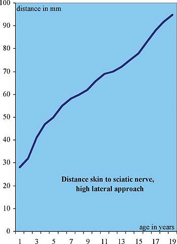
Figure 60-5. Distance skin to sciatic nerve, high lateral approach.
|
|
Table 60-4. Maximum Initial Bolus Volume of Ropivacaine 0.2%—High Lateral Approach
|
||||||||||||||||||||
|---|---|---|---|---|---|---|---|---|---|---|---|---|---|---|---|---|---|---|---|---|
|
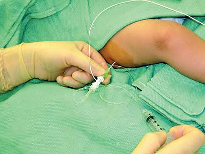
The greater trochanter of the femur is identified and a line is drawn
along the axis of the femur. In an appropriately anesthetized/sedated
child, the insulated needle, connected to a nerve stimulator (1.5 mA, 2
Hz, 0.1 ms) is introduced perpendicular to the skin, at the level of
the gluteal fold and 1 to 2 cm below the long axis of the femur. Within
2 to 10 cm, a sciatic nerve stimulation is elicited. With an
appropriate muscle response still present at a current of 0.5 mA and
after negative aspiration for blood the appropriate amount of local
anesthetic solution is slowly injected. Maintaining the introducer
needle in the same position, the catheter is threaded 2 cm beyond the
needle tip. The introducer needle is removed and the catheter is
secured in place with benzoin and a transparent adhesive dressing (Figs. 60-6, 60-7).
-
If bone contact occurs, the needle needs to be redirected posteriorly.
-
Ultrasound can be used to localize the
sciatic nerve, to position the needle, and to verify that the local
anesthetic is injected via the catheter around the nerve. -
Local stimulation of the biceps femoris
muscle can be elicited, but is not sufficient; only a plantar flexion
of the foot with flexion of the toes and/or a dorsiflexion and eversion
of the foot indicate correct needle placement. -
A stimulating catheter can be used in older children.
 |
|
Figure 60-6. Catheter placement, high lateral sciatic.
|
 |
|
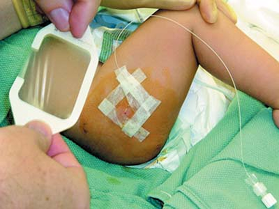
Figure 60-7. Catheter secured, high lateral sciatic approach.
|
C, Pirat Ph, Raux O, et al. Perioperative continuous peripheral nerve
blocks with disposable infusion pumps in children: A prospective
descriptive study. Anesth Analg 2003;97:687–690.
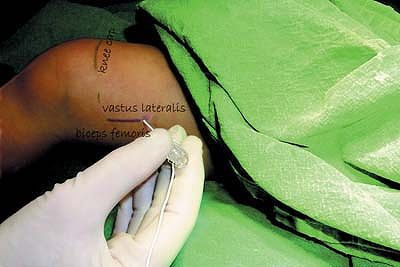
A pillow is placed under the leg. The groove between the biceps femoris
muscle and vastus lateralis muscle is identified and marked, as is the
cranial border of the patella. In an appropriately anesthetized/sedated
child, the insulated needle, connected to a nerve stimulator (1.5 mA, 2
Hz, 0.1 ms) is introduced perpendicularly to the skin, at 3 to 10 cm
cephalad from the patella border on the line along the groove. Within 2
to 7 cm, a sciatic nerve stimulation is elicited. With an appropriate
muscle response still present at a current of 0.5 mA and after negative
aspiration for blood the appropriate amount of local anesthetic
solution is slowly injected. Maintaining the introducer needle in the
same position, the catheter is threaded 2 cm beyond the needle tip. The
introducer needle is removed and the catheter is secured in place with
benzoin and a transparent adhesive dressing (Fig. 60-9).
-
If bone contact occurs, the needle needs to be redirected posteriorly.
-
Ultrasound can be used to localize the
sciatic nerve, to position the needle, and to verify that the local
anesthetic is injected via the catheter around the nerve. -
A plantar flexion of the foot with
flexion of the toes and/or a dorsiflexion and eversion of the foot
indicate correct needle placement. -
A stimulating catheter can be used in older children.
|
Table 60-5. Maximum Initial Bolus Volume of Ropivacaine 0.2%—Lateral Popliteal Approach
|
||||||||||||||||||||
|---|---|---|---|---|---|---|---|---|---|---|---|---|---|---|---|---|---|---|---|---|
|
 |
|
Figure 60-8. Lateral popliteal approach, landmarks.
|
 |
|
Figure 60-9. Catheter placed, lateral sciatic approach.
|
C, Pirat Ph, Raux O, et al. Perioperative continuous peripheral nerve
blocks with disposable infusion pumps in children: a prospective
descriptive study. Anesth Analg 2003;97:687–690.
A, Vlogka JD. A comparision of the posterior versus lateral approaches
to the block of the sciatic nerve in the popliteal fossa. Anesthesiology 1988;88:1480–1486.
JD, Hadzic A, Kitain E, et al. Anatomic considerations for sciatic
nerve block in the popliteal fossa through the lateral approach. Reg Anesth 1996;21:414–418.
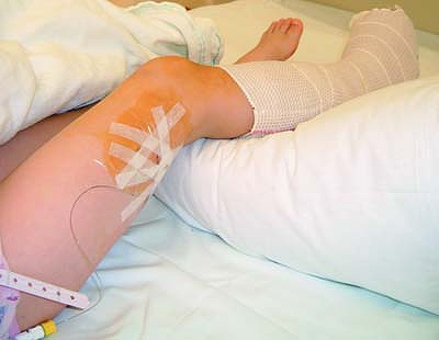
The leg is flexed to identify the popliteal crease. A line is drawn at
the level of the popliteal crease between the semitendinosus and
semimembranous tendons medially and the biceps femoris tendon
laterally. At its midpoint a perpendicular line is drawn in a cephalad
direction; this line divides the popliteal triangle. In an
appropriately anesthetized/sedated child, the insulated needle,
connected to a nerve stimulator (1.5 mA, 2 Hz, 0.1 ms) is introduced at
a 45° angle to the skin, at 2 to 5 cm proximally and 1 cm laterally to
the perpendicular line, and advanced in a cephalad direction. Within
1.5 to 6 cm, a sciatic nerve stimulation is elicited. With an
appropriate muscle response still present at a current of 0.5 mA and
after negative aspiration for blood the appropriate amount of local
anesthetic solution is slowly injected. Maintaining the introducer
needle in the same position, the catheter is threaded 2 cm beyond the
needle tip. The introducer needle is removed and the catheter is
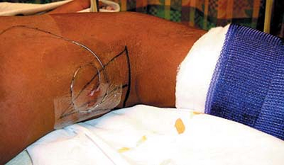
secured in place with benzoin and a transparent adhesive dressing (Figs. 60-10, 60-11, 60-12).
-
Ultrasound can be used to localize the
sciatic nerve, to position the needle, and to verify that the local
anesthetic is injected via the catheter around the nerve. -
A plantar flexion of the foot with
flexion of the toes and/or a dorsiflexion and eversion of the foot
indicate correct needle placement. -
A stimulating catheter can be used in older children.
|
Table 60-6. Maximum Initial Bolus Volume of Ropivacaine 0.2%—Posterior Popliteal Approach
|
||||||||||||||||||||
|---|---|---|---|---|---|---|---|---|---|---|---|---|---|---|---|---|---|---|---|---|
|
 |
|
Figure 60-10. Posterior popliteal sciatic approach.
|
 |
|
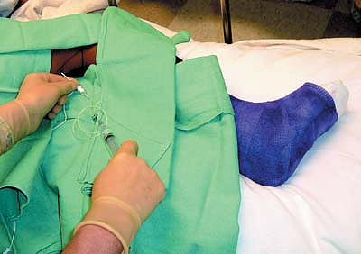
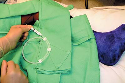
Figure 60-11. Catheter placement.
|
 |
|
Figure 60-12. Catheter secured.
|
C, Bringuier S, Nicolas F, et al. Continuous epidural block versus
continuous popliteal nerve block for postoperative pain relief after
major podiatric surgery in children: a prospective, comparative
randomized study. Anesth Analg 2006;102:744–749.
C, Motais F, Ricard C, et al. Continuous peripheral nerve blocks at
home for treatment of recurrent complex regional pain syndrome 1 in
children. Anesthesiology 2005;102:387–391.
C, Pirat Ph, Raux O, et al. Perioperative continuous peripheral nerve
blocks with disposable infusion pumps in children: a prospective
descriptive study. Anesth Analg 2003;97:687–690.
A, Vlogka JD. A comparison of the posterior versus lateral approaches
to the block of the sciatic nerve in the popliteal fossa. Anesthesiology 1988;88:1480–1486.
FJ, Gouverneur JM, Gribomont BF. Continuous popliteal sciatic nerve
block: an original technique to provide postoperative analgesia after
foot surgery. Anesth Analg 1997;84:383–386.