
Editors: Chelly, Jacques E.
Title: Peripheral Nerve Blocks: A Color Atlas, 3rd Edition
Copyright ©2009 Lippincott Williams & Wilkins
> Table of Contents > Section II – Single-Injection Peripheral Blocks > C – Miscellaneous Blocks > 21 – Bier Block
21
Bier Block
Sukhijinder Dhother
Next, wrap the upper arm with a 10-cm Webril (Kendall Co., Boston, MA)
padding band, and then apply an appropriate-sized pneumatic double cuff
to the upper arm. The lower end of the distal cuff is secured with foam
tape (10 cm) to prevent migration. Next, check the integrity of both
cuffs by sequential inflation and deflation. The arm is elevated, and
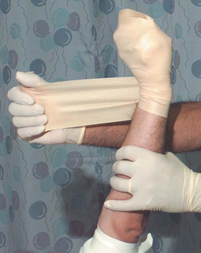
the patient is asked to close the hand firmly on a padding bandage. A
10-cm latex bandage is wrapped from distal to proximal to provide
exsanguination of the limb (Fig. 21-2).
The proximal (upper) tourniquet is inflated to 100 mm Hg above systolic
pressure, or at least 250 mm Hg, and then the latex bandage is removed.
Absence of brachial and distal pulses indicates correct function. Fifty
milliliters of 0.5% lidocaine is injected slowly. The cannula is
removed and the arm prepared for surgery. Adequate sensory anesthesia
should be achieved in approximately 10 to 15 minutes.
-
To prevent local anesthetic toxicity reactions, the tourniquet should remain in place for at least 45 minutes.P.189
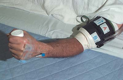 Figure 21-1. Insert and secure an intravenous cannula.
![]() Figure 21-2. A 10-cm latex bandage is wrapped from distal to proximal to provide exsanguination of the limb.
Figure 21-2. A 10-cm latex bandage is wrapped from distal to proximal to provide exsanguination of the limb. -
The patient should be asked to firmly close the hand around a 5-cm roll of tape. This allows better exsanguination of the hand.
-
It is best to provide a tourniquet effect proximal to the patient’s wrist by wrapping the operator’s hand around it.
-
Inject half the anesthetic solution and
observe the distension and backward filling of the veins. The remaining
volume is then injected after releasing the wrist to allow a proximal
spread up to the level of the proximal cuff. -
When the patient begins experiencing
tourniquet pain, it is time to inflate the distal cuff and deflate the
proximal cuff, up to an additional 30 minutes. Do not deflate the cuffs
until at least 20 minutes after injection. Be vigilant for signs of
systemic absorption during tourniquet release. -
0.2% ropivacaine or 0.125%
levobupivacaine has been suggested as an alternative to 0.5% lidocaine.
Their use prolongs the duration of the block. -
The same technique can be used for lower extremity surgery below the knee.
Suggested Readings
Atanassoff
PG, Aouad R, Hartmannsgruber MW, et al. Levobupivacaine 0.125% and
lidocaine 0.5% for intravenous regional anesthesia in volunteers. Anesthesiology 2002;97:325–328.
PG, Aouad R, Hartmannsgruber MW, et al. Levobupivacaine 0.125% and
lidocaine 0.5% for intravenous regional anesthesia in volunteers. Anesthesiology 2002;97:325–328.
Brown DL. Atlas of regional anesthesia. Philadelphia: WB Saunders, 1992.
Holmes CM. Intravenous regional anesthesia: a useful method of producing analgesia of the limbs. Lancet 1963;1:245.
Mulroy MF. Regional anesthesia: an illustrated procedural guide, 3rd ed. Philadelphia: Lippincott Williams & Wilkins, 2002.