The Elbow


9 – The Elbow

Chapter 9
The Elbow
Simon Blease
David W. Stoller
Marc R. Safran
Arthur E. Li
Russell C. Fritz
Magnetic resonance (MR) imaging is now well established as a key imaging tool in the assessment of the elbow. Superior depiction of muscles, ligaments, and tendons and the ability to directly visualize nerves, bone marrow, and hyaline cartilage are advantages of MR imaging relative to conventional imaging techniques. The origin of elbow pain may be complex, and the ability of MR to accurately depict soft-tissue pathology as well as osseous damage is critical to its usefulness. Ongoing improvements in magnet technology and surface coil design and newer pulse sequences have resulted in faster acquisition and higher-resolution images. Clinical experience has confirmed the utility of MR imaging in detecting and characterizing disorders of the elbow in a noninvasive fashion.1,2,3,4,5,6,7,8
The elbow is a complex joint, but unlike the shoulder or knee the complexity arises from highly adapted bony architecture rather than sophisticated soft-tissue support. Although arthroscopy has a well-defined role, arthroscopic studies have not added significantly to the understanding of the anatomy and pathology of the elbow.
As in the knee, many problems in the elbow arise from increased varus and valgus stress delivered in acute or, more commonly, chronic situations. Conditions amenable to characterization on MR imaging include:
  • The sequelae of medial traction and lateral compression from valgus stress, including:
    • Medial collateral ligament (MCL) injury
    • Common flexor tendon pathology
    • Medial traction spurs
    • Ulnar neuropathy
    • Osteochondritis dissecans
  • Lateral collateral ligament injury
  • Lateral epicondylitis
  • Posttraumatic osseous abnormalities such as:
    • Radiographically occult fractures
    • Stress fractures
    • Bone contusions
    • Apophyseal avulsions
  • Cartilaginous extension of fractures in children (a condition difficult to evaluate with computed tomography [CT])
  • Intra-articular loose bodies and capsular pathology, especially if fluid or contrast material is present within the elbow joint
  • P.1464


  • Biceps and triceps tendon injuries
  • Entrapment neuropathies
  • Bursitis
  • Arthropathies
  • Soft-tissue masses about the elbow
Imaging Techniques and Protocols for the Elbow
Extremity Coils and Patient Positioning
There are a variety of technical challenges to obtaining optimal images of the elbow. It is an off-center structure, which makes it difficult to perform isocenter imaging. In a conventional magnet, the elbow is scanned with the patient in a supine position with the arm at the side. Injuries commonly produce a fixed flexion deformity (due to painful limitation of motion or structural block), however, making positioning the patient in a conventional supine posture difficult. A surface coil is essential for obtaining high-resolution images. Depending on the size of the patient and the size of the surface coil relative to bore of the magnet, it may be necessary to scan the patient in a prone position with the arm extended overhead (the so-called Superman position, also used sometimes in wrist imaging). In general, the prone position is less well tolerated and results in a greater number of motion-degraded studies. The elbow should be scanned in as comfortable position as possible to avoid such motion artifact. The elbow is typically extended and the wrist is placed in a neutral position. Patients who cannot extend the elbow are more difficult to position, and obtaining optimal imaging results requires more time and skill. Taping a vitamin E capsule or other marker to the skin at the site of tenderness or at the site of a palpable mass is useful to ensure that the area of interest has been included in the study, especially when there is no pathology identified on the images. Structures that curve in the extended position, especially the biceps tendon, may be better imaged by adopting the “Superman” position but with the elbow in 90° of flexion.9
With open MR scanners, the joint can generally be placed at the isocenter regardless of patient size or joint deformity. The patient can be positioned for optimal comfort rather than having to adopt a potentially painful posture. Increasing field strength (to 3 Tesla [T] or above) and the use of microscopy surface coils10 promise a much more detailed view of the joint.
Pulse Parameters
Excellent images may be obtained with both low- and high-field strength MR systems. With high-field systems (Fig. 9.1), proton density (PD) and fat-suppressed fast spin-echo (FS PD FSE) images are typically obtained in the axial and sagittal planes. T1- and PD-weighted and FS PD FSE or STIR sequences usually are obtained in the coronal plane. Although the STIR sequence has a relatively poor signal-to-noise ratio because of the suppression of signal from fat, pathologic changes are often more conspicuous due to the effects of additive T1 and T2 contrast. FS PD FSE sequences must be added to the FS T1-weighted sequence if arthrographic gadolinium contrast has been used. FS T1-weighted images alone are not sensitive to marrow or muscle edema, tendinosis, or cysts. FS PD FSE sequences may also be helpful in visualizing hyaline cartilage defects, especially in the coronal plane.
If a low-field open scanner is used, specific pulse sequences must be adopted to achieve optimal images. The reduced cross-talk between slices and better contrast resolution allow the successful use of gradient echo (GRE) T1-weighted 3D acquisitions to provide excellent hyaline cartilage depiction. In general, FSE T1 and fast-STIR sequences are the mainstay of elbow imaging. FSE T2 sequences suffer from reduced soft-tissue discrimination at field strengths of less than 0.5 T. Higher-field-strength open systems (0.7 T and above) are better able to generate acceptable images with sequences derived from conventional higher-field-strength scanners.
In general, axial images should extend from the distal humeral metaphysis to the radial tuberosity. The common flexor and extensor origins from the medial and lateral humeral epicondyles and the biceps insertion on the radial tuberosity are routinely imaged with this coverage. Images are usually obtained with 3- or 4-mm thick slices using a sequence with a long repetition time (TR). Coronal images are angled parallel to a line through the humeral epicondyles on the axial images, and sagittal images are angled perpendicular to a line through the humeral epicondyles on the axial images.
The field of view on axial images should be as small as the signal of the surface coil and the size of the patient's elbow allow. The field of view for coronal and sagittal sequences is usually larger, to include more of the anatomy about the elbow. This is especially important when imaging a ruptured biceps tendon, which may retract 10 cm or more from the joint line. Slice thickness, interslice gap, and TR may be increased on axial sequences, just as the field of view is increased on coronal and sagittal sequences, as long as the surface coil provides adequate signal to image the entire length of the area of interest.
Additional sequences may be added or substituted, depending on the clinical problem being investigated. T2*-weighted

P.1465


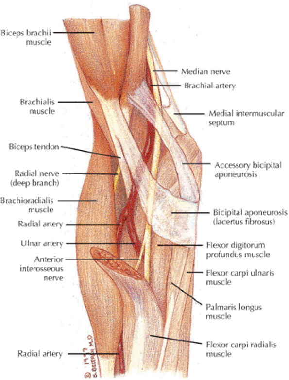
GRE sequences provide useful supplemental information for identifying loose bodies within the elbow. GRE volume sequences allow acquisition of multiple very thin axial images, which may subsequently be reformatted in any plane. In general, GRE sequences should be avoided after elbow surgery because magnetic susceptibility artifacts associated with micrometallic debris may obscure important findings and may also be mistaken for loose bodies. Artifact surrounding orthopaedic hardware is most prominent on GRE sequences because of the lack of a 180° refocusing pulse. It is least prominent on FSE sequences due to the presence of multiple 180° pulses. FSE T2-weighted sequences may be used to obtain higher-resolution images in the same amount of time as conventional spin-echo sequences or to reduce the overall time of the examination. The ability to shorten the examination with FSE is useful when scanning claustrophobic patients or patients who become uncomfortable in the prone position with the arm overhead.

FIGURE 9.1 ● Routine images obtained on a 3 T system show examples of PD-weighted (A) and FS PD FSE (B) axial images and an FS PD FSE sagittal image (C) in a patient with rupture of the distal biceps tendon.
Fat suppression may be added to various pulse sequences to improve visualization of the hyaline articular cartilage. Avoidance of chemical shift artifact at the interface of cortical bone and fat-containing marrow permits a more accurate depiction of the overlying hyaline cartilage. FS T1-weighted images are useful whenever gadolinium is administered, either

P.1466


intravenously or directly into the elbow joint as a dilute solution. Intravenous gadolinium may provide additional information in the assessment of neoplastic or inflammatory processes about the elbow. Articular injection of saline or dilute gadolinium may be useful in patients without a joint effusion to detect loose bodies, to determine if the capsule is disrupted, or to determine if an osteochondral fracture fragment is stable.
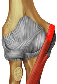
Limited true motion studies are possible within conventional magnets using phase-contrast and fast GRE techniques,11,12,13 and the increased use of higher-field-strength open systems should improve this capability.
Related Muscles
Relevant muscles about the elbow include related muscles of the arm and the volar and dorsal muscles of the forearm. The muscles of the arm include the biceps brachii (Fig. 9.2), the brachialis (Fig. 9.3), and the triceps brachii (Fig. 9.4). The superficial volar muscles of the forearm are the pronator teres (Fig. 9.5), the flexor carpi radialis (Fig. 9.6), the palmaris longus (Fig. 9.7), the flexor carpi ulnaris (Fig. 9.8), and the flexor digitorum superficialis (Fig. 9.9). The deep group of the volar muscles of the forearm are the flexor digitorum profundus (Fig. 9.10) and the flexor pollicis longus (Fig. 9.11). The pronator quadratus is covered in Chapter 10 on the wrist and hand. The superficial group of the dorsal muscles of the forearm includes the brachioradialis (Fig. 9.12), the extensor carpi radialis longus (Fig. 9.13), the extensor carpi radialis brevis (Fig. 9.14), the extensor digitorum (Fig. 9.15), the extensor digiti minimi (Fig. 9.16), the extensor carpi ulnaris (Fig. 9.17), and the anconeus (Fig. 9.18). The supinator (Fig. 9.19) is one of the deep dorsal muscles of the forearm. The others, include the abductor pollicis longus, the extensor pollicis brevis, the extensor pollicis longus, and the extensor indicis, are illustrated and discussed in Chapter 10 on the wrist and hand.
FIGURE 9.2Biceps brachii The biceps brachii is composed of two heads and is biarticulate, spanning the shoulder and elbow joints. The distal biceps brachii tendon inserts on the radial tuberosity, with superficial fibers contributing to the bicipital aponeurosis that blends with the antebrachial fascia. Most biceps brachii tears occur proximally involving the long head; however, distal avulsions are not uncommonly seen and are secondary to sudden or prolonged flexion against a heavy load. The biceps brachii acts as a flexor of both the shoulder and elbow joints. When the elbow is flexed, the biceps brachii also acts as a powerful supinator due to its slightly medial insertion on the rotating proximal radius.
FIGURE 9.3Brachialis The brachialis contributes more during isometric elbow flexion, whereas the biceps brachii is more active during dynamic elbow flexion. Isolated rupture of the brachialis muscle is a rare injury. Strains typically occur at the musculocutaneous junction.
FIGURE 9.4Triceps brachii The triceps brachii is composed of three heads. The long head of the triceps brachii is biarticulate, spanning both the shoulder and elbow joints. In addition to adduction of the shoulder, the triceps brachii acts to extend both the shoulder and elbow joints. The distal triceps tendon inserts on the olecranon, where avulsions can occur, typically following a fall on the outstretched upper extremity resulting in deceleration stress on an already contracted triceps. Midsubstance tendon and musculocutaneous injuries of the triceps brachii are less common.
FIGURE 9.5Pronator Teres The pronator teres acts synergistically with the pronator quadratus to pronate the forearm. The median nerve has variable anatomy with respect to the pronator teres. Most commonly the median nerve runs between the humeral and ulnar heads of the pronator teres, but it also can travel deep to both heads as well as perforate the humeral head. Pronator syndrome results from compression of the median nerve as it courses through the pronator teres and is manifested clinically by pain in the wrist and forearm and weakness of the thenar muscles.
FIGURE 9.6Flexor Carpi Radialis The flexor carpi radialis lies radial to the palmaris longus and ulnar to the pronator teres throughout its course. It contributes to flexion and abduction of the wrist. Along with the pronator teres it is the most common tendon involved in medial epicondylitis. Distal flexor carpi radialis tendon rupture, usually occurring after a fall on the outstretched hand, can clinically mimic scaphoid fractures.
FIGURE 9.7Palmaris longus The palmaris longus is present in approximately 85% of the population and functions to flex the wrist and tighten the palmar aponeurosis. It does not have a tendon sheath but has a paratenon. It is the most commonly used tendon graft of the hand, often used for repair of the elbow MCL in throwing athletes.
FIGURE 9.8Flexor carpi ulnaris The flexor carpi ulnaris flexes and adducts the hand. It is an important dynamic stabilizer of the pisotriquetral joint and contributes superficial fibers to the pisohamate ligament. As it is superficial and just medial to the ulnar nerve, it serves as a marker when ulnar nerve block is performed.
FIGURE 9.9Flexor digitorum superficialis The flexor digitorum superficialis tendons flex the middle phalanges of each finger and using the pulley system as a fulcrum contribute to flexion of the fingers at the metacarpophalangeal joint. The deep fibers of the flexor digitorum superficialis origin are closely apposed with the anterior bundle of the medial collateral ligament at the elbow, which is why edema and hemorrhage in the flexor digitorum superficialis are commonly seen in the setting of MCL tears. In the forearm, the median nerve lies just deep to the arch of the flexor digitorum superficialis muscle, and this is an area of potential nerve compression. The flexor digitorum superficialis divides into four musculotendinous units in the distal forearm and the tendons travel though the carpal tunnel before dividing again at the level of the proximal phalanges.
FIGURE 9.10Flexor digitorum profundus The flexor digitorum profundus tendons flex the distal phalanges at the distal interphalangeal joints and assist in flexion of the wrist and proximal phalanges. The flexor digitorum profundus divides into four musculotendinous units in the distal forearm, and the tendons travel though the carpal tunnel deep to the flexor digitorum superficialis tendons. Distal avulsions of a flexor digitorum profundus tendon, or “jersey finger,” can occur when an athlete gets a finger caught in an opposing player's jersey.
FIGURE 9.11Flexor pollicis longus The flexor pollicis longus flexes the thumb. Compression of the anterior interosseous nerve can lead to denervation of the flexor pollicis longus muscle, which may be isolated or concomitant with flexor digitorum profundus and pronator quadratus denervation.
FIGURE 9.12Brachioradialis The brachioradialis is a strong elbow flexor when the forearm is in a neutral position between supination and pronation. In forearm pronation, the brachioradialis tends to supinate as it flexes. In forearm supination, the brachioradialis tends to pronate as it flexes.
FIGURE 9.13Extensor carpi radialis longus The extensor carpi radialis longus extends and abducts the wrist. If extensor carpi ulnaris function is lost due to posterior interosseous nerve palsy, the extensor carpi radialis causes radial deviation because normally the attachment of the extensor carpi ulnaris to the ulnar aspect of the fifth metacarpal functions to neutralize the abduction movement applied by the extensor carpi radialis longus.
FIGURE 9.14Extensor carpi radialis brevis The extensor carpi radialis brevis, which provides neutral extension of the wrist, is the most common tendon involved in lateral epicondylitis. Distal ruptures of the extensor carpi radialis brevis significantly affect wrist extension.
FIGURE 9.15Extensor digitorum The extensor digitorum extends the medial four digits at the metacarpophalangeal joints and contributes to wrist extension. The proximal tendon, as part of the common extensor origin, is often involved in lateral epicondylitis. The extensor digitorum tendons are connected at the level of the metacarpal bones by fibrous bands called juncturae tendinum. Boutonnière deformity results from disruption of the central slip component of the extensor tendon at its insertion into the middle phalanx.
FIGURE 9.16Extensor digitI Minimi The extensor digiti minimi extends the proximal phalanx of the little finger at the metacarpophalangeal joint and contributes to wrist extension. Because the extensor digiti minimi tendon lies just superficial to the radioulnar articulation, it is often the first tendon to be involved in rheumatoid arthritis.
FIGURE 9.17Extensor carpi ulnaris The extensor carpi ulnaris tendon extends and adducts the wrist. It is commonly affected in tendinosis and tenosynovitis as it passes through the groove on the distal ulna. Subluxation of the extensor carpi ulnaris can also occur at this location related to disruption or insufficiency of the ligament that covers the tendon in this groove. The extensor carpi ulnaris tendon subsheath is a component of the triangular fibrocartilage complex.
FIGURE 9.18Anconeus The anconeus is located posterolateral to the elbow and functions to tighten the joint capsule and is a weak extensor of the elbow. In about 10% of the population an anomalous muscle, the anconeus epitrochlearis, arises from the medial border of the olecranon and the adjacent triceps inserting into the medial epicondyle. The anconeus epitrochlearis is thus located posteromedial to the elbow and can cause compression of the ulnar nerve in the cubital tunnel.
FIGURE 9.19Supinator The supinator is the primary supinator of the forearm when the elbow is extended. When the elbow is flexed, the supinator and the biceps brachii work synergistically to supinate the forearm. The deep branch of the radial nerve (i.e., the posterior interosseous nerve) travels between the humeral and ulnar origins of the supinator as it courses down the forearm, posterolateral to the proximal radius. The supinator is a potential site of entrapment of this nerve.

P.1467


P.1468


P.1469


P.1470


P.1471


P.1472


P.1473


P.1474


P.1475


MR Anatomic Atlas of the Elbow
The anatomic features of the elbow are best appreciated on conventional orthogonal triplane images obtained with the elbow in the fully extended position. Some structures, such as the biceps tendon, however, are better displayed if the elbow is imaged in 90° of flexion.14
Coronal Images
Coronal plane anatomy (Fig. 9.20) is key in demonstrating the collateral ligament complexes and common flexor and extensor tendon attachments:
  • The anterior bundle of the MCL, extending from the inferior margin of the medial epicondyle to the medial anterior margin of the coronoid process, is especially well demarcated, as is the lateral ulnar collateral ligament (LUCL), seen extending along the posterolateral aspect of the proximal radius to insert laterally on the tubercle of the supinator crest of the ulna.
  • The anterior articulation between the trochlea and the coronoid, the proximal radioulnar joint, and the articulation between the radius and the capitellum are all well demonstrated.
  • The rough nonarticular area at the posterior margin of the capitellum should not be mistaken for an osteochondral defect on coronal images through the posterior aspect of the radial head.
Axial Images
The axial plane (Fig. 9.21) demonstrates the distal course of the biceps and brachialis. The common flexor and extensor tendons and collateral ligaments as well as the ulnar nerve are well displayed:
  • The biceps and brachialis muscles are clearly depicted anteriorly, and the biceps tendon can be followed to the radial tuberosity and the brachialis tendon to the ulnar tuberosity.
  • P.1476


    P.1477


    P.1478


    P.1479


    P.1480


    P.1481


    P.1482


    P.1483


    P.1484


    P.1485


    P.1486


    P.1487


    P.1488


  • The bicipital aponeurosis, also known as the lacertus fibrosus, appears as a thin black line that extends from the myotendinous junction of the biceps to the fascia overlying the flexor-pronator muscle group medially.
  • The median nerve and the brachial artery and veins lie just deep to the lacertus fibrosus, at the level of the medial epicondyle.
  • The hypointense common flexor and common extensor tendons can be seen arising from the medial and lateral epicondyles, respectively.
  • The radial nerve is located between the brachialis and brachioradialis muscles, and the deep branch can be followed distally as it passes between the deep and the superficial heads of the supinator muscle to form the posterior interosseus nerve (an important site of potential impingement).
  • The annular ligament is a thin hypointense structure that lies just superficial to the articular cartilage of the radial head, which demonstrates intermediate signal intensity.
  • The insertion of the LUCL can be seen at the lateral margin of the ulna at the level of the radial neck.
  • The insertion of the anterior bundle of the MCL can be seen at the medial margin of the coronoid process, just anterior and lateral to the ulnar nerve.
  • Posteriorly, the triceps tendon can be followed to the olecranon.
  • The anconeus muscle is well seen posterolaterally.
  • The ulnar nerve and accompanying posterior ulnar recurrent artery and veins are seen posteromedially, deep to the cubital tunnel retinaculum at the level of the medial epicondyle. The ulnar nerve can be followed distally as it passes deep to the humeral and ulnar heads of the flexor carpi ulnaris muscle.
  • The proximal radioulnar joint and the posterior compartment articulation between the olecranon and the olecranon fossa are also well seen.
  • The usual site of osteochondritis dissecans—along the anterior aspect of the capitellum—is also well seen on axial images.
FIGURE 9.20 ● Normal coronal anatomy. (A) A fracture of the olecranon or a muscle strain or tear involving the muscle belly of the triceps may clinically mimic signs and symptoms of a distal triceps tendon tear. (B) The normal distal triceps tendon fibers are interspersed with intermediate to high bands of linear signal, which should not be mistaken for tears or tendinosis. (C) Loose bodies not uncommonly lodge in the posterior aspect of the joint, particularly within the olecranon fossa. (D) The lateral ulnar collateral ligament (LUCL) originates on the posterior lateral aspect of the lateral epicondyle and courses posteriorly and distally around the radial head and neck to insert on the lateral ulna. The LUCL forms a cradle around the radial head and neck and prevents posterior dislocation of the radial head. On this image, the oblique course of the distal LUCL posterior to the radial neck is visualized, and the LUCL is seen inserting on the lateral ulnar diametaphysis. (E) In cases of suspected fracture, T1-weighted images are useful in visualizing the low-signal fracture line, particularly with fractures of the radial head and coronoid process. (F) The proximal portion of the LUCL is visualized on this image, with its origin from the posterolateral aspect of the lateral epicondyle. Most tears of the LUCL occur at the origin. (G) In addition to the ulnohumeral and radiohumeral articulations, there is a third elbow articulation between the medial aspect of the radial head and the lateral aspect of the coronoid, which is covered with cartilage and susceptible to chondromalacia. (H) The distal insertion fibers of the anterior bundle of the MCL are continuous with the sublime tubercle, and any fluid interposed between the distal fibers and the medial aspect of the sublime tubercle is interpreted as a deep articular-sided partial tear with partial stripping of the distal ligament from the sublime tubercle, an appearance called the T sign. (I) Osteochondritis dissecans commonly affects the capitellum, particularly in adolescents 13 to 16 years of age. A distinct entity affecting the capitellum is Panner's disease, which is posttraumatic avascular necrosis of the capitellum; it affects a younger age group (children 5 to 11 years). (J) The radial collateral ligament (RCL) is located just anterior to the LUCL, and its fibers originate on the lateral aspect of the lateral epicondyle and insert on the lateral aspect of the annular ligament surrounding the radial head. RCL tears often accompany LUCL tears, or, in cases of posttraumatic lateral-sided blowout, the RCL, LUCL, and common extensor tendon can all be pulled off the lateral epicondyle as a single unit. (K) Fractures of the coronoid process are generally caused by hyperextension or posterior subluxation/dislocation. As a result, coronoid process fractures are frequently accompanied by fractures of the radial head and distal humerus. (L) A ruptured, retracted distal biceps tendon may manifest as fluid signal and hemorrhage at the radial tuberosity insertion site, with no tendon visualized. Viewing successive coronal images anterior to the radial tuberosity, the torn and retracted end of the distal biceps may be located and the distance of retraction measured.
FIGURE 9.21 ● Normal axial anatomy. (A) The coronoid fossa is a depression on the anterior aspect of the distal humerus that receives the coronoid during flexion, whereas the olecranon fossa is a depression on the posterior aspect of the distal humerus that receives the olecranon during extension. The coronoid fossa and olecranon fossa are common locations in which loose bodies may lodge. (B) It is not uncommon for the ulnar nerve to appear somewhat bright on MR images. This appearance may be seen in cases of ulnar neuritis but is not specific for the diagnosis. Other findings that increase the specificity for ulnar neuritis include enlargement of the ulnar nerve, thickening of the cubital retinaculum, inflammation around the ulnar nerve, and a space-occupying lesion within the cubital tunnel or an anconeus epitrochlearis. C) The cubital tunnel, formed by the posterior aspect of the medial epicondyle and the medial aspect of the olecranon, is covered by the cubital tunnel retinaculum. Scarring and thickening of the cubital tunnel retinaculum can be associated with ulnar neuritis. Also, in 11% of the population, the cubital tunnel retinaculum is replaced by the anconeus epitrochlearis muscle, which may cause ulnar nerve impingement within the cubital tunnel. (D) The common extensor tendon and common flexor tendon origins are visualized as curvilinear foci of dark tendon signal along the anterior margins of the lateral epicondyle and medial epicondyle, respectively. Tears of these tendon origins are suggested when fluid signal is visualized along the lateral or medial epicondyle in place of the normal dark tendon signal. (E) The radial nerve and its branches innervate the muscles along the anterior, radial, and posterior aspect of the elbow, including (clockwise around the elbow) the brachialis, brachioradialis, extensor carpi radialis longus, extensor carpi radialis brevis, extensor digitorum, extensor carpi ulnaris, anconeus, and triceps muscles. Evidence for denervation of these muscles suggests abnormality of the radial nerve or its branches. (F) The RCL and LUCL are visualized on this axial image as linear dark signal just deep to the common extensor tendon, with a plane of synovium separating the lateral collateral ligament complex from the common extensor tendon. Fluid signal deep to the common extensor tendon replacing the anterior fibers of the lateral collateral ligament complex represents tears of the RCL, whereas fluid signal replacing the posterior fibers suggests tears of the LUCL. (G) The ulnar nerve and its branches innervate the muscles along the posteromedial aspect of the elbow, including (clockwise around the elbow) the flexor digitorum profundus and flexor carpi ulnaris. Evidence for denervation of these muscles suggests abnormality of the ulnar nerve. (H) The median nerve is visualized on this axial image anteromedially coursing between the pronator teres and brachialis muscles. The radial nerve is visualized anterolaterally coursing just deep to the brachioradialis muscle. Bright signal in the radial and median nerve suggesting neuritis is not as commonly visualized. However, denervation of the muscles supplied by the nerves, as evidenced by high signal or atrophy of the muscles, suggests neuritis. (I) The median nerve and its branches innervate the muscles along the anteromedial aspect of the elbow, including (clockwise around the elbow) the flexor digitorum superficialis, palmaris longus, flexor carpi radialis, and pronator teres muscle. Evidence for denervation of these muscles suggests abnormality of the median nerve. (J) The LUCL is visualized on this axial image inserting distally on a ridge along the lateral aspect of the ulna known as Gerdy's tubercle. Although most tears of the LUCL occur at its proximal origin, the entire course of the LUCL from origin to insertion should be examined in the axial, coronal, and sagittal planes. (K) The radial tuberosity is the bony insertion site for the distal biceps tendon. Bony hypertrophy and bone marrow edema in the radial tuberosity are indirect signs of tendinosis and/or tearing of the distal biceps tendon. (L) A fibrous band of aponeurotic tissue called the lacertus fibrosis attaches to the distal biceps tendon. In the setting of an insertional distal biceps tendon tear, if the lacertus fibrosis is intact, the distal biceps tendon demonstrates little if any retraction. However, if the lacertus fibrosis is torn, the amount of distal biceps tendon retraction may be quite severe.
Sagittal Images
The sagittal plane (Fig. 9.22) demonstrates the radiocapitellar and humerotrochlear articulations. The triceps tendon is visualized in the longitudinal axis. The common flexor and extension tendons and deeper layer of collateral ligaments are also depicted:
  • Laterally, the components of the common extensor tendon can be followed to the lateral epicondyle.
  • Medially, the components of the common flexor tendon can be seen extending proximally to the medial epicondyle.
  • The intermediate-signal-intensity ulnar nerve is seen along the posterior margin of the medial epicondyle.
  • Near the midline, the attachment of the triceps muscle and tendon to the olecranon can be seen.
  • Normal obliteration of the subcutaneous fat is seen posterior to the olecranon at the site of the superficial olecranon bursa.
  • The posterior and anterior fat pads can be seen along the margins of the distal humerus.
  • The joint capsule appears as a thin, hypointense structure just superficial to the fat pads.
  • The brachialis muscle lies just superficial to the anterior joint capsule and can be followed distally to its insertion on the ulnar tuberosity.
  • The biceps can be followed distally to its insertion on the radial tuberosity. The adjacent low-signal-intensity brachial and ulnar arteries should not be mistaken for the biceps tendon on sagittal images.
  • The articulation of the radius and the capitellum and the articulation between the trochlea and the trochlear notch are clearly demonstrated.
  • The normal bare area of the ulna in the midportion of the trochlear notch should not be confused with an osteochondral defect. Similarly, the rough nonarticular area at the posterior margin of the capitellum should not be confused with an impaction fracture or osteochondral defect.
Imaging Checklist for the Elbow
The elbow joint can be divided into medial, lateral, anterior, and posterior compartments. In the medial compartment, structures on the MR checklist include:
  • MCL
  • Common flexor tendon
  • Proximal flexor muscles
  • Ulnar nerve
  • Trochlea and coronoid (and overlying cartilage)
  • Medial epicondyle
In the lateral compartment, checklist structures include:
  • Radial collateral ligament (RCL)
  • LUCL
  • Common extensor tendon
  • Proximal extensor muscles
  • Radius and capitellum (and overlying cartilage)
The anterior compartment structures are:
  • Biceps tendon
  • Brachialis tendon and muscle
  • Radial and median nerves
The important posterior compartment structures are the triceps tendon and muscle.
The primary stabilizers of the elbow are:
  • Ulnohumeral joint (with the coronoid process as the key stabilizer)
  • MCL (primarily the anterior bundle)
  • LUCL

P.1489


P.1490


P.1491


P.1492


P.1493


Secondary stabilizers include:
  • Radial head
  • Common flexor and extensor tendons
The severity of injuries progresses from ligament involvement alone, to involvement of the radial head, coronoid, and olecranon, to radial-ulnar dissociation (a tear of the interosseous ligament with fractures distal to the elbow).
Chronic repetitive injuries include tendinosis and chronic tearing of the common extensor tendon, which is seven times more common than chronic abnormalities of the common flexor tendon (medial epicondylitis). Although the following checklists are presented by plane, use of a multiscreen workstation with an image navigator bar is optimal for triangulating on anatomy and pathology simultaneously in multiple planes.

P.1494


Coronal Plane Checklist
The examination of the elbow in the coronal plane begins in the medial aspect of the elbow with examination of the anterior bundle of the MCL and the common flexor tendon just superficial to the MCL. Subsequently, the lateral aspect of the joint is examined and the LUCL, the RCL, and the common extensor tendon just superficial to the lateral ligaments are viewed. The radiohumeral and ulnohumeral articulations are then inspected, including examination of the cartilage surfaces and assessment for fractures as well as loose bodies in the joint space. The anterior aspect of the elbow is then examined and the insertions of the biceps tendon into the radial tuberosity and the brachialis tendon into the ulnar tuberosity are evaluated. Finally, the posterior elbow and the triceps insertion are examined, and the full length of the ulnar nerve posterior to the medial epicondyle is visualized.

P.1495


Medial Compartment
(1) Medial Collateral Ligament (Fig. 9.23)
The anterior bundle of the MCL is optimally visualized on coronal images. The anterior bundle originates at the anteroinferior surface of the medial epicondyle and courses in an oblique posterior fashion to attach firmly to the medial edge of the coronoid process at the sublime tubercle. The fibers of the anterior bundle are continuous with the sublime tubercle, and any fluid interposed between the distal fibers and the medial aspect of the sublime tubercle is interpreted as a deep articular-sided partial tear with partial stripping of the distal ligament from the sublime tubercle. The fluid signal is perpendicular to fluid within the ulnohumeral joint, resembling the letter “T,” and this appearance has been called the T sign. The anterior bundle is the primary constraint to valgus stress on the elbow

P.1496


and is one of the main stabilizers of the elbow. The posterior bundle of the MCL is thought to be less functionally important and occasionally is difficult to visualize in the coronal plane.

FIGURE 9.22 ● Normal sagittal anatomy. (A) The supinator muscle, visualized on this sagittal image, is a landmark for the division of the radial nerve into superficial and deep branches. The deep branch pierces the supinator muscle to become the posterior interosseous nerve. The tendinous origin of the supinator muscle (known as the arcade of Frohse) is a common location for posterior interosseous nerve entrapment. (B) This sagittal image demonstrates the lateral collateral ligament complex and the common extensor tendon as a triangle of dark signal with its base fanning out over the radial head. The apex of the triangle represents the proximal origin of the common extensor tendon. The anterior aspect of the base of the triangle represents the RCL, whereas the posterior aspect of the base represents the LUCL. Fluid signal within any part of this triangle of dark signal usually suggests a tear to the corresponding ligament or common extensor tendon. (C) Posterior dislocation of the radius may result in “kissing” contusions or fractures at the anterior aspect of the radial head and posterior aspect of the capitellum, at the point where the two bones impact one another during a posterior dislocation event. (D) This sagittal image demonstrates the course of the proximal to mid-lateral ulnar collateral ligament taking a turn around the posterolateral aspect of the radial head, forming a cradle that prevents the radial head from posterior dislocation. (E) Reactive bone marrow edema and hypertrophic changes in the radial tuberosity commonly accompany insertional distal biceps tendon pathology. (F) Sagittal and coronal images demonstrate the articular cartilage surfaces of the radial head, capitellum, and trochlea. In the setting of any significant chondromalacia, the remainder of the elbow joint should be scrutinized for loose bodies. (G) If fractures of the olecranon are displaced by less than 2 mm, the fracture can be treated conservatively. Fractures with greater than 2 mm of displacement are usually treated by ORIF. (H) The normal trochlear groove is located in the central portion of the articular surface of the proximal ulna and represents a bare area devoid of cartilage. This normal pseudodefect should not be mistaken for a chondral defect on sagittal images. (I) If fractures of the coronoid process involve less than 50%, the fracture may be treated conservatively. However, if more than 50% of the coronoid is involved, or if there are other fractures or valgus instability, surgical treatment is preferred. (J) The coronoid fossa and olecranon fossa are areas of bone thinning on the anterior and posterior aspect of the distal humerus, respectively. The coronoid fossa receives the coronoid on flexion, and the olecranon fossa receives the olecranon on extension. These fossae are common locations for loose bodies. In addition, hypertrophic bone can form in the olecranon and coronoid fossae due to degenerative arthritis, and the hypertrophic bone can limit flexion and extension. (K) Medial epicondylitis is 10 times less common than lateral epicondylitis. (L) The ulnar nerve courses posterior to the medial epicondyle and is visualized on this sagittal image. This is the most common location for ulnar neuritis, and findings on sagittal images can confirm abnormalities of the ulnar nerve visualized in the axial plane. (M) Little Leaguer's elbow describes avulsion injuries of the medial epicondyle, due to an acute traumatic event or chronic repetitive microtrauma. (N) Fluid signal seen on sagittal images within or replacing the dark common flexor tendon origin at the medial epicondyle is suggestive of a tear of the common flexor tendon.
FIGURE 9.23 Medial Collateral Ligament.
(2) Common Flexor Tendon (Fig. 9.24)
The common flexor tendon origin is visualized on coronal images arising medial and proximal to the MCL, coursing superficial to the MCL fibers. Tendinosis of the common flexor tendon is visualized as thickening and vague increased T2 (or fat-saturated PD) signal, whereas partial- or full-thickness tears demonstrate fluid signal disrupting tendon origin fibers. Not uncommonly, acute and chronic injuries of the common flexor tendon, often the result of valgus stress, are accompanied by evidence of ulnar neuritis and MCL injuries.
(3) Ulnohumeral Articulation (Fig. 9.25)
The articular cartilages lining the coronoid and trochlea are optimally visualized in the coronal plane, and chondral degeneration and associated subchondral edema and cystic change are evaluated. The size and location of any loose bodies within the joint secondary to chondral and osteochondral abnormalities are also identified. Fractures of the medial epicondyle, trochlea, and coronoid are also visualized in the coronal plane.
(4) Ulnar Nerve (Fig. 9.26)
The ulnar nerve courses medial to the distal humerus shaft and posterior to the medial epicondyle. Although axial images provide more detailed characterization of ulnar nerve injuries, high signal and other abnormalities of the ulnar nerve are confirmed in the coronal plane. A normal variant, the anconeus epitrochlearis (a small accessory muscle extending from the medial epicondyle to the medial ulna), occasionally causes ulnar nerve impingement by narrowing the cubital tunnel.
Lateral Compartment
(1) Lateral Collateral Ligament Complex (Fig. 9.27)
The lateral collateral ligament complex is composed of the RCL, the LUCL, and annular ligament. The RCL and LUCL are optimally visualized in the coronal plane. The RCL originates at the anterior distal aspect of the lateral epicondyle, deep to the common extensor tendon. The RCL fibers insert onto the annular ligament, which circumferentially surrounds the radial head. The origin of the LUCL is also deep to the common flexor tendon and somewhat posterior to the RCL origin along the lateral epicondyle. The LUCL swings posteromedially around the radial head, forming a posteromedial sling for the radius. The LUCL then inserts on the lateral aspect of the proximal ulna at the supinator crest. The most anterior image through the mid-radiocapitellar joint demonstrates the RCL coursing from the lateral epicondyle to the annular ligament surrounding the radial head. The common extensor tendon is superficial to the RCL and originates proximal to the RCL at this image location. On successive coronal images from anterior to posterior, the origin of the LUCL from the lateral epicondyle is visualized, the LUCL is seen swinging around the lateral aspect of the radial head, and the oblique course of the

P.1497


P.1498


LUCL posterior to the proximal radius is seen as it courses toward and attaches to the supinator crest on the ulna. Suspected tears and sprains of the RCL and LUCL are confirmed and further characterized in the axial and sagittal planes. Tears of the LUCL predispose to recurrent radial head dislocations. In cases of significant elbow trauma leading to dislocation, it is not uncommon for the RCL, LUCL, and common extensor tendon origin to completely tear off of the lateral epicondyle together as a single ligamentous/tendinous unit.

FIGURE 9.24 Common Flexor Tendon.
FIGURE 9.25 Ulnohumeral Articulation.
FIGURE 9.26 Ulnar Nerve.
(2) Common Extensor Tendon (Fig. 9.28)
The common extensor tendon originates from the anterior aspect of the lateral epicondyle and is visualized superficial to and somewhat parallel to the RCL. Common extensor tendinosis and tears are commonly referred to as lateral epicondylitis, or “tennis elbow.” The extensor carpi radialis brevis is the most commonly involved of the extensor tendons. The common extensor tendon is usually visualized on at least three consecutive coronal images from anterior to posterior.
(3) Radiohumeral Articulation (Fig. 9.29)
Cartilage covers the articular surface of the radial head and the opposing capitellum in a nearly 180° arc from anterior to posterior. Articular cartilage also extends over the medial aspect of the radial head and the lateral aspect of the coronoid, forming the third articulation of the elbow at the radioulnar joint. The cartilage covering the lateral aspect of the radial head is normally much thinner, and the absence of cartilage in this location

P.1499


P.1500


on coronal images should not be misinterpreted as a cartilage defect. Another common cartilage pseudolesion occurs at the lateral aspect of the capitellum, where a normal groove devoid of cartilage between the capitellum and lateral epicondyle may mimic an osteochondral defect. These cartilage surfaces must be examined carefully for the presence of chondral degeneration and associated stress-related subchondral edema and cystic change. Chondral degeneration is most commonly visualized on the opposing surfaces of the medial radial head and adjacent crest at the lateral margin of the trochlea. Osteochondral lesions of the capitellum commonly occur in teenage and young adult throwing athletes, whereas Panner's disease (capitellar osteochondrosis) occurs in younger children. Fractures of the radial head and neck, not uncommonly occult on plain films, are easily visualized on MR images.

FIGURE 9.27 Lateral Collateral Ligament Complex.
FIGURE 9.28 Common Extensor Tendon.
FIGURE 9.29 Radiohumeral Articulation.
FIGURE 9.30 Biceps and Brachialis.
Anterior Compartment
(1) Biceps and Brachialis Tendons (Fig. 9.30)
Successive anterior-to-posterior images in the coronal plane display the biceps tendon coursing distally to attach at the radial tuberosity along the medial inferior aspect of the proximal radial shaft. The distal tendon is examined for sprain, tendinosis, or tear. If completely torn, the length of retraction is determined. Hypertrophic bony changes at the radial tuberosity insertion site are seen as indirect evidence of chronic distal biceps tendinosis or tearing. Also, fluid around the distal biceps tendon is compatible with bicipitoradial bursitis rather than tenosynovitis, since the distal biceps tendon has no tendon sheath. The brachialis tendon runs parallel and posterior to the biceps tendon and inserts along the anterior proximal ulna along the ulnar tuberosity. The brachialis is also evaluated for signs of tears and tendinosis.
Posterior Compartment
(1) Triceps Tendon (Fig. 9.31)
The triceps tendon is visible from its origin to its insertion on the olecranon. There is normal interposition of fat between the fascicles of the distal triceps tendon, which may produce a heterogeneous appearance to the distal fibers. This should not be misinterpreted as tendinosis as long as individual fibers are still well defined. Tendinosis and strain tend to obscure distinction of individual fibers. Coronal images are particularly useful in determining whether a tear of the distal triceps tendon is complete or partial, and, if partial, the extent and medial-to-lateral location of the tear. The degree of torn tendon retraction can be determined on coronal or sagittal images.
Axial Plane Checklist
Nearly all of the ligamentous and tendinous structures seen in the coronal plane are also visualized in the axial plane. By viewing successive proximal-to-distal axial images in the medial compartment, the origins of the MCL and common flexor tendon just superficial to the MCL are identified on the medial epicondyle, the ulnar nerve is visualized within the cubital tunnel posterior to the medial epicondyle, and a normal variant anconeus epitrochlearis muscle is occasionally seen overlying the ulnar nerve and cubital tunnel. Examination of the lateral aspect

P.1501


of the elbow reveals the origins of the RCL and LUCL anteriorly along the lateral epicondyle and the origin of the common extensor tendon just posterior to the RCL and LUCL. The flexor and extensor muscles are inspected for strains, tears, or denervation injury. Proximal-to-distal images in the anterior compartment allow the biceps and brachialis tendons to be followed to their insertions. The course of the median and radial nerves is also examined anteriorly. In the posterior aspect of the joint the triceps tendon is evaluated for tears, strain, or tendinosis.

FIGURE 9.31 Triceps Tendon.
Medial Compartment
(1) Medial Collateral Ligament (Fig. 9.32)
The anterior bundle of the MCLI is most easily found on axial images by first finding the axial image through a small ridge at the most proximal and medial portion of the medial ulna, known as the sublime tubercle. The anterior bundle is seen as a vertically oriented black band of signal lining up along the medial ulna. The anterior bundle can be followed proximally on successive images to where it fans out over its origin on the medial epicondyle. Distal MCL tears are commonly associated with bone marrow edema within the sublime tubercle. Although the coronal plane is probably most useful for evaluating the MCL, the axial and sagittal planes can confirm and further characterize MCL pathology.
(2) Common Flexor Tendon (Fig. 9.33)
Common flexor tendon pathology is visualized in cross-section on axial images. The common flexor tendon origin can be

P.1502


identified along the anteromedial-most aspect of the medial epicondyle and its course can be followed distally to where it fans out into its various components making up the proximal flexor muscle group. The individual flexor muscles are also delineated and strains, tears, or denervation injuries are identified and localized.

FIGURE 9.32 Medial Collateral Ligament.
FIGURE 9.33 Common Flexor Tendon.
(3) Ulnar Nerve (Fig. 9.34)
On axial images the ulnar nerve proximal to the elbow courses medially along the medial border of the triceps. It then descends posteriorly at the level of the elbow to run posterior to the medial epicondyle in the cubital tunnel. Distal to the elbow the ulnar nerve ascends between the two heads of the flexor carpi ulnaris muscle. The entire course of the ulnar nerve through the elbow is visualized in cross-section. T2 hyperintensity within the nerve is suggestive of, but not entirely specific for, ulnar neuritis. Swelling and enlargement of the nerve, and inflammatory infiltration of the perineural fat, increase the specificity of the diagnosis. An underlying structural cause for ulnar neuritis is only occasionally found but may include thickening of the cubital retinaculum overlying the nerve, a normal variant anconeus epitrochlearis muscle, neoplasms, bony spurs, posttraumatic deformities, and ganglion cysts. Ulnar nerve compression is sometimes caused by the anconeus epitrochlearis muscle (seen in 11% of the population), which is an anomalous muscle replacing the cubital retinaculum and extending posterolaterally from the posterior medial epicondyle to the medial ulna.
(4) Ulnohumeral Joint (Fig. 9.35)
Although usually identified in the coronal or sagittal plane, fractures of the coronoid, trochlea, olecranon, and medial epicondyle are further characterized in the axial plane. Loose bodies within the joint are also confirmed axially. Salter I physeal injuries of the medial epicondyle occur in teenage throwing athletes and are known as “Little Leaguer's elbow.”
Lateral Compartment
(1) Lateral Collateral Ligament Complex (Fig. 9.36)
The origins of the RCL and LUCL are visualized in the axial plane along the anterior aspect of the lateral epicondyle. The RCL originates just anterior to the LUCL. From its origin, the short course of the RCL can be followed in cross-section to its insertion on the annular ligament. The longer, oblique course of the LUCL is more difficult to appreciate on axial images but can be identified on successive proximal-to-distal images as the LUCL turns 90° around the posterolateral aspect of the radial head to insert on the supinator crest of the ulna. Tears most commonly occur at the lateral epicondyle origin and are seen as fluid signal disrupting the normal location of origin fibers along the anterior lateral epicondyle. Axial plane images are used to confirm tears suspected on coronal or sagittal images and are sometimes helpful in localizing which components of the lateral stabilizers are involved.
FIGURE 9.34 Ulnar Nerve.
FIGURE 9.35 Ulnohumeral Joint.
FIGURE 9.36 RCL and LUCL.
FIGURE 9.37 Common Extensor Tendon.
FIGURE 9.38 Radiohumeral Articulation.

P.1503


P.1504


P.1505


(2) Common Extensor Tendon (Fig. 9.37)
Of the three lateral ligament/tendon structures arising from the lateral epicondyle, the origin of the common extensor tendon is the most posterior and proximal. Tears and tendinosis involving the common extensor tendon are evaluated in cross-section in the axial plane, as are the extensor muscles, which contribute to the common extensor tendon. Strain, tears, or denervation injuries of the extensor muscles, as well as the supinator and brachioradialis muscles, are localized on axial images.
(3) Radial-Humeral Articulation (Fig. 9.38)
Fractures or osteochondral defects of the radius and capitellum identified in other planes are further characterized in the axial plane. Loose bodies are also identified.
Anterior Compartment
(1) Biceps and Brachialis Tendons (Fig. 9.39)
The biceps and brachialis tendons are seen in cross-section, and the biceps tendon should be followed to its distal insertion on the medial inferior aspect of the proximal radius, at the radial tuberosity. In addition, radial tuberosity hypertrophy from chronic tendinosis and distal bicipitoradial bursitis are also evaluated in the axial plane. On images proximal to the elbow, the brachialis tendon begins anterolaterally, whereas the biceps is anteromedial. The brachialis runs posterior to the biceps. As the tendons are followed distally, the biceps courses posterolaterally, whereas the brachialis runs posteromedially. The two tendons cross mid-elbow, with the

P.1506


brachialis eventually inserting medially on the proximal ulna and the biceps inserting on the proximal radius.

FIGURE 9.39 Normal Biceps and Brachialis Tendon.
FIGURE 9.40 Median and Radial Nerve.
(2) Median and Radial Nerves (Fig. 9.40)
The radial nerve is seen coursing between the brachialis and brachioradialis anterolaterally, splitting into superficial and deep branches near the level of the biceps myotendinous junction. The median nerve runs just medial to the brachial artery, anterior to the brachialis muscle. Evidence of median and radial neuritis is usually seen on axial MR images as denervation (hyperintensity on FS PD FSE images) and/or fatty atrophy of the muscles innervated by the affected nerve. Occasionally, an underlying cause for the neuritis is identified, such as a mass lesion or an anatomic anomaly (e.g., severe bicipitoradial bursitis compressing the median nerve or a ganglion cyst compressing the radial nerve).
Posterior Compartment
(1) Triceps Tendon (Fig. 9.41)
The triceps tendon is also imaged in cross-section in the axial plane. It inserts in a U shape along the olecranon. Triceps tendon tears, tendinosis, or strain is evaluated in the axial plane.
Sagittal Plane Checklist
On medial-to-lateral sagittal images, the full length of the common flexor tendon is seen first on the medial-most images. The MCL is depicted next, although it is occasionally difficult to see in the sagittal plane. The ulnar nerve is visualized coursing posterior to the medial epicondyle. The ulnohumeral and radiohumeral articulations are then examined, inspecting the cartilage surfaces and looking for loose bodies. On the central images, the biceps and brachialis tendons are visualized anteriorly and the triceps tendon posteriorly. At the lateral elbow, the origins of the RCL and LUCL are evaluated. The common extensor tendon is visualized on one image lateral to the RCL and LUCL origin.
Medial Compartment
(1) Common Flexor Tendon and Ulnar Nerve (Fig. 9.42)
On medial-to-lateral sagittal images through the elbow, the common flexor tendon is seen on the most medial images as a hypointense band of signal parallel to the long axis of the ulna, coursing to insert on the medial epicondyle. Tears, strain, or tendinosis of the common flexor tendon can be characterized

P.1507


on sagittal images, while triangulating on the tendon simultaneously in the coronal and axial planes. The anterior bundle of the MCL is not as well seen on sagittal images but can occasionally be visualized on images just deep to the common flexor tendon. Strain or tears of the flexor muscles distal to the common flexor tendon insertion are also evaluated on sagittal images.

FIGURE 9.41 Triceps Tendon.
FIGURE 9.42 Common Flexor Tendon and Ulnar Nerve.
The ulnar nerve can be seen coursing just posterior to the medial epicondyle, making sagittal plane images useful for confirming abnormalities suspected on axial plane images. The ulnar nerve is often visualized on the same sagittal image as the common flexor tendon.
(2) Ulnohumeral Articulation (Fig. 9.43)
The cartilages lining the coronoid, olecranon, and trochlea are particularly well visualized and characterized on sagittal images. One common pitfall is a normal pseudodefect in the middle of the trochlear notch between the olecranon and coronoid articular surfaces, formed by a normally cartilage-free groove. Fractures of the coronoid process, olecranon, and trochlea are characterized with regard to size and displacement. The size of the coronoid process fracture fragment is important in determining potential elbow instability, and the fracture fragment is measured in the sagittal plane. On successive medial-to-lateral sagittal images near the midportion of the joint, the bone between the trochlea and the distal humeral shaft becomes thin, forming anterior and posterior fossae known as the coronoid fossa anteriorly (which receives the coronoid on flexion) and the olecranon fossa posteriorly (which receives the posterior olecranon on extension). These two fossae are common sites for loose bodies, anterior more often than posterior. In addition, in cases of degenerative arthritis, these two fossae are examined for the presence of hypertrophic bone, which may limit flexion and extension.
Lateral Compartment
(1) Common Extensor Tendon and Lateral Collateral Ligament Complex (Fig. 9.44)
On lateral-to-medial sagittal images through the lateral compartment, the common extensor tendon is seen first on the most peripheral lateral slice. It appears as a hypointense band of signal parallel to the radius and can often be visualized along its entire course on one or two images before inserting on the lateral epicondyle. The myotendinous junction of the proximal extensor muscles is also visualized on sagittal images. Strains or tears of the proximal extensor muscles and tendon are well characterized on these images.
The lateral collateral ligament complex is encountered medial to the common extensor tendon. Both the RCL and LUCL are often identified on the same image. The RCL is located just anterior to the LUCL, and the RCL courses distally from the lateral epicondyle, parallel to the long axis of the radius, to insert on the annular ligament fibers surrounding the radial head. The LUCL is located just posterior to the RCL, and the fibers diverge to run posteriorly and distally. The origins

P.1508


of the LUCL and RCL overlap somewhat on MR images, and occasionally it is difficult to completely distinguish them. The course of the LUCL, as it runs behind the radial head to insert on the supinator crest of the ulna, can be followed on the next three or four successive medial images. The sagittal plane is helpful to confirm or further characterize tears of the lateral ligaments and tendons that are suspected on images obtained in other planes.

FIGURE 9.43 Ulnohumeral Articulation.
(3) Radiohumeral Articulation (Fig. 9.45)
The cartilage surfaces covering the radial head and the nearly 180° arc of the capitellum are visualized on sagittal images, as are loose bodies displaced anteriorly or posteriorly in the joint. Fractures of the radial head and capitellum are characterized on these images as well. Similar to the coronoid fossa on the ulnohumeral side, there is a radial fossa forming a small concavity on the anterior surface of the distal humerus, which receives the radial head in flexion. The radial fossa is examined for loose bodies and osteophytes.
Anterior Compartment
Biceps and Brachialis Tendon (Fig. 9.46)
On sagittal images through the lateral aspect of the joint, the entire course of the distal biceps tendon can be visualized on one or two images coursing parallel to the distal humeral shaft before curving 90° to insert on the radial tuberosity. Complete tears, partial tears, and tendinosis are characterized with regard to the extent and the location of the involved tendon. In cases of complete tendon rupture, the amount of proximal retraction is measured.
Analogous to the biceps tendon in the lateral aspect of the joint, the full course of the distal brachialis muscle and tendon can be visualized on sagittal images through the medial joint. The brachialis courses parallel and posterior to the biceps tendon, and its distal course and insertion on the anterior proximal ulna are often visualized on the same image as the biceps tendon, or on one image medial to the biceps insertion.
Posterior Compartment
(1) Triceps Tendon (Fig. 9.47)
Sagittal plane images through the midline demonstrate the insertion of the distal triceps muscle and tendon on the olecranon. Tendinosis, strain, and tears (including the degree of retraction) can be characterized on these images.
Sample MRI Report, Elbow Injury
Clinical Information: Partial dislocation, hyperextension mechanism. Evaluate for medial ligament tear.
Technique: Axial, coronal, and sagittal T1-weighted images without fat suppression and proton density images with fat suppression.
Findings: There is a partial undersurface tear of the distal aspect of the anterior band of the medial collateral ligament (Fig. 9.48A). There is a sprain of the proximal fibers of the anterior

P.1509


P.1510


band of the medial collateral ligament (Fig. 9.48B). There is mild tendinosis of the common flexor tendon (Fig. 9.48C). There is mild tendinosis of the common extensor tendon. There is a sprain of the proximal origin of the lateral ulnar collateral ligament with bone marrow edema in the lateral aspect of the capitellum (Fig. 9.48D). There is muscle edema corresponding to the flexor digitorum superficialis muscle, indicating a muscle strain (Fig. 9.48E). There is also mild edema demonstrated in the extensor digitorum muscle (Fig. 9.48F).

FIGURE 9.44 Common Extensor Tendon and Lateral Ligament Complex.
FIGURE 9.45 Radiohumeral Articulation.
FIGURE 9.46 Biceps and Brachialis.
There is moderate edema within the brachialis muscle consistent with a grade 1 to 2 muscle strain of the brachialis muscle with hemorrhage (Fig. 9.48G). The biceps tendon and brachialis tendon are seen in continuity. Anterior soft-tissue hemorrhage is apparent and capsular distention is appreciated on sagittal images. There is elevation of the anterior and posterior fat pads secondary to a large hemorrhagic joint effusion (Fig. 9.48H).
Axial images confirm extensive edema and hemorrhage within the brachialis muscle group (Fig. 9.48I). The ulnar nerve is seen without hyperintensity.
Impression:
  • Partial undersurface tear of the distal aspect of the anterior band medial collateral ligament. There is a small focus of decreased signal intensity associated with this, which may represent a small osseous avulsion. There is a sprain of the proximal fibers of the anterior band of the medial collateral ligament.
  • There is a sprain of the origin of the lateral ulnar collateral ligament associated with subchondral marrow edema of the lateral aspect of the capitellum.
  • P.1511


  • Grade 1 to 2 muscle strain of the brachialis. Mild grade 1 strain of the extensor digitorum muscle and also grade 1 strain of the flexor digitorum superficialis muscle medially.
  • Large hemorrhagic joint effusion with elevation of anterior and posterior fat pads
  • Intact brachialis and biceps tendon distally
FIGURE 9.47 Triceps Tendon.
FIGURE 9.48 Sample Elbow Case.
Normal Anatomy of the Elbow
A thorough understanding of the anatomy and function of the elbow is essential for accurate interpretation of MR images. The anatomic structures of the elbow are depicted reliably on MR images, and knowledge of the relative functional significance of these structures allows assessment of clinically important anatomy. Focusing on the relevant anatomic structures leads to more meaningful interpretation of the images and facilitates clinical problem solving. As with other joints, new knowledge gained from arthroscopic examination usefully modifies and refines this process.14
Osseous and Articular Anatomy
The elbow is a tri-arthrodial ginglymus joint; in other words, it is composed of three articulations (the radioulnar, radiohumeral, and ulnohumeral), which are contained within a common joint cavity and together work like a hinge. These three articulations arise from three embryonic mesenchymal cavities, which in normal development merge to form one cavity. Remnants of the dividing membranes occasionally remain as plicae in the adult.15 The articulation of the radius and ulna makes up the proximal radioulnar joint. The radial head rotates within the radial notch of the ulna, allowing supination and pronation distally. The capitellum also articulates with the radius and the trochlea articulates

P.1512


with the trochlear notch of the ulna in a hinge fashion, allowing flexion and extension of the elbow joint (Fig. 9.49).

FIGURE 9.49 ● The bones that form the elbow and proximal radioulnar joints. The radial head articulates with the radial notch of the ulna and the capitellum. The trochlea articulates with the trochlear notch of the ulna. The trochlear notch is composed of the olecranon proximally and the coronoid distally.
Humerus
The distal humerus consists of medial and lateral condyles as well as the articular surfaces of the trochlea and the capitellum. The trochlea is a pulley-like surface that articulates with the trochlear notch of the ulna. A continuous surface of articular cartilage covers the trochlea and forms an arc of about 300° to 330°. The trochlear groove courses from anterolateral to posteromedial, defining medial and lateral lips of the trochlea. The capitellum, also known as the capitulum, is an anteriorly directed sphere that articulates with a depression in the radial head. The trochleocapitellar groove articulates with the medial rim of the radial head throughout the arc of flexion and extension (Fig. 9.50). The crest at the trochlear margin of the trochleocapitellar groove and the adjacent medial rim of the radial head are common sites of early articular cartilage loss.
The prominent medial epicondyle arises proximal to the trochlea and serves as the site of origin for the flexor-pronator muscle group via the common flexor tendon. The MCL also originates from the medial epicondyle, and there is a groove or sulcus for the ulnar nerve at its posterior margin. The lateral epicondyle is much less prominent than the medial epicondyle. It arises just proximal to the capitellum and serves as the site of origin for the extensor-supinator muscle group and the lateral collateral ligament. The radial fossa of the humerus lies just

P.1513


P.1514


proximal to the capitellum anteriorly and receives the radial head during flexion (see Fig. 9.50). The coronoid fossa lies just proximal to the trochlea and receives the coronoid process during flexion. Posteriorly, the olecranon fossa receives the tip of the olecranon during extension (Fig. 9.51). The olecranon and coronoid fossae are separated by a thin membrane of bone in about 90% of individuals. A supracondylar process of the humerus is found about 6 cm proximal to the medial epicondyle in 1% to 3% of individuals.16 The supracondylar process may fracture and contribute to median and ulnar nerve entrapment.

FIGURE 9.50 ● (A) Coronal section anatomy demonstrating the ulnohumeral joint and the radiocapitellar and the proximal radioulnar joint. (B) The anterior aspect of the extended elbow joint. The anterior capsule has been opened to expose the anterior articular surface. The capitellum and trochlea are separated by the trochleocapitellar groove that articulates with the medial rim of the radial head. The radial fossa of the humerus lies just proximal to the capitellum and receives the radial head during full flexion. The coronoid fossa lies just proximal to the trochlea and receives the coronoid process during full flexion. The anterior bundle of the MCL is seen extending from the anteroinferior aspect of the medial epicondyle to the medial margin of the coronoid. (C) Arthroscopic view demonstrating trochlear groove coronoid and radial head.
FIGURE 9.51 ● (A) The lateral epicondyle serves as the origin of the lateral collateral ligament complex and the supinator-extensor muscle mass. The prominent medial epicondyle serves as the origin of the MCL and the flexor-pronator muscle mass. (B) The posterior aspect of the flexed elbow joint. The capsule has been opened to reveal the olecranon fossa, which receives the olecranon during full extension. The posterior bundle of the MCL is seen extending from the posteroinferior aspect of the medial epicondyle to the medial margin of the olecranon.
Ulna
The proximal ulna comprises the olecranon and coronoid processes, which together form the articular surface of the trochlear notch. The trochlear notch is also known as the greater sigmoid notch or semilunar notch. The congruent articulation of the humeral trochlea and the trochlear notch of the ulna are largely responsible for the inherent bony stability of

P.1515


P.1516


the elbow joint. It is for this reason that any fracture of the coronoid process (which forms the anterior lip of the notch) has significant implications for the future stability and motion of the joint. The ulnohumeral articulation allows the hinge-like flexion and extension of the elbow. The trochlear notch is divided longitudinally into medial and lateral facets by a guiding ridge. In most individuals, the trochlear notch is also divided transversely by a bare area that is not covered by a continuous surface of articular cartilage (Fig. 9.52). Therefore, in most individuals there are four articular surfaces of the trochlear notch that articulate with the trochlea of the humerus. A fifth articular surface, known as the radial notch (Fig. 9.53), arises just distal to the coronoid laterally and articulates with the radial head. This radial notch, also known as the lesser sigmoid notch, consists of an arc of about 70° and is oriented perpendicular to the greater sigmoid notch.

FIGURE 9.52A) An anterior view of the proximal ulna with the humerus and radius removed shows the articular surfaces of the radial notch and the trochlear notch. The annular ligament extends from the anterior and posterior margins of the radial notch. A bare area that is normally devoid of articular cartilage extends transversely across the midportion of the trochlear notch. (B) The cortical notches create pseudodefects of the trochlear groove on sagittal MR images. These pseudodefects correspond to the medial and lateral edges of the waist of the trochlear groove. The transverse trochlear ridge is nonarticular and may be visualized as a focal convexity along the concave trochlear groove as viewed in the sagittal plane. (C) Pseudodefect seen as a focal defect in the articular surface of the trochlear groove on a T1-weighted sagittal MR arthrogram. The absence of articular cartilage corresponds to normal small cortical notches at the medial and lateral aspects of the waist of the trochlear groove (on either side of the transverse trochlear ridge). The transverse trochlear ridge is nonarticular and represents a bare area. (C reprinted with permission from

Stoller DW. MRI, Arthroscopy, and Surgical Anatomy of the Joints. Philadelphia: Lippincott-Raven, 1999.

)

FIGURE 9.53 ● Lateral perspective of the proximal ulna. The radial notch (lesser semilunar notch) articulates with the radial head and is located lateral to the coronoid process. The LUCL attaches to the supinator crest or crista supinatoris. A transversely oriented, nonarticular trochlear ridge can be visualized on sagittal MR images and varies in prominence.
The triceps muscle and tendon attach to the posterosuperior nonarticular aspect of the olecranon (Fig. 9.54). The proximal tip of the olecranon is separated from the tendon by a subtendinous olecranon bursa. The brachialis muscle and tendon insert on the anterior nonarticular aspect of the coronoid and the ulnar tuberosity distally, at the level of the radial tuberosity. The anterior bundle of the MCL inserts at the medial margin of the coronoid process. A crest along the lateral aspect of the ulna is the site of the ulnar origin of the supinator muscle. A tubercle on the supinator crest serves as the insertion site for the ulnar part of the lateral collateral ligament, also known as the LUCL. The annular ligament arises from the posterior and anterior margins of the radial notch of the ulna (see Fig. 9.52, Fig. 9.55).
Radius
The proximal radius comprises the radial head and neck as well as the radial tuberosity (Fig. 9.56). The radial head has a central depression that articulates with the capitellum. Two thirds of the outer rim of the radial head is covered with hyaline cartilage and articulates with the radial notch of the ulna

P.1517


at the proximal radioulnar joint. The anterolateral third of the radial circumference is normally devoid of articular cartilage and lacks strong subchondral bone. It is this portion of the radial head that is most commonly fractured. The radial tuberosity (see Fig. 9.56), at the distal margin of the radial neck, consists of an anterior surface for the bicipitoradial bursa and a posterior aspect where the biceps tendon attaches. The bicipitoradial bursa separates the biceps tendon from the radial tuberosity during full pronation. The pseudodefect of the capitellum (Fig. 9.57) is seen at the radial head-capitellar articulation on posterior coronal and on sagittal images. This is the result of the normal interruption of the posterior capitellar articular surface at the junction with the lateral epicondyle.

FIGURE 9.54 ● An oblique longitudinal section of the extended elbow and the proximal radioulnar joint shows the articular surfaces and relations of the joints. The triceps muscle and tendon are seen attaching to the olecranon. The posterior intracapsular fat pad is seen within the olecranon fossa.
FIGURE 9.55 ● (A) Arthroscopic view demonstrating radial head, annular ligament, and capitellum. (B) T1-weighted axial MR arthrogram showing the anterior and posterior attachments of the annular ligament at the level of the proximal radioulnar joint. (B reprinted with permission from

Stoller DW. MRI, Arthroscopy, and Surgical Anatomy of the Joints. Philadelphia: Lippincott-Raven, 1999.

)

Epiphyseal Maturation
The capitellum is the first of the six ossification centers about the elbow to appear radiographically. It generally becomes visible at 1 to 2 years of age (Fig. 9.58). The medial epicondyle appears next, at about 4 years of age. It is the last ossification center to fuse with the humerus. The medial epicondyle usually does not fuse with the humerus until age 15 or 16. The radial head ossifies shortly after the medial epicondyle, usually around 5 years of age. It often matures as one or more flat sclerotic centers that may, on radiographs, be mistaken for a fracture or avascular necrosis. At about 8 years of age the trochlea ossifies, sometimes in a multicentric fashion that on radiographs may be confused with fractures or osteochondrosis. MR imaging confirms the normal appearance of multiple trochlear ossification centers that contain high-signal-intensity yellow marrow on T1-weighted images. The olecranon ossifies at about 9 years of age, shortly after the trochlea and just before

P.1518


P.1519


the lateral epicondyle. The lateral epicondyle appears at about 10 or 11 years of age and fuses at approximately age 14. It initially appears as a thin, vertically oriented sliver that may be mistaken for an avulsion fracture.

FIGURE 9.56 ● The cylindrical radial head has a concave surface that articulates with the capitellum. The radial tuberosity is located at the distal medial aspect of the radial neck and serves as the insertion site for the biceps tendon. The radial neck shaft angle (approximately 15°) is formed in the direction opposite to the radial tuberosity.
FIGURE 9.57 ● (A) The normal pseudodefect of the capitellum is seen at the level of the radial head capitellar joint. Posterior to the lateral capitellar articular margin is a normal groove at the junction of the capitellum and the distal humerus (lateral epicondyle). (B) MR arthrogram. The pseudodefect may be accentuated in extension, during which the lateral aspect of the radial head projects lateral and posterior to the articular surface of the capitellum. The normal groove between the capitellum and the lateral epicondyle produces the pseudodefect on sagittal MR images. (Reprinted with permission from

Stoller DW. MRI, Arthroscopy, and Surgical Anatomy of the Joints. Philadelphia: Lippincott-Raven, 1999.

) (C) Prominent pseudodefect on a posterior coronal FS PD FSE image.

FIGURE 9.58 ● Epiphyseal maturation. (A) Ossification of the capitellum on a T1-weighted coronal image of the left elbow in a 5-year-old child. There is also early ossification (arrows) of the radial head (RH). The trochlea (T) remains unossified. (B) Ossification of the capitellum on a T1-weighted coronal image of the right elbow in a 2-year-old child. The radial head, trochlea, and medial epicondyle (M) remain unossified. The cartilaginous templates of the epiphyses are well seen.
Awareness of the normal orderly sequence of epiphyseal maturation about the elbow is important in recognizing a medial epicondyle that has been avulsed and trapped in the medial aspect of the joint. The trochlear ossification center is normally not seen radiographically before the appearance of the medial epicondyle. Therefore, the finding of an apparent trochlear ossification center in a child 4 to 8 years of age without visualization of the medial epicondyle suggests avulsion and displacement of the medial epicondyle.
Joint Capsule and Collateral Ligaments
The anterior and posterior portions of the joint capsule are relatively thin, whereas the medial and lateral portions are thickened to form the collateral ligaments. The fibers of the anterior capsule have a cruciate orientation, which results in significant strength. The anterior capsule is normally lax in flexion and taut in extension. The synovial membrane lines the joint capsule. The normal capacity of the fully distended joint is 25 to 30 mL. Intracapsular, but extrasynovial, fat pads normally occupy the coronoid fossa anteriorly and the olecranon fossa posteriorly (see Fig. 9.54). Displacement of these fat pads is a well-known radiographic sign of an elbow effusion or hemarthrosis. A synovial fold (Fig. 9.59) extending from the posterior fat pad is a frequent finding. As in the knee, these folds occasionally cause symptoms requiring arthroscopic treatment.
Medial Collateral Ligament Complex
The MCL complex consists of anterior and posterior bundles as well as an oblique band, which is also known as the transverse ligament (Fig. 9.60). The posterior bundle and the transverse ligament lie at the deep margin of the ulnar nerve and make up the floor of the cubital tunnel. The functionally important anterior bundle of the MCL extends from the inferior aspect of the medial epicondyle to the medial aspect (sublime tubercle)

P.1520


P.1521


P.1522


P.1523


of the coronoid process (see Fig. 9.50B). The anterior bundle, which is clearly displayed on coronal images (Fig. 9.61), provides the primary constraint to valgus stress and commonly is damaged in throwing athletes (Fig. 9.62).17,18,19 However, the anterior band has a variable insertion that should not be mistaken for pathology.20

FIGURE 9.59 ● (A) Arthroscopic view of intraarticular plica. Lateral plica (fold or fringe) may be symptomatic with a presentation of pain, inflammation, and snapping. (B) Interior exposure of the elbow joint shows the articular surfaces of the capitellum, trochlea, radial head, radial notch, and trochlear notch. A bare area that is normally devoid of articular cartilage extends transversely across the midportion of the trochlear notch. (B reprinted with permission from

Stoller DW. MRI, Arthroscopy, and Surgical Anatomy of the Joints. Philadelphia: Lippincott-Raven, 1999.

)

FIGURE 9.60 ● The medial collateral ligament complex includes the anterior and posterior bands and the transverse ligament (oblique band). The posterior bundle and the transverse ligament lie at the deep margin of the ulnar nerve and make up the floor of the cubital tunnel. The functionally important anterior band (bundle) extends from the inferior aspect of the medial epicondyle to the medial aspect of the coronoid process. The anterior, posterior, and transverse bundles of the medial collateral ligament are shown. (A) Sagittal color illustration. (B, C) FS T1-weighted sagittal MR arthrograms (the image in B is more medial than that in C). (B, C reprinted with permission from

Stoller DW. MRI, Arthroscopy, and Surgical Anatomy of the Joints. Philadelphia: Lippincott-Raven, 1999.

)

FIGURE 9.61 ● (A) MR arthrography. A T1-weighted FS coronal image obtained after intra-articular injection of dilute gadolinium reveals the normal anterior bundle of the MCL attaching to the medial margin of the coronoid process (small black arrows). The inferior margin of the annular ligament (open arrows) is seen at the inferior margin of the radial head. Contrast extends through a defect in the lateral capsule, secondary to detachment of the RCL and the extensor carpi radialis brevis tendon from the anterior aspect of the lateral epicondyle (curved arrow). The anterior (B) and posterior (C) bundles of the medial collateral ligament complex are shown on T1-weighted coronal MR arthrograms. (B, C reprinted with permission from

Stoller DW. MRI, Arthroscopy, and Surgical Anatomy of the Joints. Philadelphia: Lippincott-Raven, 1999.

)

FIGURE 9.62 ● Primary ligamentous stabilizers of the elbow joint. A T1-weighted coronal image reveals a midsubstance rupture of the anterior bundle of the MCL (open arrow). The normal LUCL (arrowheads) is also well seen extending along the posterolateral aspect of the radial head from the lateral epicondyle to the lateral aspect of the ulna. The anterior bundle of the MCL is the primary restraint to valgus stress, whereas the LUCL is the primary restraint to varus stress and posterolateral rotatory stress.
Lateral Ligament Complex
The lateral ligament complex (Fig. 9.63) consists of:
  • RCL (Fig. 9.64)
  • Annular ligament (see Fig. 9.64)
  • Variably present accessory lateral collateral ligament
  • LUCL (see Fig. 9.64, Fig. 9.65)
The RCL proper arises from the lateral epicondyle anteriorly and blends with the fibers of the annular ligament, which surrounds the radial head. The annular ligament is the primary stabilizer of the proximal radioulnar joint and is evaluated best on axial images. The annular ligament is tapered distally to form a funnel about the radial head. The anterior fibers of the annular ligament become taut in supination, whereas the posterior fibers become taut at the extreme of pronation. Disruption of the annular ligament results in proximal radioulnar joint instability. The accessory lateral collateral ligament is an inconstant structure that extends from the annular ligament to the supinator crest along the lateral aspect of the ulna. When present, it acts to further stabilize the annular ligament during varus stress. A more posterior bundle, known as the ulnar part of the lateral collateral ligament or the LUCL, arises from the lateral epicondyle and extends along the posterior aspect of the radius to insert on the tubercle of the supinator crest of the ulna. Anatomic dissection has shown that the LUCL is invariably present.13,16 The LUCL acts as a sling or guy-wire that provides the primary ligamentous constraint to varus stress.19,20,21,22 Disruption (or division) of the LUCL results in a pivot shift phenomenon and posterolateral rotatory instability of the elbow.23 Both the RCL proper and the LUCL are clearly displayed on coronal images progressing from anterior to posterior. They should be considered separately because of the difference in functional significance of these structures. Anatomic dissection has shown that appearances may be variable and arthrography may be needed to aid visualization.22,23
Muscles and Tendons
The muscles of the elbow are divided into anterior, posterior, medial, and lateral compartments:
  • The anterior compartment contains the biceps and brachialis muscles (Fig. 9.66), which are evaluated best on sagittal and axial images. The brachialis extends along the anterior joint capsule and inserts on the ulnar tuberosity. The biceps lies superficial to the brachialis and inserts on the radial tuberosity. At the level of the joint line, the biceps is visualized only as a small anteriorly placed tendon.
  • The posterior compartment contains the triceps (see Fig. 9.66) and anconeus (Fig. 9.67) muscles, and they are evaluated best on sagittal and axial images. The triceps inserts on the proximal aspect of the olecranon. The anconeus arises from the posterior aspect of the lateral epicondyle and inserts more distally on the olecranon. The anconeus provides dynamic support to the lateral collateral ligament in resisting varus stress.
  • The medial and lateral compartment muscles are seen best on coronal and axial images:
    • The medial compartment structures (Fig. 9.68) include the pronator teres and the flexors of the hand and wrist that arise from the medial epicondyle as the common flexor tendon. The common flexor tendon provides dynamic support to the MCL in resisting valgus stress.
    • The lateral compartment structures (Figs. 9.69 and 9.70) include the supinator, the brachioradialis, the extensor carpi radialis longus, as well as the extensors of the hand and wrist that arise from the lateral epicondyle as the common extensor tendon.
As in other joints there is variant muscular anatomy, most notably the anconeus epitrochlearis, which lies posterior to the common flexor origin and may be a potential cause of ulnar nerve compression.
FIGURE 9.63 ● (A) The lateral ligament complex consists of the RCL, the annular ligament, a variably present accessory lateral collateral ligament, and the LUCL. (B) Components of the lateral collateral ligament complex. The LUCL origin from the lateral epicondyle is close to the axis of rotation of the elbow, permitting the ligament to remain taut throughout the range of elbow motion. Gross dissection identifies the anterior (C), posterior (D), and transverse (E) bundles of the MCL. (CE reprinted with permission from

Stoller DW. MRI, Arthroscopy, and Surgical Anatomy of the Joints. Philadelphia: Lippincott-Raven, 1999.

)

FIGURE 9.64 ● Normal lateral collateral ligament complex structures on T1-weighted sagittal (A) and coronal FS PD FSE (B) images. (A reprinted with permission from

Stoller DW. MRI, Arthroscopy, and Surgical Anatomy of the Joints. Philadelphia: Lippincott-Raven, 1999

)

FIGURE 9.65 ● (A) Coronal FS PD FSE image of LUCL origin. (B) The LUCL, which originates more posteriorly than the RCL proper, courses superficial to the annular ligament and attaches onto the supinator crest of the ulna.
FIGURE 9.66 ● Sagittal section through the ulnohumeral joint.
FIGURE 9.67 ● Sagittal section of the articulation of the radial head and capitellar joint.
FIGURE 9.68 ● The medial compartment structures include the pronator teres and the flexors of the hand and wrist, which arise from the medial epicondyle as the common flexor tendon. The common flexor tendon provides dynamic support to the medial collateral ligament complex in resisting valgus stress. (Reprinted with permission from

Stoller DW. MRI, Arthroscopy, and Surgical Anatomy of the Joints. Philadelphia: Lippincott-Raven, 1999.

)

FIGURE 9.69 ● The extensor carpi radialis brevis, the extensor digitorum, the extensor digiti minimi, and the extensor carpi ulnaris arise from the lateral epicondyle as the common extensor tendon. The anconeus and supinator muscles are also shown. The anconeus arises from the posterior aspect of the lateral epicondyle and inserts more distally on the olecranon. The anconeus provides dynamic support to the lateral collateral ligament in resisting varus stress. (Reprinted with permission from

Stoller DW. MRI, Arthroscopy, and Surgical Anatomy of the Joints. Philadelphia: Lippincott-Raven,1999.

)

FIGURE 9.70 ● The muscles of the elbow are divided into anterior, posterior, medial, and lateral compartments. The anterior compartment contains the biceps and brachialis muscles. The posterior compartment contains the triceps and anconeus muscles. The medial compartment contains the pronator teres and the flexors of the hand and wrist, which originate on the medial epicondyle. The lateral compartment contains the supinator, the brachioradialis, the extensor radialis longus, and the extensors of the hand and wrist, which originate on the lateral epicondyle. (Reprinted with permission from

Stoller DW. MRI, Arthroscopy, and Surgical Anatomy of the Joints. Philadelphia: Lippincott-Raven, 1999.

)

P.1524


P.1525


P.1526


P.1527


P.1528


P.1529


P.1530


Neurovascular Structures
The brachial artery and veins descend along the anteromedial aspect of the brachialis muscle in the arm. The brachial artery (Fig. 9.71) branches into the radial and ulnar arteries (see Fig. 9.71) along the anteromedial aspect of the biceps tendon at the level of the radial head. The radial artery (see Fig. 9.71) has a more proximal origin in up to 15% of individuals. The ulnar, median, musculocutaneous, and radial nerves are subject to entrapment in the elbow region. These nerves normally are surrounded by fat and are seen best on axial images. Prominent veins may accompany the median or ulnar nerves and should not be mistaken for swollen, edematous nerves on T2-weighted or STIR sequences.
Pathology of the Elbow
Medial Collateral Ligament Injury
Degeneration and tearing of the MCL, with or without concomitant injury of the common flexor tendon, is a common injury in throwing athletes.
Diagnosis, Etiology, and Clinical Features
Damage to the medial stabilizing structures is usually caused by chronic microtrauma from repetitive valgus stress. The anterior band of the MCL (Fig. 9.72) is the primary soft-tissue restraint to valgus stress, and it is most at risk during the acceleration phase of throwing (Fig. 9.73).22,23,24 The MCL may also be injured in posterior dislocation of the elbow. Complete rupture of the anterior bundle of the MCL usually occurs as a sudden event. There may be an acute popping sensation followed by pain and limitation of extension. Clinically, there may be crepitus on palpation of the ligament, and Tinel's sign may be positive.
FIGURE 9.71 ● Brachial artery and anastomosis about the elbow. The brachial artery courses in the anterior compartment of the arm and gives off collateral arterial branches proximal to the elbow joint. The brachial artery bifurcates into the radial and ulnar arteries at the level of the radial head.
FIGURE 9.72 ● (A) The anterior bundle of the MCL is the primary restraint to valgus stress. The anterior bundle can be subdivided into an anterior and posterior band, not to be confused with the posterior bundle. The anterior portion of the anterior bundle tightens during extension and the posterior portion tightens during flexion. Medial perspective sagittal color illustration. (B) The anterior bundle consists of parallel collagen bundles in two layers. One layer is between two synovial layers of the joint capsule. A second layer is superficial to the joint capsule and blends with the deep surface of the flexor mass. Coronal color illustration with medial epicondyle sectioned. (C) Origin and course of the anterior bundle on a coronal MR arthrogram.
FIGURE 9.73 ● (A) In the throwing arm, valgus stress results in compressive forces on the lateral aspect of the elbow and tension forces are produced over the medial aspect of the elbow. (B) A chronically thickened anterior bundle of the MCL in a symptomatic pitcher with medial complex instability. Coronal T1-weighted image.

P.1531


P.1532


Midsubstance MCL ruptures (Fig. 9.74) can be differentiated from proximal avulsions (Fig. 9.75) and distal avulsions (Fig. 9.76). In one study of a large series of surgically treated throwing athletes,17 midsubstance ruptures of the MCL were the most common (87%), and distal (10%) and proximal (3%) avulsions were found less frequently. In other reports, midsubstance ruptures were not as commonly found.25,26 Since the fibers of the flexor digitorum superficialis muscle blend with the anterior bundle of the MCL,16 associated strains of the flexor digitorum superficialis muscle commonly occur when the MCL is injured (see Fig. 9.76). In addition, ulnar traction spurs at the insertion of the MCL on the coronoid process (caused by repetitive valgus stress) have been found in 75% of professional baseball pitchers.27 Chronic degeneration of the MCL is characterized by thickening of the ligament secondary to scarring, often accompanied by foci of calcification or heterotopic bone (Fig. 9.77).17 The findings are similar to those seen after healing of MCL sprains in the knee, in which the development of heterotopic ossification has been termed the Pellegrini-Stieda phenomenon.
A number of different conditions may occur secondary to the repeated valgus stress to the elbow that occurs with throwing (Fig. 9.78). Medial tension overload typically produces extra-articular injury such as flexor-pronator strain, MCL sprain, ulnar traction spurring, and ulnar neuropathy. Lateral compression overload typically produces intra-articular injury such as osteochondritis dissecans of the capitellum or radial head, degenerative arthritis, and loose body formation. All of these related pathologic processes associated with repeated valgus stress can be assessed with MR imaging.28 The additional information provided by MR imaging can be helpful in formulating a logical treatment plan, especially when surgery is being considered.
Rupture of the MCL is also commonly encountered as a result of posterior dislocation of the elbow.29,30 After the shoulder, the elbow is the second most common joint to be dislocated.30 The mechanism of posterior elbow dislocation usually involves falling on an outstretched arm. Typically there is rupture of the medial and lateral collateral ligaments as well as the anterior and posterior capsule during posterior elbow dislocation.22 Associated rupture of the common extensor tendon or the common flexor tendon may also occur. The extent of injury secondary to elbow dislocation is well delineated with MR imaging.
MR Appearance
Acute injury of the MCL (Fig. 9.79) can be detected, localized, and graded with MR imaging. The status of the functionally important anterior bundle of the MCL complex is best assessed on axial and coronal images (Fig. 9.80). Partial tears of the MCL, which may also occur in pitchers with medial elbow pain, characteristically spare the superficial fibers of the anterior bundle and are therefore not visible with an open surgical approach unless

P.1533


P.1534


P.1535


the ligament is incised to inspect the torn capsular fibers.31,32 MR imaging, therefore, is particularly important in localizing these partial tears, which are treated with repair or reconstruction. Detection of partial detachment of the deep undersurface fibers of the anterior bundle (Fig. 9.81) may require intra-articular contrast and MR or CT arthrography.33 The capsular fibers of the anterior bundle of the MCL normally insert on the medial margin of the coronoid process at the sublime tubercle. Undersurface partial tears of the anterior bundle are characterized by distal extension of fluid or contrast along the medial margin of the coronoid, producing the so-called T sign.33 FS images may show marrow edema due to a stress response at the humeral origin or the coronoid attachment. It is important to differentiate partial tears from an anatomically variant distal MCL insertion (Fig. 9.82).

FIGURE 9.74 ● MCL rupture. (A) T1-weighted and (B) STIR coronal images, as well as (C) a PD axial image, reveal a midsubstance rupture of the anterior bundle of the MCL (arrows). A strain of the adjacent flexor digitorum superficialis muscle is also noted.
FIGURE 9.75 ● Proximal anterior bundle tear on coronal (A) and sagittal (B) FS PD FSE images.
FIGURE 9.76 ● Distal avulsion of the anterior bundle of the MCL (A) and disruption of the posterior bundle of the MCL (B) on coronal FS PD FSE images.
FIGURE 9.77 ● Proximal anterior bundle avulsion and heterotopic ossification on T1-weighted coronal image.
FIGURE 9.78 ● (A) The MCL, the radiocapitellar joint, and the olecranon resist forces acting across the elbow joint. In the overhead athlete the olecranon is subjected to medial shearing forces with valgus stress. Valgus laxity (with resulting valgus extension overload) is associated with osteophyte formation and loose bodies. (B) Valgus stress with tear of the anterior bundle of the MCL and subchondral capitellar and lateral epicondylar edema. Coronal FS PD FSE image.
Typical MR findings include:
  • Heterotopic ossification indicated by increased fat signal of bone marrow in the ligament (Fig. 9.83) or hypointensity in sclerotic ossification
  • Increased signal within the ligament, usually in the anterior bundle on FS PD FSE images (Fig. 9.84)
  • A chronically thickened ligament (anterior bundle), hyperintense on T1- or PD-weighted images and hypointense on FS PD FSE images (Fig. 9.85)
  • Epicondylar avulsion, including visualization of corticated structures, the donor site on the humerus, surrounding edema and Little Leaguer's elbow, and a hyperintense sublime tubercle from stress response avulsive stress
  • Stress response or fracture at the humeral origin site or attachment site on ulnar coronoid
  • Synovitis, intermediate to hyperintense on FS PD FSE images
  • A traction spur (hyperintense on T1- and PD-weighted images if it contains marrow and hypointense if it is calcified)
  • Associated lateral impaction injury, on T1- and PD-weighted images indicated by hypointense (if edematous) capitellum, hypointense subchondral cysts (in chronic injury), and hypointense subchondral sclerosis, hypointense loose bodies (especially in osteoarthritis)
  • Possible visualization of discontinuity of fibers in a complete tear
  • Possible hypertrophy of sublime tubercle
  • Hyperintense associated flexor-pronator (FPG) strain or tear on FS PD FSE images (Figs. 9.86 and 9.87)
FIGURE 9.79 ● Acute rupture of the distal anterior bundle associated with a brachialis muscle strain. (A) Sagittal color illustration, medial perspective. (B) Coronal FS PD FSE image. (C) Axial FS PD FSE image.
FIGURE 9.80 ● Assessment of acute MCL injury with involvement of both anterior and posterior bundles. There are proximal tears of both the anterior and posterior bundles. In the acceleration phase of overhead throwing, the increased valgus stress to the elbow joint is associated with injury to the MCL. At 90° of flexion the MCL provides more than 50% of the resistance to valgus stress. (A) Coronal FS PD FSE image. (B) Axial FS PD FSE image.C) Sagittal FS PD FSE image. (D) Sagittal color illustration.
FIGURE 9.81 ● Tear of the deep portion of the anterior bundle from its ulnar insertion. The T sign is characterized by the extension of fluid or contrast between the distal anterior bundle undersurface and the adjacent bone. Coronal FS PD FSE image.
FIGURE 9.82 ● (A) Distal attachment of the anterior bundle MCL to the coronoid process without extension of contrast. This may be seen as a normal variant or secondary to degenerative changes in the ligament. Coronal FS T1-weighted MR arthrogram. (B) Normal flush sublime tubercle attachment of the anterior bundle shown for comparison. Coronal FS T1-weighted MR arthrogram.
FIGURE 9.83 ● Chronic anterior bundle MCL proximal tear with heterotopic ossification within the substance of the ligament. (A) Coronal T1-weighted FSE image. (B) Axial T1-weighted FSE image.
FIGURE 9.84 ● Hyperintensity in a partial tear of the anterior bundle and anterior fibers of the posterior bundle of the MCL. (A, B) Coronal FSE PD FSE images. (C) Sagittal FS PD FSE image.
FIGURE 9.85 ● Acute or chronic injury with an incompetent thickened anterior bundle of the MCL. Note the thin posterior bundle morphology in comparison. (A) Coronal FS PD FSE image. (B) Axial FS PD FSE image.
FIGURE 9.86 ● Flexor digitorum superficialis (A) and pronator teres muscle (B) strain in association with acute proximal tear of the anterior bundle of the MCL. Coronal FS PD FSE images.
FIGURE 9.87 ● Flexor digitorum superficialis strain in association with an anterior bundle tear of the MCL. Coronal FS PD FSE image.

P.1536


P.1537


P.1538


P.1539


P.1540


P.1541


After contrast administration, T1-weighted images demonstrate enhancement of the inflamed or injured MCL, and MR arthrograms display the following:
  • Extravasation of contrast in complete ruptures
  • Extension of contrast around the sublime tubercle in distal deep partial tears
  • “T sign” on coronal images in distal partial tears that spare the superficial fibers (caused by fluid tracking between the torn fibers and the medial aspect of the coronoid process)
Treatment
Patients with symptomatic MCL insufficiency usually are treated with reconstruction using a palmaris tendon graft (Fig. 9.88). Graft failure, although unusual, can be evaluated with MR imaging. Other complications include damage to the ulnar nerve and medial antebrachial cutaneous nerve. Lateral compartment bone contusions often are seen in association with acute MCL tears and may provide useful confirmation of recent lateral compartment impaction secondary to valgus instability.
FIGURE 9.88 ● Traditionally MCL avulsions were treated by reattaching the ligament to bone through drill holes and midsubstance ruptures were treated with primary repair. Current recommendations now suggest MCL reconstruction for both acute and chronic injuries. MCL reconstruction uses a graft (e.g., palmaris longus) through tunnels in the ulna at the sublime tubercle and in the humeral epicondyle. Divergent drill holes in the medial epicondyle originate from the isometric point of the MCL. Coronal T1-weighted image of MCL graft reconstruction.

P.1542


Medial Epicondylitis
Medial epicondylitis, also known as golfer's elbow, pitcher's elbow, or medial tennis elbow, is less common than lateral epicondylitis.34,35 It is caused by degeneration of the common flexor tendon secondary to overload of the flexor-pronator muscle group that arises from the medial epicondyle (Fig. 9.89).36,37,38 The most common location is the interval between the pronator teres (Fig. 9.90) and flexor carpi radialis. The spectrum of damage to the muscle-tendon unit includes muscle strain injury, tendon degeneration (tendinosis), and macroscopic tendon disruption (Fig. 9.91). Caused by chronic valgus stress, it occurs mainly in individuals 30 to 50 years of age, with an equal sex distribution. In golfer's elbow it is seen most commonly in the dominant hand, and it is also associated with throwing sports such as baseball.
Although tears of a normal muscle-tendon unit may occur at the myotendinous junction, failure of a muscle-tendon unit through an area of tendinosis is a much more common clinical entity.39 Tendon degeneration, or tendinosis, is common about the elbow,34,38 and concurrent medial and lateral epicondylitis secondary to flexor and extensor tendinosis is not unusual.
FIGURE 9.89 ● Muscle strain and partial tear of the MCL. T2*-weighted sagittal (A) and coronal (B) images reveal increased signal within the flexor digitorum superficialis muscle (large arrow), as well as partial tearing of the anterior bundle of the MCL from the coronoid (open arrow). The common flexor tendon (curved arrow) is intact. (C) Sagittal color illustration, medial perspective. In the evaluation of medial epicondylitis, especially in the throwing athlete, the MCL should be assessed for instability secondary to excessive valgus forces.
FIGURE 9.90 ● (A) Microtear of the humeral head origin of the pronator teres with tendon degeneration and associated inflammatory synovitis. Angiofibroblastic hyperplasia is characterized by gray and friable pathologic tissue. (B, C) Medial epicondylitis with tendinosis of the common flexor tendon and pronator teres muscle strain. (B) Coronal T1-weighted image. (C) Coronal FS PD FSE image.
FIGURE 9.91 ● (A) Findings in medial epicondylitis findings include macroscopic partial or complete tearing of the flexor-pronator origin and tears of the MCL. Overuse tendinopathy of the flexor-pronator group is due to chronic valgus stress and is therefore often associated with throwing sports. (BD) Tendinosis with partial tendon tearing and muscle strain affecting the flexor-pronator musculature. Associated anterior bundle pathology is also demonstrated. Muscle strain may contribute to increased transmission of forces to the medial collateral ligament. (B, C) Coronal FS PD FSE images. (D) Axial FS PD FSE image.

P.1543


P.1544


P.1545


MR Appearance
MR imaging is useful for detecting and characterizing acute muscle injury as well as for following its resolution.40 Coronal FS sequences are the most sensitive for detecting muscle pathology. The common flexor tendon and MCL should be evaluated carefully for associated tearing when there is evidence of medial muscle strain injury on MR images (see Figs. 9.89 and 9.91). However, abnormal signal intensity within a muscle may simply be due to the effect of a therapeutic injection for epicondylitis, rather than an indication of muscle strain. Increased signal intensity on FS PD FSE sequences may be seen after an intramuscular injection and may persist for as long as a month.41 Ideally, therefore, steroid injections should be administered after MR imaging to avoid the confounding appearance of the injection on the structures about the elbow.
With MR imaging it is possible to differentiate tendinosis (Fig. 9.92) (secondary to degeneration, microscopic partial tearing, and repair) from macroscopic partial tearing or complete rupture (Fig. 9.93). This distinction is made by identifying fluid signal intensity delineating the presence or absence of tendon fibers on FS PD FSE images. The appearance of medial and lateral epicondylitis about the elbow is similar to the appearance of other common degenerative tendinopathies that involve the attachment of tendons to bone. Similar MR criteria can be used to evaluate the common flexor and common extensor tendons in the elbow, the supraspinatus tendon in the shoulder, the patellar tendon in the knee, and the plantar fascia in the foot. In each of these conditions, there is degenerative tendinosis and a failed healing response that precedes rupture.39,42,43
The best MR diagnostic clue is thickening and increased signal intensity within the flexor-pronator group (FPG) at the level of the medial epicondyle. In medial tension overload there is often increased signal intensity within the common flexor tendon origin at the medial epicondyle, and the tendon is often thickened as well. Discrete to large irregular tears may also be seen, in which case there is fluid signal intensity within the tendon. Associated common flexor muscle belly strains are also characterized by increased signal intensity. Lateral compression may cause osteochondral injuries of the humeral capitellum with chondromalacia and underlying bone marrow edema or cysts. Loose bodies may also be seen. In skeletally immature individuals there may be avulsion of the medial epicondyle (see discussion of Little Leaguer's elbow below).
Characteristic MR findings include:
  • Intermediate-signal-intensity tendon with or without reactive epicondylar edema (hypointense on T1- or PD-weighted images and hyperintense on FS PD FSE images)
  • Partial MCL tear
  • Synovitis, intermediate to hyperintense on FS PD FSE images
  • Thickened tendon (see Fig. 9.92)
  • Discontinuous fibers possibly seen in tendon tear, hyperintense MCL on FS PD FSE images if the tear is acute (see Fig. 9.93)
  • Flexor-pronator group (FPG) swelling and edema
  • Associated lateral impaction injury (see Fig. 9.93B) with subchondral cysts (in chronic injury), subchondral sclerosis, and loose bodies (especially in osteoarthritis)
  • Associated ulnar neuritis, indicated by hyperintensity on FS PD FSE images and thickening of the nerve, usually within the cubital tunnel
FIGURE 9.92 ● Common flexor tendinosis characterized by tendon degeneration. Coronal FS PD FSE image.
FIGURE 9.93 ● (A) Complete tear of flexor-pronator origin from the medial epicondyle. Medial epicondylitis is the result of a repetitive stress or overuse of the flexor-pronator musculature. Chronic repetitive concentric and eccentric contractile loading of the flexor-pronator group results in degenerative musculotendinous changes, including tendinous failure. The pronator teres and the flexor carpi radialis muscles are frequently involved in this degenerative process. Larger diffuse tears with tendon rupture may also involve the palmaris longus, flexor digitorum superficialis, and flexor carpi ulnaris. Coronal color graphic. (B, C) Flexor digitorum superficialis tendon tear with pronator teres strain. Associated laxity in the torn distal anterior bundle is demonstrated. (B) Coronal FS PD FSE image. (C) Axial FS PD FSE image.

P.1546


P.1547


Treatment
Treatment for medial epicondylitis may be conservative, including limitation of activity, physical therapy, and steroid injections, or surgical. Surgical options include release of the common flexor origin and extirpation of affected tissue; tendon repair; and, in the presence of ulnar neuritis and common flexor tendinosis, transposition or decompression of the ulnar nerve. MR imaging facilitates surgical planning by delineating and grading tears of the common flexor tendon as well as evaluating the underlying MCL and adjacent ulnar nerve. Coronal, sagittal, and axial plane sequences are all useful for assessing the degree of tendon injury. Ulnar neuritis associated with common flexor tendinosis (found in 25% to 50% of patients undergoing surgery for medial epicondylitis) may be difficult to identify clinically. Patients with concomitant ulnar neuropathy have a significantly poorer prognosis after surgery than do patients with isolated medial epicondylitis44,45 and, as mentioned, require transposition or decompression of the ulnar nerve in addition to débridement and repair of the abnormal flexor tendon.37,44,45,46 The availability of improved preoperative information from MR studies may reduce the need for extensive surgical exploration in cases in which the MCL is clearly intact on MR imaging. In addition, MR imaging may be useful for problem solving in patients who develop recurrent symptoms after surgery for medial or lateral epicondylitis.
Little Leaguer's Elbow
In skeletally immature individuals, valgus stress overload may cause the flexor muscle-tendon unit to fail at the unfused apophysis of the medial epicondyle, causing injury variously called Little Leaguer's elbow (Fig. 9.94), extension overload injury, medial epicondylar avulsion, medial epicondylitis, medial epicondyle apophysitis, and thrower's elbow. The most commonly used of these terms, Little Leaguer's elbow, is derived from the fact that stress fracture, avulsion, or delayed closure of the medial epicondylar apophysis not infrequently occurs in young baseball players secondary to overuse.47 The valgus stress results in tension injuries on the medial aspect of the elbow and compressive injuries on the lateral aspect.
MR Appearance
With MR imaging, particularly FS sequences, it is possible to identify soft-tissue or marrow edema about the medial epicondylar apophysis, useful in detecting these injuries before complete avulsion and displacement occur.6 The characteristic appearance of the medial muscle-tendon unit varies depending on the age of the patient. In a child, the ossification nucleus of the medial epicondyle may be displaced into the joint (Fig. 9.95), whereas in an adolescent there is more likely to be a crescentic flake of bone avulsed from the margin of the epicondyle (Fig. 9.96).
FIGURE 9.94 ● Little Leaguer's elbow is an extension overload injury (valgus stress) with medial epicondylar avulsion. The childhood injury pattern is microtrauma to the apophysis and ossification center of the medial epicondyle. The adolescent pattern is an avulsion of the medial epicondyle and possible nonunion injury.
Key MR findings include:
  • A zone of separation through the epicondylar region with or without epicondylar edema
  • Presence of a discontinuous MCL, indicating a tear
  • Variable signal within the fragment, depending on whether there is sclerosis or edema
  • Marrow edema in the parent bone (e.g., humerus)
  • Fluid signal in MCL tears (tears may be complete or incomplete)
Although STIR images have a lower spatial resolution, they have a high sensitivity for edema, and characteristic findings include high signal within the fragment and in the parent bone and high-signal edema in the MCL complex, with or without defects.
Because this an overuse injury, a careful search for associated pathology, including capitellar osteochondritis, radial head osteochondritis, olecranon apophysitis, MCL sprain, and ulnar neuropathy, should be made.
Treatment
Treatment approaches are either conservative (rest, ice, temporary splinting, and training modification) or surgical. Surgical procedures include closed reduction or open repair with internal fixation. Complications of treatment include instability, early osteoarthritis, and epicondylar under- or overgrowth giving rise to varus and valgus deformities.
FIGURE 9.95 ● Little Leaguer's elbow in a 10-year-old. In a type 1 medial epicondylar avulsion the entire apophysis separates and rotates. In a type 2 avulsion there is a smaller “flake” avulsion. Coronal FS PD FSE image.

P.1548


Lateral Epicondylitis
Lateral epicondylitis, also known as tennis elbow, is caused by degeneration and tearing of the common extensor tendon (Fig. 9.97).48,49 The tendon is formed by the origins of the extensor carpi radialis brevis, the extensor digitorum communis, and the extensor carpi ulnaris muscles and is subject to chronic microtrauma (overuse) injury, often caused by repetitive sports-related trauma, although it is seen far more commonly in non-athletes.35,38,50 Although approximately 50% of tennis players develop this condition, 95% of cases occur in the general population. In the typical case, the extensor carpi radialis brevis tendon is the first component of the common extensor origin to be affected (Fig. 9.98). On examination it is found to be degenerated

P.1549


P.1550


and partially avulsed (Fig. 9.99) from the lateral epicondyle.49 Scar tissue, formed in response to the partial avulsion, is susceptible to further tearing with repeated trauma. Histologic studies of surgical specimens of patients with lateral epicondylitis show angiofibroblastic tendinosis with a lack of inflammation, suggesting that the abnormal signal seen on MR images is secondary to tendon degeneration and repair rather than to inflammation.34,43 Signal alteration in the region of a local steroid injection should not be confused with primary muscle pathology on MR imaging.

FIGURE 9.96 ● Flake avulsion of the medial epicondyle in a 14-year-old baseball pitcher. (A) Axial PD FSE image. (B) Axial FS PD FSE image.
FIGURE 9.97 ● (A) Anatomic relationship of the extensor carpi radialis longus, extensor carpi radialis brevis, brachioradialis, and extensor digitorum communis to the underlying radiocapitellar joint. Common extensor tendinosis on coronal T1-weighted (B) and FS PD FSE (C) MR arthrograms.
FIGURE 9.98 ● (A) Partial tear with granulation tissue (angiofibroblastic tendinosis) at the origin of the extensor carpi radialis brevis. There is also tendinosis of the extensor digitorum communis. Lateral color graphic. (B) Coronal FS PD FSE image with partial tear of the extensor carpi radialis brevis. This is a degenerative and not an inflammatory process.
The typical patient is an adult who presents with lateral elbow pain and who participates in tennis or other activities that result in chronic, repeated varus stress. The elbow is tender to palpation over the insertion of the conjoined tendon, just distal to the lateral epicondyle. The pain is aggravated by resisted wrist extension and passive flexion with full elbow extension.
MR Appearance
Tendon degeneration (tendinosis) is manifested by normal to increased tendon thickness with increased signal intensity on all pulse sequences. MR arthrography does not add significant benefit to the imaging of tendinosis.51 Partial tears are characterized by thinning of the tendon, which is outlined by adjacent fluid on T2-weighted images (including FS PD FSE images).52

P.1551


Tendinosis and tearing typically involve the extensor carpi radialis brevis portion of the common extensor tendon anteriorly. In complete tears, there is a fluid-filled gap separating the tendon from its adjacent bony attachment site.

FIGURE 9.99 ● Partial tearing of the common extensor tendon. MR arthrography with injection of dilute gadolinium into the elbow joint. There is contrast delineating high-grade tearing of the extensor carpi radialis brevis from the lateral epicondyle (arrows) on this FS T1-weighted coronal image.
Typical findings are:
  • Increased thickening and signal intensity changes of the common extensor tendon origin at the lateral epicondyle (see Fig. 9.99)
  • Loose bodies
  • Edema and/or fracture lines after trauma
  • Synovitis and a thickened synovium
  • Variable disruption of fibers
  • Macroscopic tear of the extensor carpi radialis brevis, with or without tears of the extensor digitorum communis (Fig. 9.100)
  • Strains (Fig. 9.101) and tears (Fig. 9.102) of the LUCL in advanced cases
  • Associated extensor muscle belly strain (Fig. 9.103)
  • Osteochondral injuries, including fracture/trabecular injury in posterior dislocation, capitellar injury, humeral trochlear injury, injury of the coronoid process, and injury of the radial head/neck
STIR images, which provide improved contrast resolution but with loss of spatial resolution, are necessary at low field strengths.
Treatment
Conservative treatment includes restriction of activity, physical therapy, and anti-inflammatory agents. Some investigators believe that local steroid injections, commonly used to treat lateral epicondylitis, may actually increase the risk of tendon rupture.53,54,55 Although the majority of patients respond to conservative measures, 4% to 10% of cases of lateral epicondylitis are resistant to conservative therapy.35,48 MR imaging is useful in staging the degree of tendon damage in such cases. Clinically, the condition has four grades:
  • Grade I: reversible tendon changes
  • Grade II: nonreversible changes
  • Grade III: tendon rupture
  • Grade IV: additional fibrosis and calcification
At surgery for lateral epicondylitis, 97% of the tendons appear scarred and edematous and 35% are shown to have macroscopic tears (see Figs. 9.98, 9.99, and 9.100).48,49 MR imaging is useful in identifying high-grade partial tears and complete tears that are unlikely to improve with rest and repeated steroid injections. In addition to determining the degree of tendon damage, MR imaging also allows a more global assessment of the elbow, thereby facilitating detection of additional pathologic conditions that may explain the lack of a therapeutic response. For example, unsuspected ruptures of the lateral collateral ligament complex may occur in association with tears of the common extensor tendon (see Fig. 9.102).56,57 Iatrogenic tears of the LUCL may occur secondary to an overaggressive release of the common extensor tendon.57 Operative release of the extensor tendon may further destabilize the elbow when rupture of the LUCL and subtle associated instability is not recognized clinically (see Fig. 9.103). Arthroscopic release of lateral epicondylitis has comparable results to open surgery58,59 and does not destabilize the elbow if performed correctly.60,61,62 Capsular linear tears and complete capsular ruptures with retraction can be identified at arthroscopy in association with a frayed extensor carpi radialis brevis tendon (Fig. 9.104). MR imaging can reveal concurrent tears of the LUCL and common extensor tendon in patients with lateral epicondylitis, as well as isolated LUCL tears in patients with posterolateral rotatory instability.62
Lateral Collateral Ligament Injury
The lateral collateral ligament complex includes the RCL, the LUCL, the annular ligament, and the accessory lateral collateral ligament. Elbow stability requires contributions from both static and dynamic constraints. The key static restraints are:
  • The ulna-humerus-radius articulation
  • The MCL complex
  • The lateral collateral ligament complex
  • The capsule
The dynamic restraints include muscles and their fascial bands, which contribute to compressive forces about the elbow.
Posterolateral rotatory instability (PLRI) is the result of injury to both the static ligamentous and dynamic stabilizers about the posterolateral elbow.
The LUCL is functionally important because it forms one of the main soft-tissue restraints against posterior and rotatory shift of the forearm relative to the arm (Fig. 9.105). The LUCL, however, is not the single essential lesion in PLRI of the elbow.63 Both the RCL and the LUCL together are important in preventing PLRI of the elbow. Injuries to the entire lateral collateral ligament complex are associated with a positive lateral pivot shift test. In addition, proximal tears of the RCL and the LUCL may be more significant than distal lesions in leading to symptomatic PLRI.
Tears of the LUCL usually occur at the humeral attachment (Figs. 9.106 and 9.107). Ligamentous disruption allows the radius and ulna to move posteriorly relative to the humerus, resulting in the so-called perched elbow of PLRI. Eventually, the MCL, which becomes the main soft-tissue restraint to further posterior shift of the forearm, may also rupture (Fig. 9.108).
FIGURE 9.100 ● In lateral epicondylitis, angiofibroblastic tendinosis (disorganized, immature collagen formation with immature fibroblastic and vascular elements) is secondary to eccentric or concentric overloading of the extensor muscle mass. The extensor digitorum communis may also be involved in addition to the preferentially affected extensor carpi radialis brevis. (A) Sagittal color illustration of lateral epicondylitis. (B) Coronal T1-weighted image. C) Coronal FS PD FSE image. (D) Sagittal T1-weighted image.
FIGURE 9.101 ● Common extensor tendon tear with discontinuity of the radial collateral ligament and degeneration of the proximal origin of the LUCL on coronal (A) and sagittal (B) T1-weighted MR arthrograms.
FIGURE 9.102 ● (A) A complete, massive tear of the common extensor tendon and lateral collateral ligament complex except for the annular ligament. Lateral epicondylitis is a degenerative tendinopathy. The term “epicondylitis” is misleading as inflammation is present only in the initial stages of the disease. Angiofibroblastic tendinosis is present with partial to complete tearing of the extensor carpi radialis brevis tendon. Lateral color illustration. (B) Massive tear of the common extensor tendon and underlying lateral collateral ligament. MR arthrography with injection of dilute gadolinium into the elbow joint. There is contrast extending through a large defect in the lateral capsule and common extensor tendon (arrows) on this FS T1-weighted coronal image.
FIGURE 9.103 ● (A) Extensor carpi radialis brevis muscle overloading is seen with a grade 1 muscle strain distal to the elbow joint on an axial FS PD FSE image. (B, C) Torn LUCL in a patient whose symptoms of instability and pain worsened after extensor tendon release. T1-weighted (B) and FS T1-weighted (C) coronal images obtained after intravenous injection of gadolinium reveal complete absence of the common extensor tendon and LUCL adjacent to the lateral epicondyle (curved arrow). Micrometallic artifact is noted from prior surgical release (open arrow).
FIGURE 9.104 ● Arthroscopic view of a capsular rent or linear tear in association with lateral epicondylitis.

P.1552


P.1553


P.1554


P.1555


The most important MR finding is a disruption in the normal continuous low-signal-intensity ligament. Discontinuous fibers may be identified, and there may be surrounding hypointense edema or hemorrhage on T1-weighted images. On FS PD FSE images of a perched elbow, the coronoid can be seen perched on the dorsal aspect of the trochlea. There is disruption of the anterior and posterior capsule, and high-signal-intensity edema can be seen surrounding the injury. Signs of associated elbow dislocation and/or fractures may also be seen (see discussion below).
Other Causes of Lateral Elbow Pain
Patients who present with lateral elbow pain but no significant abnormality involving the common extensor or LUCL on MR imaging should be examined for disorders such as radial nerve entrapment or a plica, which may mimic or accompany lateral epicondylitis.64,65,66
At the elbow the radial nerve branches and gives off the posterior interosseous nerve, which enters the forearm deep to the extensor carpi radialis brevis to pass into the supinator via a neurovascular channel called the arcade of Frohse. This channel becomes a potential site of nerve entrapment if swelling and edema of the extensor carpi radialis (as part of lateral epicondylitis) restrict the arcade of Frohse, impinging the posterior interosseous nerve against the tough epimysium of the supinator. (More detailed discussions of radial nerve entrapment can be found later in this chapter and in Chapter 12 on entrapment neuropathies of the upper extremity.)
Another potential source of lateral elbow pain is the radiohumeral meniscus, a normal variant that has an MR appearance similar to the meniscal homolog in the wrist or the glenoid labrum in the shoulder.67 Chronic trauma and fibrosis of this meniscus-like invagination of the lateral capsule are thought to be a possible cause of lateral elbow pain that may mimic symptoms of a loose body or lateral epicondylitis (Fig. 9.109). The term “radiohumeral meniscus” is not particularly accurate, since pathologic analysis has not identified fibromyxoid components to confirm meniscal origin. The structure seen on MR imaging is probably the same structure that has been termed a symptomatic lateral synovial plica of the elbow at arthroscopy.68 The lateral synovial plica of the elbow, like the more familiar symptomatic medial patella plica in the knee, may be encountered at arthroscopy as a thickened fold or fibrotic fringe of synovial tissue at the lateral margin of the radiocapitellar articulation.69 The fold can be 1 to 5 cm long, 1 to 10 mm wide, and up to 4 mm thick. If impingement occurs, neovascularity and ingrowth of new nerve fibers may result in pain. In addition, areas of contact chondromalacia may be seen in the radial head or capitellum. Relief of symptoms has been described in several cases after arthroscopic resection of the plica.70
A much rarer cause of lateral elbow pain is a snapping annular ligament.71 A variable extension of ligament-like tissue may be found interposed in the radiocapitellar joint in extension, but it snaps out in flexion. Both hereditary and activity-related etiologies have been proposed, and it is effectively cured by arthroscopic resection.
Posterior Dislocation Injury and Instability
Although posterior dislocation of the elbow is a relatively unusual event, it is the second most common major joint dislocation (after the shoulder) in adults, and it is the most common dislocation in children under 10 years of age.57 Children are predisposed to elbow dislocation due to the relative lack of congruity of the immature cartilaginous articulation compared with the constrained bony articulation in adults (Fig. 9.110). Recurrent complete dislocation of the elbow is unusual, but when it does occur it is more frequent in children and adolescents

P.1556


P.1557


P.1558


P.1559


P.1560


than in adults.72,73 Many dislocations in children go unrecognized because there is spontaneous reduction and the only finding is a swollen tender elbow.57 MR imaging in such cases usually shows an effusion as well as a contusion or strain of the brachialis muscle. Bone contusions may be seen at the posterior margin of the capitellum as well as at the radial head and coronoid process (Fig. 9.111).

FIGURE 9.105 ● Coronal T1-weighted MR arthrogram (A) and a coronal T1-weighted FSE image (B) showing the intact LUCL. Elbow dislocation is the most common cause of lateral collateral ligament complex insufficiency. Sports-related (usually contact sports) injuries of the lateral collateral ligament complex occur secondary to a collision or fall on an outstretched hand. Acute varus stress may also cause lateral collateral ligament complex disruption but is less common than elbow dislocation.
FIGURE 9.106 ● (A) Tear of the LUCL of the lateral collateral ligament complex of the elbow. The other components of the lateral collateral ligament complex include the radial collateral ligament, the annular ligament, and the accessory lateral collateral ligament. Lateral instability of the elbow ranges from mild laxity to recurrent dislocation. (B) LUCL insufficiency with high-grade partial tear of the common extensor tendon and partial tear at the origin of the LUCL with resultant ligamentous laxity. Coronal FS PD FSE image. C) In posterolateral rotatory instability (PLRI), the proximal ulna and radial head externally rotate about the distal humerus when the forearm is positioned in supination and slight flexion. On this sagittal T1-weighted MR image, the radial head is translating posterior to the capitellum.
FIGURE 9.107 ● Tear of both the RCL (A) and LUCL (B) proximally. Coronal FS PD FSE images.
FIGURE 9.108 ● External rotation of the ulna on the humerus plus posterolateral radius-humerus subluxation is a common pathway to elbow dislocation. The pathoanatomy of injury, as demonstrated on this coronal FS PD FSE image, involves a circle of soft-tissue disruption from the LUCL to the anterior bundle of the MCL.
FIGURE 9.109 ● Radiohumeral meniscus. (A) An FS PD FSE coronal image shows a prominent meniscus-like structure (arrow) that extends into the lateral margin of the radiohumeral joint. (B) A thin lateral synovial fringe is seen in a different patient on an FS T1-weighted coronal image obtained after intra-articular injection of gadolinium. (C) The lateral synovial fringe or radiocapitellar meniscus is not a true fibrocartilaginous meniscus. Located posterolaterally, the synovial fringe may become inflamed or thickened. Symptoms of thickening and inflammation may mimic tennis elbow.
FIGURE 9.110 ● Anterior dislocation associated with a direct blow to the posterior aspect of a flexed elbow in a child. The olecranon is forced anterior to the distal humerus. The radial head is shown anterior to the capitellum on a T1-weighted sagittal image (A) and an axial FS PD FSE image (B).
Diagnosis, Clinical Features, and Staging
The usual mechanism of dislocation involves a fall on the outstretched hand.74 Classically, a hyperextension force has been proposed to explain posterior dislocation of the elbow, but seminal work by O'Driscoll et al.21,75 resulted in a clearer understanding of how the flexed elbow may subluxate posterolaterally

P.1561


and may then dislocate. This mechanism involves hypersupination, valgus stress, and axial compressive loading of the elbow, all of which may occur during a fallK on the outstretched hand.

FIGURE 9.111 ● (A) Contusions and fractures associated with elbow dislocations may involve the radial head, the coronoid process, and occasionally the humeral epicondyles. The “terrible triad” complex dislocation that occurs is associated with the lateral collateral ligament complex and capsular tear involves a radial head fracture, a coronoid fracture, and an MCL tear. (B, C) Elbow dislocation demonstrating anterior radial head fracture and posterior capitellar contusion in stage 1 posterolateral instability. (B) Sagittal PD FSE image. (C) Sagittal FS PD FSE image.
Elbow instability occurs as a spectrum from subluxation to dislocation that has been divided into three stages,76 each of which is associated with progressive soft-tissue injury that extends from lateral to medial, the so-called circle of Horii (Fig. 9.112):
  • Stage 1: There is posterolateral subluxation of the ulna and radius relative to the humerus, with disruption of the LUCL. Ruptures of both the LUCL and RCL, however, are considered essential for the development of PLRI. If the RCL and annular ligament are intact (Fig. 9.113), however, the LUCL may be injured without instability. Thus, a positive pivot shift may require the entire lateral collateral ligament complex to be disrupted (Fig. 9.114).
  • Stage 2: There is incomplete dislocation, so that the coronoid appears perched on the trochlea. There is further disruption of the lateral ligamentous structures in stage 2 as well as tearing of the anterior and posterior joint capsule (see Fig. 9.114).
  • Stage 3: There is complete posterior dislocation with progressive disruption of the MCL complex. Stage 3 is further subdivided into two categories:
    • Stage 3A: Disruption of the posterior bundle of the MCL only; the elbow is stable to valgus stress (Fig. 9.115)
    • Stage 3B: Disruption of the anterior bundle of the MCL so that the elbow is unstable in all directions (Fig. 9.116)
Complete disruption of the MCL (stage 3B) is the most common finding after complete dislocation (see Fig. 9.116). In addition, the common flexor and extensor tendons are often disrupted when there is complete posterior dislocation of the elbow.75,77,78 Disruption of the LUCL is commonly seen in patients with severe tennis elbow who also have tears of the common extensor tendon. Iatrogenic causes of LUCL disruption resulting in PLRI include overaggressive extensor tendon release for lateral epicondylitis (common extensor tendinosis) and radial head excision for comminuted fractures of the radial head.57
Clinical assessment of elbow instability is difficult, in part because the physical examination is often compromised by guarding and pain. The pivot shift test of the elbow (analogous to the widely known pivot shift test of the knee) is designed to test for PLRI of the elbow due to insufficiency of the LUCL and the RCL.21 In this test a supination/valgus moment is applied during flexion, causing the radius and ulna to subluxate posteriorly. Further flexion produces a palpable and visible clunk as the elbow reduces. This subluxation/reduction maneuver creates apprehension, however, and it is usually not possible to perform it in the awake patient. Thus, clinical confirmation of recurrent instability of the elbow may require examination under anesthesia to elicit a pivot shift maneuver.75 The entire lateral collateral ligament complex may require disruption to demonstrate both varus-valgus laxity and rotational instability.
On radiographs the semilunar notch of the ulna is empty and there is abnormal alignment of the humeral epicondyles with the radial head. A careful search should be made for associated fractures of the coronoid, radial head, and humeral epicondyles. If the injury caused capsular rupture, the fat pad signs of fracture may be absent.
MR Appearance
MR imaging is quite reliable for the detection of rupture of the LUCL. This ligament usually tears proximally, at the lateral margin of the capitellum, and is best evaluated on coronal and axial images. LUCL tears may appear as an isolated finding in patients with PLRI in stage 1, or they may be detected in association with rupture of the MCL in stage 3B. MR is also useful in the assessment of the cubital tunnel retinaculum in posterior dislocations (Fig. 9.117).
Displacement of the ulna and radius is usually seen on sagittal MR images, and there is disruption of the normal anatomy and low signal of the MCL on coronal views. On T1-weighted images there is decreased marrow signal indicating areas of bone stress, a low-signal joint effusion, and low-signal fracture lines with or without bony displacement. FS PD FSE images improve the visualization of changes, and findings include:
  • Increased marrow signal in areas of edema
  • High-signal-intensity joint effusions with or without periarticular leakage with capsular rupture
  • Disruption of the MCL anatomy with high-signal tears
  • High-signal-intensity stress lesions at the bony attachments of torn ligaments
  • Edema/disruption of neurovascular structures
  • Disruption of the LCL (RCL/LUCL) anatomy with high-signal-intensity tears
  • Associated medial epicondylar avulsion (in children) with hyperintense surrounding edema
  • Associated fractures include “kissing” fractures of the radial head and capitellum and coronoid fractures.
  • Associated muscle strains (Fig. 9.118)
Treatment
Conservative treatment includes reduction, bracing in pronation, and protected mobilization. Surgery, however, is required for recurrent instability, which involves a common pathway of posterolateral rotatory subluxation due to insufficiency of the LUCL.78 Surgical correction includes reattachment of the avulsed LUCL to the humerus or reconstruction with a tendon graft placed isometrically through tunnels in the ulna and the humerus.57,79 MCL repair may also be necessary. The radial head is preserved if possible, and interposed soft tissue or an osteochondral fragment within epicondylar fractures is excised. The coronoid process is also reconstructed. Failed reduction may be caused by an entrapped medial epicondyle fracture fragment, an inverted cartilaginous flap, or an osteochondral fragment. Late complications include MCL

P.1562


P.1563


P.1564


P.1565


P.1566


P.1567


P.1568


laxity, posterolateral instability, heterotopic ossification, flexion contractures, and median and ulnar nerve palsies. Volkmann's ischemic contracture and brachial artery injury may also complicate posterior elbow dislocation.

FIGURE 9.112 ● The circle of Horii representing the progression of structures injured in elbow dislocation. The lateral ligamentous complex is torn first, followed by the anterior and posterior capsule, with the dislocation completed by disruption of the MCL. The MCL is the final structure to be injured and may be intact in the presence of posterolateral instability. (A) Coronal color illustration with anterior perspective. (B) Superior view of the trochlear notch, radial head, collateral ligaments, and capsule with the humerus removed.
FIGURE 9.113 ● Disruption of the LUCL and common extensor tendon associated with a posterior dislocation. If the annular ligament and RCL are intact, the LUCL may be torn without resultant instability.
FIGURE 9.114 ● (A, B) Coronal FS PD FSE images showing rupture of the RCL proper with abnormal laxity of the LUCL in posterolateral instability (PLRI). Both the radial collateral ligament and the LUCL have key roles in preventing PLRI of the elbow. (C) Lateral color illustration of “perched elbow” in PLRI. In stage 1 PLRI there is posterolateral subluxation of the ulna and radius relative to the humerus in association with a tear of the LUCL. In stage 2 PLRI the coronoid process is perched on the trochlea. In stage 3A there is complete dislocation with tear of the posterior bundle of the MCL. In stage 3B PRLI there is a complete dislocation with tear of the entire medial collateral ligament complex with the elbow unstable in all directions. (D, E) Stage 2 instability in a 13-year-old child with a history of instability and recurrent elbow dislocation.
FIGURE 9.115 ● Stage 3A posterolateral instability with complete dislocation and tearing of the entire lateral collateral ligament complex, including the annular ligament. The posterior bundle of the MCL was torn but the anterior bundle was still intact. (A, B) Coronal PD FSE images. (C) Sagittal FS PD FSE image.
FIGURE 9.116 ● Complete posterior dislocation with disruption of the entire medial collateral ligament complex in stage 3B posterolateral instability. (A) Sagittal color illustration. (B) Coronal FS PD FSE image. (C) Sagittal FS PD FSE image.
FIGURE 9.117 ● When the cubital tunnel retinaculum is torn, the ulnar nerve may undergo subluxation. The ulnar nerve is more commonly compromised than the anterior interosseous branch of the median nerve in posterior dislocations. Neurapraxia may occur in up to 20% of cases and primarily involves the ulnar nerve. Axial FS PD FSE image.
FIGURE 9.118 ● Pronator teres strain in association with a posterior dislocation. (A) Sagittal FS PD FSE image. (B) Axial FS PD FSE image.
Fractures
Radiographically occult or equivocal fractures about the elbow may be assessed with MR imaging. Moreover, MR imaging can be performed with the patient in a cast and does not suffer significant degradation of image quality. If the cast is large, a larger surface coil, such as the head coil, may be needed. In general, the findings of bone injury are somewhat subtle on PD, FS PD FSE, and T2*-weighted sequences and are more conspicuous on T1-weighted, FS PD or FS PD FSE images, and STIR sequences. Fractures of the supracondylar and condylar regions of the humerus are very much more common in children and are considered later in the section on pediatric elbow fractures.
Coronoid Fractures
The coronoid is an important structure for stability of the elbow. Coronoid process fractures (also called Regan/Morrey fractures or fractures of the sublime tubercle) may be subtle on standard radiographs and require MR evaluation, especially when small or nondisplaced (Fig. 9.119). They are highly associated with previous posterior dislocation or subluxation of the elbow.80 The coronoid is fractured in up to 15% of elbow dislocations, and 40% of coronoid fractures are associated with elbow dislocation (Fig. 9.120).
Diagnosis, Etiology, and Clinical Features
Coronoid process fractures occur as a result of direct shear injury by the trochlea during posterior dislocation or subluxation, usually the result of a fall on the outstretched hand.75 They are not hyperextension-avulsion injuries, as the tip of the coronoid is an intra-articular structure that does not have a capsular attachment. The anterior capsule and the brachialis muscle insert further distally on the ulna.
These fractures may predispose to recurrent posterior instability, depending on the size of the fracture fragment and the presence of associated collateral ligament rupture.81 Anterior capsular injury and contusion or strain of the adjacent brachialis muscle, as well as medial and lateral collateral ligament injury, are also commonly seen after posterior elbow dislocation.22,78,82 Frequently, the common flexor and extensor tendons also rupture with posterior dislocation. Rarely, if the common flexor tendon is torn or the medial epicondyle is avulsed, the median nerve may become entrapped within the elbow joint during posterior dislocation.83,84,85 It is important, therefore, that each of these structures be carefully

P.1569


P.1570


evaluated when a coronoid process fracture is identified on MR examination.

FIGURE 9.119 ● (A) Lateral color illustration and (B) Sagittal FS PD FSE image of a coronoid process fracture. (B) A coronoid (Regan/Morrey) fracture is a distraction injury due to bony avulsion of the brachialis insertion by posterior dislocation of the ulna. The etiology is related to a fall on an outstretched arm and hyperextension of the elbow.
FIGURE 9.120 ● (A) Sagittal color illustration shows that a coronoid process fracture occurs as a shear injury from trochlear contact during posterior dislocation. (B, C) Coexisting coronoid and radial head fractures and MCL injury as part of the “terrible triad” are associated with a posterior dislocation event. Sagittal FS PD FSE images.
Classification
Fractures of the coronoid process have been classified into three types by Regan and Morrey:80,81
  • Type I fractures: Type I fractures are small shear fractures that do not destabilize the joint. They should be recognized, however, as an indicator of posterior elbow dislocation/subluxation injury that may be associated with significant soft-tissue disruption.
  • Type II fractures: Type II fractures involve less than 50% of the coronoid. Fixation is necessary if the joint remains dislocated or subluxed, but type II fractures may otherwise be managed conservatively with early mobilization.
  • Type III fractures: Type III fractures involve more than 50% of the coronoid and have a poor prognosis.
Type III fractures, as well as malunions and nonunions of the coronoid in patients with instability, also require fixation.75,81 When type III fractures are diagnosed, a diligent search for other fractures about the elbow should be made.
MR Appearance
On T1-weighted images the MR appearance of coronoid fractures includes decreased marrow signal intensity (indicating bone stress), hypointense lines indicating fracture, and a hypointense joint effusion. Sagittal views are best for depiction of bony displacement, and coronal views are used to depict disruption of normal anatomy and signal of the MCL.
FS PD FSE images are very sensitive for bony injury, which is depicted as high-signal-intensity areas in the marrow space. They also provide a graphic illustration of joint effusion or leakage and are the most sensitive for demonstrating MCL injury. Other findings include increased marrow signal indicating edema and low-signal fracture lines, possibly with interposed fluid.
Radial Head Fractures
The most common injury associated with a coronoid fracture is a radial head fracture. Both fractures may be associated with MCL disruption, resulting in the significantly unstable “terrible triad” injury complex of the elbow (see Figs. 9.111 and 9.120).
Diagnosis, Etiology, Clinical Features, and Classification
Approximately 10% of elbow dislocations result in fractures of the radial head, and approximately 10% of patients with a radial head fracture have an elbow dislocation.57 The mechanism of fracture is axial overloading of the lateral column of the joint. Radial head fractures can be difficult to diagnose radiographically, but a high index of suspicion is needed if the fat pad sign is positive. Fractures typically occur across the anterolateral edge of the head, as this overhangs the supporting radial shaft more than the posteromedial edge. Radial head fractures account for approximately 30% of adult elbow fractures, more than any other type. Conversely, radial head fractures are uncommon in children, in whom the radial neck is more likely to be injured.
Mason graded radial head fractures into four subtypes:
  • Grade I: fracture with less than 2 mm of displacement (Fig. 9.121)
  • Grade II: fracture with more than 2 mm of displacement
  • Grade III: comminuted fracture (Fig. 9.122)
  • Grade IV: comminuted and dislocated fracture
As would be expected, the prognosis worsens with increasing grade, as it does with the presence of associated fractures elsewhere in the elbow or delayed mobilization. If a radial head fracture is associated with dislocation of the distal radioulnar joint, the injury complex is known as the Essex-Lopresti fracture. In this situation, careful examination for associated carpal injuries should be made.
CT is the technique of choice when additional information about fracture morphology or degree of comminution is needed. MR imaging is useful for detection and characterization of radial head fractures and is also helpful in the exclusion of associated collateral ligament injury that may contribute to instability. The integrity of the MCL is especially important if excision of the radial head is being considered. When there is ligamentous disruption and instability, displaced fractures of the radial head are best treated with internal fixation.75,81 Prosthetic replacement may be necessary to maintain stability when the radial head is comminuted and cannot be repaired.
MR Appearance
On MR images radial head fractures are indicated by linear decreased signal intensity within the radial head surrounded by edema. Changes in marrow signal intensity indicate bone stress or contusion and edema, and there may be bony displacement as well. Joint effusions, which demonstrate low signal intensity on T1-weighted images and high signal intensity on FS PD FSE images, may be accompanied by periarticular leakage of fluid or hemorrhage with capsular rupture. On coronal images disruption of the normal anatomy and signal of the collateral ligaments is displayed. Synovitis may also be seen.
FIGURE 9.121 ● (A) Radial head fracture as a result of an applied axial load from a fall on an outstretched hand. Grade I fractures of the radial head are nondisplaced or minimally displaced (less than 2 mm of offset). Type II are displaced but reconstructible. Type III fractures are severely comminuted and unreconstructible. Fractures that involve less than 30% of the radial head are stable unless they are associated with more than 2 mm of incongruity. (B, C) Radial head fracture. T1-weighted coronal (B) and axial (C) images reveal a minimally displaced, comminuted fracture of the radial head. The ligaments about the elbow are normal.

P.1571


Capitellar Fractures
Although lateral axial shearing forces usually fracture the radial head, if there is a coronal shearing component the capitellum may fracture. This is a rare injury, seen in less than 6% of all elbow fractures, and hardly ever occurs under the age of 10 years.
Capitellar fractures have been classified into four types:
  • Type I injuries (Hahn-Steinthal fractures): coronal fractures without trochlear involvement
  • P.1572


  • Type II injuries (Kocher-Lorenz fracture): coronal fractures with a sleeve avulsion of the articular cartilage (Fig. 9.123)
  • Type III injuries (Broberg-Morrey fracture): comminuted type I injuries
  • Type IV injuries (McKee fracture): type III injuries with trochlear involvement
FIGURE 9.122 ● Radial head fracture with comminution. These fractures may be associated with posterior elbow dislocation. The anterolateral aspect of the radial head is more vulnerable to injury because of a relative lack of subchondral bone.
Radiographs are usually sufficient to make the diagnosis in acute injury, and MR is primarily used to be sure that a subtle fracture is not mistaken for a capitellar pseudodefect or osteochondrosis. Because of the coronal orientation of the fracture line, it is best evaluated on sagittal images. MR examination shows edema, low-signal fracture lines, effusion, and sometimes a displaced fragment. Arthroscopic reduction with fixation offers good results with less potential for soft-tissue damage than open repair.86
Olecranon Fractures
Olecranon fractures are relatively common injuries, accounting for 20% of all adult elbow fractures. They are much rarer in children, accounting for only 6% of pediatric elbow fractures.87 The fracture is intra-articular (Fig. 9.124) and results in a positive fat pad sign on radiographic examination (unless there has been a capsular rupture). The most common mechanism is a distraction injury due to resisted triceps contraction (Fig. 9.125) or a direct blow to the “point” of the elbow. Because the olecranon is strong, fractures usually result from high-energy injuries, and a search for other upper limb injuries, especially coronoid fractures (which as discussed have a critical impact on elbow stability), should be undertaken. Olecranon fractures resulting from minor trauma should raise suspicion about the metabolic status of the bone.
Sagittal FS and T1-weighted images are best suited for appreciation of the olecranon injury and evaluation of the status of the coronoid. Typical findings include changes in marrow signal (decreased on T1-weighted images and increased on FS PD FSE images) indicating bone stress and edema, low-signal fracture lines (which may or not show displacement on coronal images), and joint effusion. Hemorrhage may also be seen interposed between the fracture lines, and there may be associated disruption of the normal anatomy and signal of the triceps tendon.
Injuries in adolescents require particularly careful assessment since the olecranon ossification center normally fuses at 16 to 18 years of age. Failure of fusion results in an os supratrochleare dorsale, which should not be mistaken for an injury. The location of the fracture line should be identified with respect to the midpoint of the trochlear notch of the ulna, since this will determine the stability of healing. The relationship of the fracture line to the ulnar nerve should be carefully documented on axial scans.
FIGURE 9.123 ● Type II Kocher-Lorenz fracture with a superficial layer of subchondral bone and attached articular cartilage. This type of fracture may be difficult to identify on conventional radiographs. Lateral color illustration.
FIGURE 9.124 ● (A) Transverse fracture originating in the middle third of the olecranon fossa. (B) Coronoid fractures are associated with olecranon fractures in transolecranon fracture dislocations. Sagittal FS PD FSE image.

P.1573


Colton88 described a classification system for these injuries, but in practical terms the most important factor is the extent of displacement. The need for open reduction with internal fixation (ORIF) is determined by the degree of displacement, and it should be considered if displacement exceeds 2 mm. Most fractures show little displacement and have a good outcome with conservative treatment.
Pediatric Elbow Fractures (Condylar and Supracondylar Fractures)
Fractures of the supracondylar and condylar regions of the humerus occur most frequently in children. Clinical and radiographic evaluation of these injuries commonly underestimates the extent and severity of the bony and soft-tissue damage, and MR demonstrates bony, cartilage, and soft-tissue injury better than any other modality.89,90 Advances in management techniques, such as arthroscopic reduction,91 have made it increasingly important to have a thorough preoperative evaluation, and MR is well suited to this task because of its ability to display the involvement of nonossified cartilage and all soft-tissue components at risk.
Medial Condylar Fractures
Medial condylar fractures most commonly occur in children between 9 and 15 years of age.
Diagnosis, Etiology, and Clinical Features
Frac-tures of the medial epicondyle and condyle are usually secondary to a valgus stress or sudden contraction of the forearm flexors that produces traction and avulsion at the unfused apophyseal growth plate (Fig. 9.126). This mechanism is frequently seen in a fall on the outstretched arm. A fall onto the

P.1574


point of the elbow in flexion, with impact on the olecranon, is another mechanism of injury. A varus stress or axial load may also cause a fracture through the trochlea that extends to the medial condyle, although this mechanism is seen more typically in adults. If the injury involves only the epicondyle it may be displaced inferiorly, due to the pull of the attached flexor muscles (Fig. 9.127). T1- or PD-weighted images demonstrate marrow fat signal intensity of the avulsed medial epicondyle in the older child and adolescent pattern of injury (Fig. 9.128).

FIGURE 9.125 ● Transverse olecranon stress fractures are related to triceps traction with an extension force. Olecranon stress fractures are associated with pain during the acceleration phase of throwing. (A) Sagittal T1-weighted image. (B) Sagittal FS PD FSE image.
With associated rupture of the MCL and opening of the joint space at the time of valgus stress, the medial epicondyle may become entrapped within the medial aspect of the joint. Entrapment of the medial epicondyle is typically associated with posterior dislocation of the elbow and rupture of the collateral ligaments.92,93 The diagnosis can ordinarily be made on plain films, but it is important to remember the sequential appearance of the ossification centers about the elbow so that the entrapped medial epicondyle is not mistaken for a normal trochlear ossification center (see discussion on epiphyseal maturation earlier in this chapter). MR imaging may be needed to evaluate this complication when the medial epicondyle has not yet ossified. The ulnar nerve is commonly displaced within the joint along with the medial epicondyle, and MR imaging may be used to evaluate the status of the ulnar nerve in such cases. Moreover, chronic tension stress injury and avulsion fractures in young baseball pitchers (Little Leaguer's elbow) may also be evaluated with MR imaging.6
Extra-articular fracture of the medial epicondyle should be differentiated from the more complicated intra-articular fracture of the medial humeral condyle. Intra-articular fractures of the medial humeral condyle are unusual but can be confused with avulsion fractures of the medial epicondyle in younger (less than 6 years of age) children, in whom the trochlea may be unossified.92 The presence of an effusion and significant displacement of the medial epicondyle are plain film clues to the diagnosis of a fracture of the medial humeral condyle.94

P.1575


P.1576


If this diagnosis is missed and the injury is treated nonsurgically, a poor functional result can be anticipated.95 MR imaging, rather than arthrography, can be used to exclude involvement of the unossified trochlear cartilage. This type of injury is most common in adolescents, accounting for 10% of all elbow fractures.

FIGURE 9.126 ● Medial epicondylar fracture involving the ossification center of the medial epicondyle in a 7-year-old. These injuries are often associated with repetitive throwing activity. Acute valgus stress is a less common mechanism of injury. Coronal PD FSE image.
FIGURE 9.127 ● Avulsion fracture of the medial epicondyle in a 5-year-old boy with medial elbow pain after a fall on his outstretched arm. Radiographs (not shown) revealed partial ossification of the capitellum, radial head, and medial epicondyle with apparent distal displacement of the medial epicondyle compared with the uninjured elbow. T1-weighted (A) and STIR (B) coronal images reveal distal avulsion of the medial epicondylar apophysis (arrows) that is just beginning to ossify. The fracture (curved arrows) is well delineated on the STIR sequence. C, capitellum.
FIGURE 9.128 ● Medial epicondylar avulsion fractures are the most common fracture in the juvenile or adolescent throwing athlete. The mechanism of medial epicondylar apophyseal failure is medial traction from an acute valgus stress associated with forceful mass muscle contraction of the flexor-pronator group muscles. (A) Coronal T1-weighted image. (B) Axial FS PD FSE image.
Classification
Two classification schemes have been proposed for medial condylar fractures. The Milch system divides fractures into two types, depending on involvement of the lateral trochlear ridge:
  • Type I: The lateral trochlear ridge is intact and mediolateral stability is preserved (Salter-Harris type II fracture).
  • Type II: The fracture originates in the capitotrochlear sulcus and the lateral trochlear ridge is part of the fracture. These fractures are unstable (Salter-Harris type IV fracture).
The Kilfoyle classification system has three subtypes:
  • Type I: Type I fractures are incomplete and nondisplaced.
  • Type II: The fracture is through the epiphysis.
  • Type III: The lateral condyle rotates or is displaced.
MR Appearance
MR imaging reveals marrow signal changes that indicate bone stress (decreased signal intensity on T1-weighted images) or edema (increased signal intensity on FS PD FSE images). Low signal-intensity fracture lines are also evident and may be accompanied by bony displacement, which can be assessed on axial views. High-signal cartilage fractures are sometimes seen on FS PD FSE images, and joint effusions and alteration of ulnar nerve signal may also be visible.
Treatment
Treatment of medial epicondyle fractures is controversial and depends on the degree of displacement as well as the functional requirements of the patient.92,96,97 Milch type I fractures, which do not involve the lateral trochlear ridge, can be treated conservatively (splinting in elbow and wrist flexion) if they are not displaced. Both function and range of motion of the elbow were uniformly good in one series of 56 children followed for 21 to 48 years after conservatively treated fractures of the medial epicondyle.96
Displaced type I and type II fractures (which do include the lateral ridge of the trochlea and so are unstable) require ORIF. Transposition of the ulnar nerve, which is at risk from both the injury and any active intervention, may be necessary to safeguard its future status. Young athletes who require a stable elbow for throwing are also generally treated surgically.
Complications are relatively common, occurring in 33% of cases, and include late ulnar palsy and loss of flexion/extension or cubitus varus/valgus. Late development of ulnar neuropathy is a common complication in conservatively treated cases.96
Supracondylar Fractures
Supracondylar fractures, also known as Gartland fractures, typically occur in children under 10 years of age. MR imaging is useful in identifying them when a fracture is not visualized but there is radiographic evidence of a joint effusion. Supracondylar fractures that do not involve the physis are more common than all physeal injuries about the elbow combined, accounting for 60% of all pediatric elbow injuries (Fig. 9.129).57,95 This injury is rare in adults. The mechanism of injury is a bending force in hyperextension (as would occur in a fall onto the extended forearm) or, more rarely, a posteriorly directed force on a flexed elbow (as might occur in a fall onto the point of the elbow).
In up to 25% of cases the fracture line is radiographically invisible. On lateral views the fat pad sign and anterior humeral line displacement are more reliable indicators of injury and are positive in over 90% of patients. It is important to identify the injury at an early stage because of the risk of damage to the median nerve and the brachial artery as they pass over the jagged edge of an angulated distal fracture fragment. MR imaging usually reveals edema-related changes in bone marrow signal (decreased on T1-weighted images and increased on FS PD FSE images) and a low-signal-intensity fracture line. Sagittal views may depict rotation and displacement of the fracture fragment. An associated joint effusion is often seen, sometimes with periarticular extension of fluid with capsular rupture and vascular injury.
Gartland has proposed a classification system of three grades based on the degree of displacement and the integrity of the posterior humeral cortex:
  • Type I: Type I fractures, which account for 30% of cases, are nondisplaced.
  • Type II: Type II fractures, which account for 24% of cases, are displaced but the posterior cortex is intact.
  • Type III: Type III fractures, which account for 45% of cases, are displaced posterior fractures with complete cortical disruption.
Type I fractures can be managed conservatively (splinting in 90° of flexion), but type II or type III fractures require either percutaneous pinning or ORIF. Prompt treatment is required to avoid disabling complications such as cubitus varus (the most common complication), median nerve injury (occurring in up to 5% of patients), or Volkmann's contracture (the most severe complication).
FIGURE 9.129 ● Nondisplaced supracondylar fracture as a simple extra-articular fracture of the distal humerus. (A) Coronal color graphic. (B) Coronal FS PD FSE image.

P.1577


Lateral Condylar Fractures
The elbow is a relatively common site of physeal injury,89 with only distal radial and distal tibial physeal fractures being more common.1 Fractures of the lateral humeral condyle are the most common specific type of physeal injury about the elbow. They occur in children between the ages of 2 and 14 years but are most common between 6 and 10 years of age. The typical mechanism is an avulsion injury caused by contraction of the forearm extensor/supinator muscles, as might occur in a fall onto an outstretched hand, with impaction caused by a transmitted load through the radial head. Varus stress, with the elbow flexed and supinated, may also cause avulsion by common extensor action.
Milch's classification of medial condylar injuries, based on involvement of the lateral trochlear ridge, can also be applied to lateral condylar fractures:
  • Type I: The lateral trochlear ridge is intact and mediolateral stability is preserved (Salter-Harris type II fracture).
  • Type II: The fracture line is medial to the trochlear groove and the lateral trochlear ridge is part of the fragment. These are usually unstable (Salter-Harris type IV) fractures, but stability may be preserved by an articular cartilage hinge even in the presence of a complete physeal injury.
Another classification system has been proposed by Jakob and is based on displacement:
  • Type 1: Fracture is incomplete and nondisplaced.
  • Type 2: Fracture extends through the epiphysis and the fragment is laterally displaced.
  • Type 3: The lateral condyle rotates or is completely displaced.
Since longitudinal Salter-Harris type IV fractures of the lateral humeral condyle are both intra-articular and transphyseal,95 the intra-articular fracture line is usually entirely through cartilage and is therefore not visible on radiographs or CT. Injury to the physis and the unossified epiphyseal cartilage may be assessed with arthrography or MR imaging in these cases.98 Typical MR findings include changes in marrow signal, fracture lines (sometimes with bony displacement), and joint effusion.

P.1578


MR imaging with GRE technique may be useful in depicting physeal bars and growth arrest after growth plate injuries.103

Early identification and appropriate treatment of Salter-Harris type IV fractures of the lateral humeral condyle are important because these fractures require surgical intervention, whereas Salter-Harris type II fractures can be treated successfully with closed reduction (Fig. 9.130). Unrecognized Salter-Harris type IV fractures of the lateral humeral condyle are frequently complicated by malunion and nonunion that result in deformity, loss of motion, degenerative arthrosis, and tardy ulnar neuropathy.95 Milch type I fractures may be managed conservatively or with percutaneous pinning, but type II injuries require ORIF.
Capitellar Osteochondritis Dissecans
Overuse and resultant chronic lateral impaction may lead to osteochondritis dissecans of the capitellum99 or radial head, most commonly seen in adolescent pitchers or gymnasts.100,101,102 Repeated valgus stress and a relatively tenuous blood supply within the capitellum have been proposed to explain the frequent occurrence of osteochondritis dissecans in this location (Fig. 9.131).103 Increased rotatory, compressive, or axial loads generated during the acceleration and deceleration phases are important in throwing athletes and cause radiocapitellar compressive and shearing forces. The condition is most commonly seen in adolescents 12 to 16 years of age (after ossification of the capitellum) and is bilateral in 20% of patients. It accounts for 6% of all cases of osteochondritis. Loose body formation and necrosis of the bone followed by a healing response and reossification may lead to residual deformity.
FIGURE 9.130 ● Radiographically subtle elbow fracture. T1-weighted coronal image reveals a Salter-Harris type II fracture (arrows) with mild lateral displacement of the lateral humeral condyle (curved arrow). The fracture does not extend into the capitellum (C). This 5-year-old child did well with closed reduction. R, unossified radial head cartilage; T, unossified trochlear cartilage.
MR Appearance
MR imaging can reliably detect and stage osteochondritis dissecans,104 although the accuracy of staging is improved with the use of MR arthrography and contrast enhancement.105,106 On T1-weighted images there is a hypointense low-signal zone of separation around the developing fragment and low signal within the fragment. Intermediate-signal-intensity synovial thickening and synovitis may also be present. On FS PD FSE images, unstable lesions are characterized by fluid or contrast encircling the osteochondral fragment and by variable signal within the fragment, depending on the degree of sclerosis. Unstable lesions are larger than 1 cm and demonstrate an increased zone of separation (Fig. 9.132). STIR protocols are also helpful in displaying the high-signal rim of a loose fragment and high signal within the fragment and in deeper bone indicating edema. Intravenous gadolinium administration results in enhancement of fluid within the joint as well as enhancement of granulation tissue in the defect, which is not as bright as the joint fluid. Synovial enhancement indicates synovitis. Loose in situ lesions also may be diagnosed by identifying a cyst-like lesion beneath the osteochondral fragment.104 At surgery, these cyst-like lesions are typically found to contain loose granulation tissue, explaining why they may enhance after contrast administration.
It is important to differentiate osteochondritis dissecans from the pseudodefect of the capitellum (Fig. 9.133). Pseudodefects are related to the normal anatomy of the radiocapitellar articulation,107 and familiarity with their typical MR appearance and location is important to avoid an erroneous diagnosis of an osteochondral defect or an impaction fracture of the capitellum. The capitellum is an anteriorly directed prominence that arises from the lateral aspect of the distal humerus and resembles half of a sphere. The articular cartilage of the capitellum extends through an arc of approximately 180° from superior to inferior.57 Osteochondritis dissecans and osteochondral defects typically involve the anterior aspect of the capitellum,108 whereas the pseudodefect of the capitellum occurs at the abrupt transition between the posterolateral margin of the capitellum and the adjacent nonarticular portion of the lateral humeral condyle. Fluid or contrast in the posterior aspect of the lateral compartment further highlights this abrupt transition between the normal overhanging margins of the capitellum and the rough nonarticular portion of the humerus that simulates an osteochondral defect. This pseudodefect is also conspicuous because of the normal incomplete articulation between the capitellum and the radial head that occurs when the elbow is extended. The absence of contact between

P.1579


P.1580


P.1581


P.1582


the posterolateral aspect of the radial head and the capitellum is a normal feature of the extended elbow joint. However, this further creates the illusion of an osteochondral defect as the cartilage of the radial head opposes the rough nonarticular portion of the humerus.

FIGURE 9.131 ● (A) Osteochondritis dissecans is seen with chronic valgus stress and lateral impaction in gymnasts and adolescent pitchers. The end arteries of the capitellum are vulnerable to necrosis and there are increased rotatory and axial loading forces in the throwing athlete. Radiocapitellar compressive and shearing forces occur during the acceleration and deceleration phases of overhead throwing. (CD) Osteochondritis dissecans with complete fragment separation as a loose body in the olecranon fossa. (B) Coronal T1-weighted image. (C) Coronal FS PD FSE image. (D) Axial FS PD FSE image.
FIGURE 9.132 ● Unstable lesions tend to be greater than 1 cm and are associated with a loose fragment or hyperintense fluid interface insinuating beneath or undermining the fragment. (A) Sagittal FS PD FSE image. (B, C) Separate case of detached osteochondral fragment in an unstable osteochondritis dissecans lesion. (B) Sagittal T1-weighted image. (C) Sagittal FS PD FSE image.
FIGURE 9.133 ● Pseudodefect of the capitellum should not be mistaken for an osteochondral defect or osteochondritis dissecans. There is a normal transition between the capitellum and lateral epicondyle. (A) Sagittal T1-weighted MR arthrogram. (B) Coronal T1-weighted MR arthrogram.
The capitellum has a tapered appearance from anterior to posterior that accounts for the lack of contact with the posterolateral radius and the appearance of the pseudodefect. Sagittal images through the lateral aspect of the capitellum may have the appearance of a posteroinferior defect, whereas coronal images through the posterior aspect of the capitellum may have the appearance of a inferolateral defect. It is important to note that the variable conspicuity of the pseudodefect may depend on the presence of fluid and the angle of the sagittal and coronal images.107 The pseudodefect is present and more conspicuous when coronal images are truly parallel to the plane of the humeral epicondyles and sagittal images are truly perpendicular to these “oblique” coronal images.
Grading and Treatment
Osteochondritis dissecans lesions are classified as grades 1 to 4:
  • Grade 1: The hyaline cartilage covering the lesion remains intact.
  • Grade 2: There is an enhancing zone of separation, but these are considered stable lesions.
  • Grade 3: Fluid is seen in the zone of separation. These lesions are unstable.
  • Grade 4: There is a loosened and displaced fragment of bone.
Stable osteochondral lesions are usually treated with rest and splinting, whereas unstable lesions and loose bodies are usually excised.109,110,111 Unless a large, acutely displaced defect is encountered (in which case internal fixation or bone grafting may be necessary), unstable lesions are often treated with an abrasion chondroplasty or microfracture to stimulate a healing response.109,112,113,114 Arthroscopic fragment excision may be used if there is exposed bone with a fixed osteochondral fragment or loose, nondisplaced fragment. Nonthrowing athletes have been successfully treated with osteochondral autografts.115 Long-term follow-up has shown that osteochondritis dissecans of the capitellum ultimately leads to osteoarthritis in more than half of patients.116
Panner's Disease
Osteochondrosis of the capitellum, known as Panner's disease, needs to be distinguished from osteochondritis dissecans (Fig. 9.134). Age is an important factor in making this distinction: osteochondritis dissecans typically is seen in 13- to 16-year-olds, whereas Panner's disease typically is seen in 5- to 11-year-olds, before ossification of the capitellum is complete. Loose body formation and significant residual deformity of the capitellum are concerns in osteochondritis dissecans but are not usually seen in Panner's disease.117,118 On T1-weighted images, Panner's disease is characterized by fragmentation and abnormally decreased signal intensity within the ossifying capitellar epiphysis, similar in appearance to Legg-Calvé-Perthes disease in the hip (Fig. 9.135). Panner's disease is believed to represent avascular necrosis of the capitellar ossification center that occurs secondary to trauma. Even without treatment, follow-up MR studies show normalization of these changes, with little or no residual deformity of the capitellar articular surface. The articular surface typically remains intact and does not undergo fragmentation or loose body formation (Fig. 9.136).
Loose Bodies
Loose bodies are osteochondral fragments within the joint capsule (Fig. 9.137). After the knee, the elbow is the second most common site of loose bodies.57 They are thought to arise from a small nidus of bone or cartilage within the joint. The nidus itself is thought to result from fragmentation of articular cartilage associated with osteoarthritis or from an osteochondral fracture. The nidus grows in a laminar fashion for as long as it is exposed to synovial fluid, which provides nutrition.119 Loose bodies may be attached to the synovium or they may float freely within the joint space (Fig. 9.138). A change in position of a loose body over sequential imaging studies indicates that it is freely mobile. Similarly, movement of a loose body with changes in position of the elbow joint on a particular imaging study also indicates that the loose body is not firmly attached to the synovium.
Loose bodies may lie anywhere within the elbow joint but are most commonly seen anteriorly.120 In throwing athletes, loose bodies are typically found in the posterior compartment as a result of the incongruity of the olecranon and the olecranon fossa that develops from chronic valgus stress.121,122 Loose bodies may also lodge in the midportion of the trochlear notch of the ulna (Fig. 9.139). The predilection of loose bodies for this location may be explained by the normal anatomy of the trochlear notch (also known as the greater sigmoid

P.1583


notch of the ulna), the midportion of which contains a variably sized bare area that is normally devoid of articular cartilage. A deep groove with a strip of synovium and fat extends transversely across the trochlear notch at the junction of the olecranon and the coronoid portions of the proximal ulna (Fig. 9.140). Loose bodies may lodge in this transverse groove or at the margins of this groove, where the ulna is normally somewhat constricted. This normal waist or constriction in the midportion of the trochlear notch should not be mistaken for an osteochondral defect (see Fig. 9.140). A thin transverse bony ridge may also extend across this portion of the trochlear notch, which lacks articular cartilage and should not be mistaken for an intra-articular osteophyte or a loose body on a single sagittal MR image.123

FIGURE 9.134 ● Panner's disease is seen in a younger patient population (5- to 11-year-old patients) compared to osteochondritis dissecans. Loose body formation and residual deformity are not usually seen in Panner's disease. Panner's disease is associated with avascular necrosis secondary to trauma.
Loose bodies may become quite large and result in mechanical symptoms such as locking and limitation of motion. Most patients present with loss of motion, usually extension. Pain is variably present and usually occurs with a sensation of grating or locking.120
MR Appearance
Symptomatic loose bodies are usually arthroscopically removed because in addition to their effect on joint function, they may lead to premature degenerative arthritis.124 Patients benefiting most from elbow arthroscopy are those with loose bodies,125,126 and therefore accurate diagnosis of loose bodies is important prior to arthroscopy to avoid the unnecessary expense and potential complications of a surgical procedure. Although plain films are routinely obtained prior to arthroscopy, they may be unreliable. Indeed, in one recent study, there was no radiographic demonstration of loose bodies in 7 of 23 patients treated with arthroscopic removal.125 In addition, osteophytes and periarticular ossification may be mistaken for intra-articular loose bodies on radiographs. Noncalcified chondral loose bodies cannot be visualized on CT or radiographs.
Although both calcified and noncalcified chondral loose bodies can be identified with MR scanning, and calcified loose bodies are quite conspicuous, especially on GRE T2*-weighted images, the overall results for MR evaluation are variable.127 Loose bodies are more easily seen when an effusion is present.128 Characteristic findings include the following:
  • Low-signal round structures with variable internal high signal equivalent to fat (marrow) and adjacent moderate-signal synovial thickening on T1-weighted images
  • A high-signal joint effusion, low-signal loose bodies and synovial folds or hypertrophy, and intermediate-signal fragment on FS PD FSE images
  • Increased signal within loose bodies on STIR images
  • May appear slightly larger than their actual size on T2* GRE sequences because of magnetic susceptibility effects that are normally dampened by the 180° refocusing pulse on spin-echo images
Small loose bodies may be more difficult to detect and differentiate from other foci of signal void, such as thickened synovium (see Fig. 9.139). Air bubbles may also mimic loose bodies on MR images. Small air bubbles may arise naturally from the vacuum phenomenon or may be introduced iatrogenically during aspiration or injection of fluid (Fig. 9.141). The vacuum phenomenon is unusual in the elbow joint, but small bubbles are commonly seen with MR arthrography. Even with good arthrographic technique, it is not uncommon

P.1584


P.1585


P.1586


P.1587


P.1588


to inject several small air bubbles into the joint that may mimic loose bodies on MR images. Air bubbles can be recognized by a characteristic margin of high signal adjacent to the signal void (due to a magnetic susceptibility artifact), which is not found along the margins of a real loose body. Multiple foci of magnetic susceptibility artifact have a similar appearance and may also be seen at the site of micrometallic deposition associated with prior surgery. These foci of magnetic susceptibility artifact are most prominent on GRE T2*-weighted images.

FIGURE 9.135 ● Panner's disease (osteochondrosis) in a 9-year-old girl. (A) Sclerosis within the capitellar ossification center is seen on this AP radiograph. (B) A T1-weighted coronal image reveals decreased signal throughout the capitellum (arrow). (C) A T1-weighted axial image reveals decreased signal throughout the capitellum (large arrow). The overlying articular cartilage is intact (small arrows). C, capitellum.
FIGURE 9.136 ● Panner's disease in a 10-year-old girl. T1-weighted coronal (A) and GRE T2*-weighted sagittal (B) images show irregular ossification of the anteromedial aspect of the capitellum (arrows). A STIR axial image (C) shows a smooth articular surface in this portion of the capitellum (arrowheads). There is insufficient contrast between the signal intensity of fluid and the unossified epiphyseal cartilage on the T1-weighted (A) and the T2*-weighted (B) scans to determine if the surface of the cartilage is intact. C, capitellum.
FIGURE 9.137 ● Loose bodies form from a nidus of cartilage or an osteochondral fragment. The loose body may be unattached or pedunculated with a tether of synovium. (A) Sagittal color illustration. (B) Sagittal PD-weighted image. (C) Sagittal FS PD FSE image.
FIGURE 9.138 ● Loose bodies. The loose body in the anterior compartment (curved arrow) is apparently attached to the synovium since it does not appear to respect gravity and sink posteriorly toward the humerus. A posterior compartment loose body (open arrow) is also noted on this GRE T2*-weighted axial image.
FIGURE 9.139 ● Loose body and synovitis. PD-weighted sagittal image shows a small loose body in the central aspect of the trochlear notch of the ulna (curved white arrow). Curvilinear foci of synovial thickening (small white arrows) should not be mistaken for loose bodies.
FIGURE 9.140 ● A variably sized nonarticular groove is seen in the midportion of the trochlear notch and should not be mistaken for an osteochondral defect of the ulna. Loose bodies may lodge in this groove, which is normally devoid of articular cartilage. Sagittal T1-weighted MR arthrogram.
FIGURE 9.141 ● Air bubbles inadvertently injected at MR arthrography. An FS T1-weighted coronal image performed after dilute gadolinium was injected into the elbow joint. Multiple small air bubbles are noted anteriorly that should not be mistaken for loose bodies (small black arrows). Fewer air bubbles, as a consequence of a diagnostic aspiration or therapeutic injection, may be more confusing than this case, especially if the radiologist is unaware that such a procedure was performed. C, capitellum.
Os Supratrochleare Dorsale
The os supratrochleare dorsale is an accessory ossicle that lies within the olecranon fossa of the humerus (Fig. 9.142).57,129,130 Because of its intra-articular location, the precise origin of this ossicle is controversial.131 It is generally considered an accessory bone that arises from a separate ossification center that is partially within the olecranon fossa. It is almost always found in the dominant arm and is more common in males. Although the os supratrochleare dorsale may be subject to trauma and impaction during forced elbow extension or hyperextension, it is regarded as a congenital or developmental lesion rather than posttraumatic.132
Os supratrochleare dorsale may be asymptomatic and discovered as an incidental finding on radiographs or MR imaging, or it may be associated with pain and progressive loss of elbow extension. In asymptomatic cases, it tilts to allow the olecranon to enter the olecranon fossa during full extension. In symptomatic cases, there is progressive enlargement of the ossicle through synovial nutrition, resulting in loss of elbow extension. Deepening and remodeling of the olecranon fossa may occur as the ossicle enlarges (see Fig. 9.142, Fig. 9.143). If fragmentation and sclerosis of the ossicle occur as a result of forced extension or hyperextension of the elbow, MR imaging of the olecranon fossa may be helpful in differentiating posttraumatic osteochondral loose bodies from a fragmented os supratrochleare dorsale.130 Differentiation of these entities is not clinically significant, however, since the treatment is the same whether the loose body was caused by trauma or not, or was an os supratrochleare dorsale that was subjected to trauma. If an os supratrochleare dorsale is painful owing to a direct impact, the symptoms should resolve with conservative treatment. If persistent pain, progressive loss of extension, locking, or catching is present, the ossicle can be removed arthroscopically or through a limited arthrotomy.
MR imaging is also used to confirm the intra-articular location of a symptomatic os supratrochleare dorsale prior to arthroscopic removal. Posterior and superior displacement of the posterior intra-articular fat pad of the elbow and joint fluid along the margins of the ossicle on sagittal images confirm its location within the joint. The relatively large size of these ossicles at the time of presentation allows accurate MR characterization. In general, small loose bodies are more difficult to accurately identify because foci of synovitis, scarring, and small loose bodies all appear as foci of signal void on FS PD FSE images. Routine radiographic correlation may be useful when attempting to identify loose bodies and to differentiate foci of synovitis.128 CT is occasionally useful as a problem-solving examination when there is uncertainty regarding the presence of a small loose body on MR imaging. Arthrography (CT or MR) may also help to differentiate periarticular ossification from intra-articular loose bodies by clearly delineating the joint space with contrast. In general, when there is a high clinical suspicion of loose bodies and the initial radiographs are negative or equivocal, conventional MR imaging is the next best study. MR arthrography is then used if there is little fluid in the elbow on conventional MR imaging and the results are negative or uncertain.
Idiopathic Synovial Osteochondromatosis
Synovial osteochondromatosis is an uncommon disorder characterized by metaplasia of the subsynovial soft tissues resulting in cartilage formation within the synovium (Fig. 9.144).57,133 This benign proliferative process may involve any joint but is predominantly seen in the knee, hip, and elbow. It is almost invariably a monoarticular process that occasionally arises within a bursa or tendon sheath. It may be sharply localized, multifocal, or diffusely present within the synovium of a particular joint. The average age of patients with synovial osteochondromatosis is about 40 years, and it occurs more commonly in men than in women (in a ratio of 2:1).
Diagnosis, Etiology, and Clinical Features
The disease process is typically predictable and self-limiting. In the elbow, there is usually a several-year history of pain and

P.1589


P.1590


swelling with progressive limitation of motion.134 Gross distention of the joint space may occasionally cause entrapment of various nerves about the elbow.135,136,137,138 Malignant degeneration has been rarely reported.57

FIGURE 9.142 ● Os supratrochleare dorsale is an accessory ossicle located within the olecranon fossa. The os supratrochleare dorsale may be symptomatic and associated with pain and loss of elbow extension. It is typically between 1 and 2 cm in size. (A) Coronal color graphic with sagittal inset. (B) Sagittal T1-weighted image. (C) Sagittal FS PD FSE image.
FIGURE 9.143 ● Symptomatic os supratrochleare dorsale and synovitis. PD- (A) and T2-weighted (B) sagittal images reveal a large intraarticular ossicle (large white arrows). There is enlargement and remodeling of the olecranon fossa (small black arrows), probably caused by gradual enlargement of the adjacent os supratrochleare dorsale. Foci of synovial thickening (small white arrows) should not be mistaken for loose bodies.
This process may be called synovial chondromatosis or synovial osteochondromatosis, depending of the presence of enchondral bone formation within the multiple cartilaginous nodules. Some clinicians use the term “synovial osteochondromatosis” to encompass each of the progressive stages of this condition. The terms “idiopathic synovial osteochondromatosis” and “primary synovial osteochondromatosis” are used to differentiate this condition from the more common secondary causes of loose bodies, such as osteochondral fractures and osteoarthritis with degenerative fragmentation of the articular surface.57,139
Three progressive phases of idiopathic or primary synovial osteochondromatosis have been identified:140
  • In the initial phase, there is active intrasynovial disease without loose body formation.
  • In the transition phase, there is both active intrasynovial proliferation and multiple loose bodies, which may or may not be ossified.
  • In the final phase, the process may apparently become quiescent with multiple osteochondral loose bodies and no active intrasynovial disease.
The latter stages of primary synovial osteochondromatosis frequently result in destruction of the articular surfaces and secondary osteoarthritis.139
Radiographically, there is usually calcification or ossification of the chondromatosis within the elbow that allows recognition of this condition. Widening of the joint space, bony erosions, and displacement of the intra-articular fat pads may also be seen on plain films. Secondary degenerative changes are frequently visible in the latter stages of the disease. In as many as one third of reported cases, there is no calcification or ossification present, making radiographic diagnosis difficult.134,141 In these cases, CT reveals an intra-articular mass of approximately the same density as water, contributing to the diagnostic difficulty and the erroneous impression of a large effusion. Arthrography or MR imaging is useful in identifying the mass of uncalcified chondromatosis in these cases.
FIGURE 9.144 ● (A) Synovial osteochondromatosis on a sagittal color section. (B, C) Diffuse primary synovial osteochondromatosis. T1-weighted sagittal (B) and T2-weighted axial (C) images reveal multiple osteochondral loose bodies (arrows) throughout the elbow joint. Thirty loose bodies were arthroscopically removed.

P.1591


P.1592


MR Appearance
The MR imaging appearance of idiopathic synovial osteochondromatosis reflects the variable gross pathologic appearance of this condition.128,142 The most difficult cases to recognize are those that do not have visible calcification or ossification. The closely packed nodules of cartilage demonstrate increased signal intensity on FS PD FSE images and may mimic fluid, especially if the images are not properly windowed. Thin septations of decreased signal and the somewhat decreased, heterogeneous signal intensity of chondromatosis allow differentiation from a simple effusion. Foci of signal void are present on all pulse sequences when there is calcification within the osteochondromatosis.142 These foci of signal void are most prominent on GRE T2*-weighted images. Multiple bony loose bodies with a low-signal-intensity cortical rim and central marrow fat are visible when there is ossification of the synovial chondromatosis in the more advanced stages of this condition. Inhomogeneous enhancement of synovial osteochondromatosis may be seen after intravenous gadolinium administration.
Treatment
Patients with disease in the initial or transition stages of primary synovial osteochondromatosis are usually treated with complete synovectomy to prevent recurrence. Focal recurrence after surgery is not uncommon, however, as nests of synovium may be left behind.143 Patients with disease in the final phase may not require synovectomy and may simply be treated with removal of the multiple osteochondral loose bodies. Patients with loose bodies secondary to degeneration or trauma also do not require synovectomy, since the nidus for loose body formation is unrelated to metaplasia within the synovium. Arthroscopic removal is becoming an effective alternative to open synovectomy.144
Biceps Tendon Injury
Rupture of the distal biceps tendon was once thought to be an unusual injury. Recently, however, it has become more commonly diagnosed and reported.57,145,146,147,148,149,150 The vast majority of distal biceps ruptures occur in men, with the injury involving the dominant arm in 86%.150 The average age at rupture is 55 years, although body builders and weight lifters usually present at a younger age. Anabolic steroid abuse has been implicated in some of these younger patients.54
Diagnosis, Etiology, and Clinical Features
The biceps brachii is a long fusiform muscle that has two heads proximally and one tendon distally. The long head arises from the supraglenoid tubercle and the superior labrum at the shoulder joint, whereas the short head arises from the coracoid process in a conjoined fashion with the coracobrachialis. The two heads join to form a common muscle belly that ends in a flattened, horizontal distal tendon at the elbow. The distal biceps tendon is approximately 7 cm in length and rotates laterally about 90° before inserting on the radial tuberosity (Fig. 9.145).151,152 At the radial tuberosity the tendon is coronally oriented proximally and sagittally oriented distally (Fig. 9.146). The bicipital radial bursa separates the distal tendon from the anterior aspect of the radial tuberosity just proximal to the tendon insertion. The distal tendon also has a flattened aponeurotic attachment known as the lacertus fibrosus (Fig. 9.147) that extends from the myotendinous junction to the medial deep fascia of the forearm. The lacertus fibrosus covers the median nerve and brachial artery that lie medial to the distal biceps

P.1593


tendon. The primary function of the biceps brachii is flexion of the elbow and supination of the forearm. Flexion of the elbow is assisted by the brachialis, which lies just posterior to the biceps. Supination of the forearm is assisted by the supinator muscle.

FIGURE 9.145 ● Normal distal biceps tendon. The biceps tendon (arrows) undergoes a 90° rotation as it extends approximately 7 cm from the myotendinous junction to the radial tuberosity (RT). GRE T2*-weighted sagittal image.
FIGURE 9.146 ● Sagittal orientation of the distal course of the biceps on an axial T1-weighted MR arthrogram.
Complete rupture of the tendon from its insertion on the radial tuberosity is most commonly observed (Fig. 9.148). Complete tears of the distal biceps are thought to be much more common than partial tears.153,154 Rupture of the tendon of the long head of the biceps also occurs commonly. It either avulses from the superior labrum or tears within the bicipital groove of the proximal humerus. Rupture of the short head of the biceps is rare. Ruptures of the biceps muscle belly or the myotendinous junction, as well as midsubstance ruptures of the distal tendon, are considered rare injuries that may occur secondary to direct trauma.155,156,157
The mechanism of distal biceps tendon injury usually involves eccentric contraction of the biceps against resistance, as typically occurs in weight lifters or manual laborers who are attempting to lift a heavy object. Tendon ruptures may also be caused by trauma, as in a fall, and are seen in patients who participate in sports such as snowboarding and football. The bicipital aponeurosis (lacertus fibrosus) is usually damaged to varying degrees at the time of biceps tendon rupture, but it may remain intact (Fig. 9.149). In some cases, the distal biceps tendon ruptures in stages, first with avulsion of the tendon, followed by secondary tearing of the lacertus fibrosus, which allows proximal retraction of the biceps.153,154 In cases where the lacertus fibrosus remains intact, there is minimal retraction of the muscle and clinical diagnosis can be difficult.57 Although flexion power at the elbow may be preserved with an intact lacertus fibrosus, supination of the forearm usually is weakened due to the biceps tendon detachment from the radial tuberosity.
Spontaneous tendon ruptures may be associated with and/or preceded by distal biceps tendinosis, a relatively common disorder.158 Tendinosis of the distal biceps is probably a multifactorial process that involves repetitive mechanical impingement of a poorly vascularized distal segment of the tendon

P.1594


(Fig. 9.150). Irregularity of the radial tuberosity and chronic inflammation of the adjacent radial bicipital bursa may also contribute.159,160,161 There is a zone of relatively poor blood supply within the distal biceps tendon, approximately 10 mm from its insertion on the radial tuberosity.152 This hypovascular zone may be impinged between the radius and the ulna during pronation. CT and MR studies of pronation in asymptomatic volunteers showed that the space between the radius and ulna progressively narrows (by 50%) during pronation. On average, this space is approximately 8 mm in supination, 6 mm in neutral position, and 4 mm in pronation.152 Repetitive impingement during pronation, coupled with an intrinsically poor blood supply to the distal biceps tendon, may result in a failed healing response and degenerative tendinosis. Enlargement of the degenerated tendon, as well as irregularity and hypertrophy of the radial tuberosity, may lead to inflammation of the adjacent bursa. Each of these factors may contribute to worsening impingement between the radial tuberosity and the ulna, leading to further degeneration of the distal biceps tendon. This process may ultimately result in complete tendon rupture or, less commonly, partial tendon rupture or bursitis.

FIGURE 9.147 ● Lacertus fibrosus tear associated with a brachialis muscle strain. The lacertus fibrosus is usually injured in conjunction with biceps tendon ruptures. (A) Axial FS PD FSE image. (B) Sagittal FS PD FSE image.
FIGURE 9.148 ● (A) Coronal color illustration of a distal biceps tendon rupture from the radial tuberosity. The intact lacertus fibrosus limits proximal retraction of the tendon. The lacertus fibrosus originates from the medial aspect of the biceps muscle belly and inserts onto the dorsal aspect of the ulna. (B, C) Biceps tendon rupture at the radial tuberosity. (B) Sagittal FS PD FSE image. (C) Coronal FS PD FSE image.
The distal biceps tendon is covered by an extrasynovial paratenon and is separated from the radial tuberosity by the bicipital radial bursa. The bursa may communicate with the elbow joint via a channel that lies deep to the annular ligament. Inflammation of this cubital bursa (cubital bursitis) may accompany tendinosis and tearing of the distal biceps (Fig. 9.151). Enlargement of the bicipital radial bursa occasionally presents as a nonspecific antecubital fossa mass as large as 5 cm in diameter.159,161,162 Intravenous gadolinium is helpful in the recognition of this enlarged bursa on MR imaging and may allow differentiation of this benign entity from a solid neoplasm.160 Cubital bursitis, tendinosis, and partial tendon rupture may coexist to differing degrees and may be impossible to distinguish clinically.153,162 Cubital bursitis and partial tendon

P.1595


P.1596


rupture may both cause irritation of the adjacent median nerve, further complicating the clinical findings.162 An inflamed bursa may occur in inflammatory arthropathies, especially rheumatoid arthritis. A rheumatoid bursa may also contain rice bodies.

FIGURE 9.149 ● High-grade partial tear of the distal biceps tendon. PD- (A) and T2-weighted (B) sagittal images reveal a lax and redundant-appearing biceps tendon (curved arrows) with surrounding edema. (C) A T2-weighted axial image reveals a thin strand of the biceps tendon (curved arrow) that remains attached to the radial tuberosity. Increased signal delineates rupture of the lacertus fibrosus (small black arrows).
FIGURE 9.150 ● Intact biceps tendon associated with intrasubstance tendon degeneration with mild tendon heterogeneity and adjacent fluid. Axial FS PD FSE image.
Clinically, patients usually present with ecchymosis anterior to the elbow and acute inflammation and swelling that limit elbow motion. In partial rupture there may be palpable crepitus with pronation and supination. On palpation of the antecubital fossa there is absence of the biceps tendon and the biceps muscle belly may be “balled up” in the proximal forearm (the “Popeye” sign).
MR Appearance
MR imaging is useful in identification of biceps tendon rupture and in differentiating degenerative tendinosis, partial tears, and complete ruptures.146,163,164,165 It also provides useful information for preoperative planning, including the degree of tearing, the size of the gap, and the location of the tear. The tendon typically tears from its attachment on the radial tuberosity as a result of attempted elbow flexion against resistance.166 Other injuries that may occur via the same mechanism include avulsion and strain of the brachialis as well as disruption of the annular ligament with anterior dislocation of the radial head. These rare injuries also may be identified with MR imaging.164,167
The characteristic appearance on MR includes:
  • Fluid surrounding the tendon, which is thickened and may or may not be retracted (Fig. 9.152)
  • A retracted biceps tendon (see 9.152)
  • A frayed and hyperintense distal tendon end if there is preexisting degeneration
  • Hypertrophy of the radial tuberosity
  • A fluid-filled bicipital bursa
  • P.1597


  • Discontinuous signal intensity within the tendon, indicating a tear
  • Hemorrhage adjacent to the torn tendon
FIGURE 9.151 ● Bicipital radial bursitis may present as an antecubital mass or as pain associated with supination. Associated abnormalities include biceps tendinopathy tears and radial tuberosity hypertrophy. (A) Coronal color illustration. (B) Axial FS PD FSE image.
FIGURE 9.152 ● Proximal biceps tendon retraction with tendon redundancy. (A, B) Axial FS PD FSE images. (C) Sagittal FS PD FSE image.
Hyperintense signal on FS PD FSE images in the common extensor tendon origin is seen in associated lateral epicondylitis or lateral collateral ligament damage. Synovial thickening may also be seen in associated synovitis.
Axial and sagittal FS PD FSE images are commonly used to determine the degree of tendon tearing. Axial images should extend from the musculotendinous junction to the insertion of the tendon on the radial tuberosity. Axial images also are useful for evaluating the lacertus fibrosus. The status of the lacertus on MR imaging is usually not a critical issue because this structure is typically not included in surgical repair of a ruptured biceps tendon. However, surgical repair of a symptomatic lacertus fibrosus rupture has been reported along with repair of a partial tear of the biceps tendon.154 More information on the status of the tendon can be obtained using the flexed abducted supinated (FABS) view.9 For this maneuver the elbow is placed in 90° of flexion, which straightens the biceps tendon and allows easier visualization of pathology in the sagittal plane without the magic-angle artifact. In a conventional scanner, the patient has to be placed in a modified “Superman” posture, but positioning is easier in an open magnet. The brachialis muscle and distal tendon attachment should always be evaluated in the differential diagnosis of biceps injuries (Fig. 9.153).
Treatment
The goal of treatment is to restore elbow supination and flexion. In mild strains or partial tears, conservative treatment (rest, ice, immobilization, physical therapy, and nonsteroidal anti-inflammatory drugs) may be sufficient. However, rupture of the distal biceps tendon is generally treated with prompt surgical repair and reattachment to the radial tuberosity.168,169 Early diagnosis of biceps tendon rupture is important because the surgical outcome is improved in patients treated during the first several weeks after injury.170 After several months, the tendon retracts into the substance of the biceps muscle, making retrieval and reattachment more complicated. In such cases MR imaging may help to confirm the clinical diagnosis and to plan reconstructive surgery. Reattachment of a chronically retracted biceps to the radial tuberosity has a significantly higher risk of radial nerve injury.57,148,166 In chronic ruptures, therefore, the retracted distal tendon may be attached to the brachialis muscle or the ulnar tuberosity to avoid injury to the radial nerve. These delayed repairs restore some flexion strength but do not improve supination weakness. Surgical repair (Fig. 9.154) may be complicated by radial nerve injury or heterotopic bone formation, resulting in radioulnar synostosis.148,171
FIGURE 9.153 ● The brachialis is susceptible to hemorrhage where it crosses the elbow joint as a muscle belly and not as tendinous tissue. Myositis ossificans with scar formation is a potential complication of brachialis muscle strains. Sagittal T1-weighted (A) and FS PD FSE (B) images.
FIGURE 9.154 ● Distal biceps tendon repair on (A) a coronal color illustration, (B) an axial PD-weighted image, and (C) an axial FS PD FSE image. Complications of operative repair include radial nerve palsy, posterior interosseous nerve palsy, heterotopic ossification, radioulnar synostosis, and elbow flexion contractures.

P.1598


P.1599


Triceps Tendon Injury
Rupture of the distal triceps tendon is one of the least common tendon injuries, with under 100 cases reported in the literature to date. Nonetheless, triceps tendon injury is being reported with increasing frequency,172 particularly in athletes who participate in professional high-impact sports. The injury typically occurs in males with a previous history of elbow trauma. Partial tears of the triceps tendon may also occur but are generally considered to be even less common than complete ruptures.166,172
Diagnosis, Etiology, and Clinical Features
The triceps muscle lies in the posterior compartment of the arm and is composed of three heads:
  • Long head, which arises from the infraglenoid tubercle of the scapula
  • Lateral head, which arises from the lateral and posterior aspect of the humerus
  • Medial head, which arises further distally, from the medial and posterior aspect of the humerus
The triceps is innervated by the radial nerve, which passes between the lateral and medial heads in a bony groove along the posterior aspect of the humerus. The distal triceps tendon begins in the middle of the muscle and is initially composed of a small superficial layer and a more substantial deep layer. These layers then combine to form the tendon that inserts on the posterior superior surface of the olecranon.
Typically, the triceps tendon tears at or adjacent to its insertion on the olecranon (Fig. 9.155) and usually retracts with a small fleck of bone imbedded in it. This small avulsion fracture may be detected radiographically in approximately 80% of reported cases.173,174,176 Partial tears usually involve the central third of the tendon, adjacent to the olecranon. Rupture of the muscle belly or rupture at the myotendinous junction (Fig. 9.156) of the triceps may also occur but is relatively rare.177,178
The mechanism of injury in triceps tendon rupture usually involves forced flexion of the elbow against the resistance of a contracting triceps muscle. Such eccentric contraction of the triceps, which most commonly occurs during a fall on an outstretched arm, results in avulsion of the tendon at its bony attachment to the olecranon. Rupture of the triceps tendon may also occur secondary to a direct blow.179,180,181 The mechanisms of injury in sports-related triceps ruptures include a fall on an outstretched arm, a direct blow to the tendon, a decelerating counterforce during active extension, or some combination of these factors.172 The triceps tendon may also rupture during sustained extreme concentric contraction while weight lifting.182,183,184 Both anabolic steroid abuse and local steroid injections have been implicated in rupture of the triceps tendon.185 The detrimental effects of either systemic anabolic steroids or local corticosteroid injections on the strength of tendons have been documented. Athletes who use anabolic steroids are at risk for tendon ruptures as their excessive muscle strength is exerted on tendons that have become stiffer and absorb less energy prior to failure.
The triceps tendon may rupture secondary to minor trauma if there is preexisting degenerative tendinosis (Fig. 9.157). Spontaneous rupture of the triceps has been reported in association with chronic renal failure and secondary hyperparathyroidism, Marfan's syndrome, and osteogenesis imperfecta, as well as in patients treated with oral corticosteroids for systemic lupus erythematosus or rheumatoid arthritis.177,186,187
Olecranon bursitis (see discussion below) may mimic or accompany triceps tendon tears (Fig. 9.158). It has also been suggested that olecranon bursitis may predispose to triceps rupture.186 The presence of underlying triceps tendinosis may explain the association between olecranon bursitis and triceps tendon rupture. An additional factor may be the frequent use of local steroid injections in the treatment of olecranon bursitis. Both complete and partial tears of the triceps tendon may occur in patients who have previously undergone surgery for olecranon bursitis.
In patients who have fallen on an outstretched arm, there may be other associated injuries in addition to rupture of the triceps. In one group of 16 patients, concomitant radial head fractures and triceps tendon ruptures were noted.188 Posterior compartment syndrome may accompany more proximal triceps muscle injury. An interesting association of triceps tendon rupture and ulnar neuritis has been reported. Delayed surgery resulted in scar formation about the ulnar nerve.183
The consequences of overloading the extensor mechanism of the elbow depend largely on the age of the patient and the presence of preexisting tendon degeneration. Most often, the tendon ruptures at a site of degenerative tendinosis. In skeletally immature individuals, separation of the olecranon growth plate may occur and require internal fixation. The growth plate fuses in an anterior-to-posterior direction, usually at about 14 years of age in girls and 16 years of age in boys. Closure of the olecranon physis may be delayed in pitchers, secondary to overuse and the repetitive traction of the triceps tendon.189 This painful persistence of the olecranon growth plate in the throwing elbow usually responds to rest but may require internal fixation. Acute overload of the extensor mechanism in an adolescent with a partially closed olecranon growth plate may result in a Salter-Harris type II fracture. These fractures are often radiographically subtle, and MR imaging may be useful in this setting to evaluate the extensor mechanism and detect occult injury to the growth plate.
FIGURE 9.155 ● Triceps tendon rupture occurs with deceleration stress on a contracted triceps muscle or eccentric contraction against resistance. Triceps tears occur in motorcycle accidents and football, soccer, and rugby injuries. Systemic disease, including hyperparathyroidism, and steroid use are associated etiologies. (A) Coronal color illustration. (B) Sagittal FS PD FSE image. (C) Axial PD FSE image.
FIGURE 9.156 ● Direct trauma with myotendinous strain of the triceps on sagittal (A) and axial (B) FS PD FSE images.
FIGURE 9.157 ● Tendinosis represents collagen degeneration without the influx of inflammatory cells. (A) Coronal posterior view color illustration. (B) Sagittal T1-weighted image. (C) Sagittal FS PD FSE image.

P.1600


P.1601


Patients present with pain and swelling at the posterior aspect of the elbow at the level of the olecranon insertion of the triceps that limit the physical examination.190 Assessment focuses on the loss of extension power, which is seen with complete rupture.181,191 Other elements in the clinical profile include a palpable depression proximal to the olecranon, posterior ecchymosis, and a history of either previous olecranon bursectomy or trauma (seen in sports such as football or in motorcycle accidents). Unlike the biceps, there are no other muscles that substantially assist the triceps in extending the elbow, so a missed rupture may result in severe functional impairment.57
MR Appearance
Injuries of the triceps tendon and muscle are clearly seen with MR imaging.192,193 The normal triceps tendon often appears lax and redundant when the elbow is imaged in full extension or mild hyperextension. This appearance resolves when the elbow is imaged in mild degrees of flexion and should not be mistaken for pathology. Degenerative tendinosis is characterized by thickening and signal alteration of the distal tendon fibers (see Fig. 9.157).
Partial or complete triceps tendon disruption from the olecranon is indicated by interrupted fibers. Specific findings may include:
  • Discontinuous signal intensity in the tendon
  • Tendon retraction (see Figs. 9.155 and 9.158)
  • Tendinosis, with a variably thickened tendon
  • Fluid gap, highlighting avulsion of the tendo-osseous attachment (with or without an osseous fleck or fragment) or a musculotendinous tear
  • Bursal fluid and bursitis
  • Soft-tissue fluid and/or edema
  • Fluid signal intensity in an anterior-to-posterior surface defect through the tendon (in partial tears)
  • Reactive hyperemia in the olecranon (see Fig. 9.158)
  • Hyperemic bone trabecular injury/fracture, especially in trauma
  • Intramuscular hemorrhage in a myotendinous junction or muscle tear (see Fig. 9.156)
FIGURE 9.158 ● Triceps tendon rupture from the insertion site in the olecranon with posttraumatic olecranon bursitis and olecranon osseous marrow edema. Sagittal FS PD FSE image.

P.1602


On FS PD FSE images acute ruptures are clearly delineated by surrounding fluid. Partial tears are much less common than complete rupture and are more difficult to diagnose clinically.57 MR imaging is useful for differentiation between complete tears that require surgery and partial tears that may heal well with protection and rehabilitation. MR imaging can also help delineate the degree of tendon retraction and muscular atrophy that is present when rupture of the triceps has been missed and a more extensive reconstruction of the defect is required.
Treatment
Although immediate surgery is considered the treatment of choice for complete ruptures, partial rupture may heal with rest, ice, and temporary splinting. However, conservatively treated partial tears must be followed closely to ensure that complete disruption and retraction of the tendon does not develop. Cases that present late with a large gap in the triceps require reconstruction. Reconstruction has less reliable results compared with simple repair at the time of injury.166,190
Entrapment Neuropathies
The elbow is a common site of neural injury because of the very peripheral and relatively exposed position of the three major nerves of the forearm and hand (the ulnar, radial, and median nerves) and their proximity to bony structures that are frequently fractured in the very mobile upper limb. MR imaging may be complementary to electromyography and nerve conduction studies in evaluation of nerve entrapment about the elbow.194,195,196 In subacute denervation, the affected muscles have prolonged T1 and T2 relaxation times, secondary to muscle fiber shrinkage, and associated increases in extracellular water.197 Therefore, entrapment of a nerve about the elbow may cause increased signal within the muscles innervated by that nerve on FS PD FSE images. These changes may be followed to resolution or progressive atrophy and fatty infiltration.198 Moreover, the site and cause of entrapment may be discovered with MR imaging by following the nerve implicated from the distribution of the abnormal muscles.199 More detailed discussions of nerve injuries about the elbow can be found in Chapter 12 on entrapment neuropathies of the upper extremity.
Ulnar Neuropathy (Cubital Tunnel Syndrome)
Ulnar neuropathy is one of the most common nerve injuries of the upper extremity. Although there are several sites of potential injury, the cubital tunnel is the most frequent. The ulnar nerve is clearly displayed on axial MR images as it passes through the cubital tunnel (Fig. 9.159).200 The roof of the cubital tunnel is formed by the deep fibers of the flexor carpi ulnaris aponeurosis distally (Fig. 9.160) and the cubital tunnel retinaculum proximally (see Fig. 9.159).201,202,203 The flexor carpi ulnaris aponeurosis is a triangular, tendinous arch that extends between the humeral and ulnar heads of the flexor carpi ulnaris muscle and forms the roof of the cubital tunnel just distal to the medial epicondyle and the cubital tunnel retinaculum. The flexor carpi ulnaris aponeurosis is also sometimes referred to as the arcuate ligament (see Fig. 9.160), although this term may be confused with the cubital tunnel retinaculum. During normal elbow flexion the flexor carpi ulnaris aponeurosis tenses as the MCL relaxes and bulges superficially.204 These changes result in decreased volume and increased pressure within the cubital tunnel during flexion (Fig. 9.161).
The cubital tunnel retinaculum (sometimes referred to as the epicondylo-olecranon ligament, or the Osborne ligament or

P.1603


P.1604


band) is normally a thin fibrous structure that extends from the medial epicondyle to the olecranon.201,205 Anatomic variations of the cubital tunnel retinaculum may contribute to ulnar neuropathy (Fig. 9.162).123,203,206,207 These variations, and the appearance of the ulnar nerve itself, can be identified with MR imaging. A thickened cubital tunnel retinaculum (sometimes called the Osborne lesion) (Fig. 9.163) results in dynamic compression of the ulnar nerve during elbow flexion and can be found in 22% of the population.57 In 11% of the population, the cubital tunnel retinaculum may be replaced by an anomalous muscle, the anconeus epitrochlearis (Fig. 9.164), which results in static compression of the ulnar nerve (Fig. 9.165).208,209,210 Even more rarely an enlarged medial head of the triceps may be the compressive agent.211 In 10% of the population, the cubital tunnel retinaculum may be absent, allowing anterior dislocation of the nerve over the medial epicondyle during flexion, with subsequent friction neuritis (Fig. 9.166).201,212

FIGURE 9.159 ● Ulnar nerve at the level of the medial epicondyle and cubital tunnel retinaculum on an axial T1-weighted MR arthrogram.
FIGURE 9.160 ● The flexor carpi ulnaris aponeurosis or arcuate ligament distal to the medial epicondyle and thus distal to the cubital tunnel retinaculum. Axial T1-weighted MR arthrogram.
FIGURE 9.161 ● Narrowing of the cubital tunnel with flexion and secondary compression of the ulnar nerve. Axial color section in extension (A) and flexion (B).
FIGURE 9.162 ● Compression of the ulnar nerve at the level of the cubital tunnel retinaculum. Posterior view color coronal illustration.
The floor of the cubital tunnel is formed by the capsule of the elbow and the posterior and transverse portions of the MCL. Thickening of the MCL and medial bony spurring may undermine the floor of the cubital tunnel and result in ulnar neuropathy.213,214,215 Heterotopic ossification in the MCL, underlying loose bodies, tumors, scarring, displaced fracture fragments, or ganglion cysts also may result in ulnar nerve entrapment.216,217 The nerve may also be compressed 5 to 8 cm proximal to the medial epicondyle as it passes through a myofascial intermuscular septum called the arcade of Struthers.218 Ulnar compression can complicate medial epicondylitis seen in throwing athletes who may experience valgus overload of the elbow or repeated traction injuries to the nerve (Fig. 9.167).219
Patients with ulnar nerve injuries present with transient paresthesias in the ring and small fingers or more serious signs of denervation such as intrinsic muscle atrophy. Pain, locally or radiating to the shoulder or wrist, may also be a presenting feature. The differential diagnosis includes brachial plexitis (Parsonage-Turner syndrome), leprosy, and engorged perineural veins.
MR imaging signs of ulnar neuritis and entrapment include:
  • Displacement and flattening of the nerve adjacent to a mass
  • Swelling and enlargement of the nerve proximal or distal to a mass
  • Infiltration of the perineural fat
  • Intraneural increased signal intensity on FS PD FSE and FS images (Fig. 9.168)200
Peripheral nerves are normally intermediate in signal intensity on FS PD FSE images. The ulnar nerve must be followed carefully to avoid mistaking it for enlargement of the adjacent veins. The posterior ulnar recurrent artery and the deep veins that accompany it are normally small structures that course with the ulnar nerve through the cubital tunnel. Enlargement of a deep vein may appear as a bright tubular structure on FS PD FSE or GRE sequences, mimicking an edematous ulnar nerve.123
Surgical procedures for ulnar nerve entrapment include medial epicondylectomy, decompression of the nerve, and translocation of the nerve.220,221,222,223,224 Translocation or transfer of the nerve may be subcutaneous (Fig. 9.169), intramuscular, or submuscular (Fig. 9.170). After surgery, low-signal-intensity scarring may be seen along the margins of the translocated ulnar nerve. This finding also has been observed in patients with ulnar nerve subluxation and friction neuritis.
Median Nerve Entrapment (Anterior Interosseous Nerve Syndrome/Pronator Syndrome)
The most common cause of median nerve entrapment at the elbow is the pronator syndrome. However, there are a variety of uncommon anatomic variations about the elbow that may also be associated with median neuropathy, including the presence of a supracondyloid process with the ligament of Struthers,225 anomalous muscles (e.g., Gantzer's muscle), an accessory bicipital aponeurosis (the lacertus fibrosus),226 and hypertrophy of the ulnar head of the pronator teres.227,228 These anatomic variants and pathologic mass lesions (such as an enlarged radial bicipital bursa), which may entrap the median nerve, can be identified with MR imaging. Patients may present with pain

P.1605


P.1606


P.1607


P.1608


P.1609


P.1610


along the course of the median nerve. If the anterior interosseous nerve is involved, there is weakness or paralysis of the flexor pollicis longus and the flexor profundus to the index and middle fingers.229 This implies a compression between 5 and 8 cm distal to the lateral epicondyle, where the anterior interosseous nerve divides from the superficial branch of the median nerve. The clinical picture can be complicated by the presence of the Martin-Gruber anastomosis. This variation, occurring in 15% of the population, involves a communication between the median and ulnar nerves in the forearm.230,231 Thus, median nerve compression at the elbow can result in weakness of the intrinsic hand muscles. Localized compression of the median nerve results in the pronator syndrome, which presents with pain and paresthesia in the proximal volar forearm.228,232

FIGURE 9.163 ● Thickened cubital tunnel retinaculum in a patient with ulnar neuritis. (A) A T1-weighted axial image reveals the ulnar nerve (black arrow) deep to a thickened cubital tunnel retinaculum (white arrows). (B, C) T1-weighted sagittal images show the course of the ulnar nerve (small arrows) passing beneath the thickened cubital tunnel retinaculum (white arrows) at the level of the medial epicondyle. Proximally, the ulnar nerve is just posterior to the medial intermuscular septum (curved arrow). The ulnar nerve then passes between the two heads of the flexor carpi ulnaris.
FIGURE 9.164 ● The anconeus epitrochlearis muscle (also called the fourth head of the triceps muscle) decreases the cubital tunnel volume and, combined with aggravation from prolonged elbow flexion, may produce ulnar nerve compression neuropathy. The muscle may also produce a vascular steal phenomenon. Axial T1-weighted MR arthrogram.
FIGURE 9.165 ● The accessory or anomalous anconeus epitrochlearis muscle replaces the cubital tunnel retinaculum and may cause compression ulnar neuropathy. (A) Color coronal illustration. (B) Axial T1-weighted FSE image. (C) Axial FS PD FSE image. (D) Posterior coronal T1-weighted FSE image.
FIGURE 9.166 ● (A) Subluxation of the ulnar nerve with friction neuritis on a posterior perspective color coronal illustration. (B, C) Subluxation of the ulnar nerve after cubital tunnel release. The ulnar nerve demonstrates persistent hyperintensity in this symptomatic patient. (B) Axial PD FSE image. (C) Axial FS PD FSE image.
FIGURE 9.167 ● Traction on the ulnar nerve at the shoulder and wrist and compression of the ulnar nerve at the elbow and forearm in the throwing arm. In the cocking position the flexor carpi ulnaris generates compressive force, increasing pressure in the cubital tunnel up to six times greater than the resting pressure.
FIGURE 9.168 ● Edematous indurated ulnar nerve. There may be a variable number of inflammatory cells associated with the compression neuritis. Chronic compression is associated with pain, muscle weakness, and/or atrophy. (A) Axial FS PD FSE image. (B) Coronal FS PD FSE image. (C, D) In a separate case there is a shorter segment of ulnar neuritis with a thickened edematous ulnar nerve. (C) Axial FS PD FSE image. (D) Coronal FS PD FSE image.
FIGURE 9.169 ● Residual ulnar nerve hyperintensity after anterior subcutaneous transfer on axial PD-weighted (A) and FS PD FSE (B) images.
FIGURE 9.170 ● Normal ulnar nerve signal intensity after an anterior submuscular transfer on axial PD-weighted (A) and FS PD FSE (B) images. The cubital tunnel retinaculum has been divided and the ulnar nerve has been transferred deep to the pronator teres muscle anteriorly. The aponeurosis between the humeral and ulnar heads of the flexor carpi ulnaris is released distally to provide further decompression of the nerve.
MR imaging findings include:
  • Denervation hyperintensity on FS PD FSE images (Fig. 9.171)
  • Fatty atrophy of affected muscles in chronic cases
  • Distal humerus fracture (supracondylar)
  • Swelling of the flexor pollicis longus and profundus flexor
  • P.1611


  • Varying degrees of hemorrhage
  • Fibrosis (streaky decreased signal intensity on all pulse sequences) in the entrapment region
FIGURE 9.171 ● (A) Color illustration demonstrating an inflamed median nerve as it courses between the two heads of pronator teres before passing deep to the flexor digitorum superficialis. (B) Axial FSE T2-weighted image demonstrates abnormal increased signal consistent with denervation involving the flexor digitorum superficialis, the flexor pollicis longus, and the flexor digitorum profundus. Signal changes correspond to compression of median nerve branches in the region of the pronator teres, including the anterior interosseous nerve and the muscular branch to flexor digitorum superficialis.
If symptoms do not resolve with rest, activity modification, and nonsteroidal anti-inflammatory agents, surgical decompression may be necessary.
Radial Nerve Entrapment (Posterior Interosseous Nerve Syndrome)
Radial nerve entrapment (Fig. 9.172) may occur before, at, or distal to the elbow and presents differently depending on the site of entrapment:
  • Precubital entrapment usually occurs at a fibrous arch of the long head of the triceps,233 although it may also be caused by a humeral diaphyseal fracture (Holstein-Lewis fracture) (Fig. 9.173).234 In nerve entrapment at this location the presentation is reduced power of forearm supination/flexion and weak wrist extension. There is also a loss of sensation in the dorsal and radial aspects of the wrist.
  • Postcubital entrapment is more common and involves the posterior interosseous branch of the nerve, resulting in posterior interosseous nerve syndrome (PINS). Entrapment at this location presents as a loss of extension of the metacarpophalangeal joints and radial deviation of the wrist with extension. The usual cause of PINS is impingement related to thickening of the tendinous origin of the supinator, the arcade of Frohse.235,236,237,238 Parosteal lipomas arising from the proximal radius239 and ganglion cysts arising from the anterior margin of the elbow joint240,241,242,243 may also compress the radial nerve. PINS may be subdivided into two types depending on whether the medial or lateral subdivision of the nerve is involved. In medial branch compression, the extensor carpi ulnaris, the extensor digiti minimi, and the extensor digitorum communis are affected. In lateral branch compression, weakness affects the abductor pollicis longus, the extensor pollicis brevis, the extensor pollicis longus, and the extensor indicis.
  • Cubital causes of entrapment, including lateral epicondylitis or soft-tissue masses from the elbow, result in the radial tunnel syndrome, which presents with vague forearm pain but no loss of power.235,244,245 The most common compression site is the arcade of Frohse.
The MR findings in both PINS and the radial tunnel syndrome are similar, demonstrating denervation changes in the muscles (increased fat content or increased signal on FS PD FSE images).
Surgical intervention is usually required for median nerve entrapment. Treatment of PINS is often more reliable than treatment of the radial tunnel syndrome, but the general principle of surgical decompression is followed once the site of compression is confirmed.246
Olecranon Bursitis
The bursae of the human body are generally divided into deep and superficial types. Although most of the deeper bursae are present at birth, superficial bursae, like the olecranon bursa, do not form until childhood in response to movement and function.247 The subcutaneous olecranon bursa is the most common superficial site of bursitis in the body.57 Deeper bursae have also been described in the olecranon region, and an intratendinous bursa (in the substance of the distal triceps tendon) may possibly be involved in tears of the triceps tendon and degenerative tendinosis. An association has been also been suggested between subcutaneous olecranon bursitis and triceps tendon rupture.186 Although a subtendinous bursa (between the triceps tendon and the posterior joint capsule) has also been described, pathology of this deep bursa is rarely identified.
Olecranon bursitis, also known as miner's elbow or student's elbow, is most commonly due to trauma, either acute or repetitive (Fig. 9.174).248,249,250 Traumatic olecranon bursitis is a common football injury, usually associated with artificial turf, and is also associated with ice hockey and wrestling. Olecranon bursitis may also be secondary to systemic diseases such as rheumatoid arthritis, gout, hydroxyapatite deposition, and calcium pyrophosphate deposition.251,252,253 It is also commonly seen in patients undergoing hemodialysis.254,255 In patients with rheumatoid arthritis, the bursa may communicate with the joint, may rupture, or may dissect into the forearm'similar to a popliteal cyst about the knee.
About 20% of patients presenting with acute bursitis have infection, most commonly due to Staphylococcus aureus. Trauma frequently precedes septic olecranon bursitis, and steroid injections are implicated in about 10% of cases.256 The source of infection may be hematogenous, or it may result

P.1612


P.1613


from direct spread from abrasions and cellulitis. MR imaging is useful for the identification of osteomyelitis, which sometimes develops in the underlying olecranon; however, this is thought to be uncommon. Septic olecranon bursitis can be excluded in the absence of bursal or local soft-tissue enhancement after gadolinium administration.257

FIGURE 9.172 ● (A) Color illustration demonstrating the superficial and deep branches of the radial nerve. The deep branch of the radial nerve is inflamed as it courses posteriorly between the ulnar and humeral (not seen) origins of the supinator muscle. (B) Axial and (C) sagittal FS PD FSE images demonstrate abnormal increased signal within the anconeus, extensor carpi ulnaris, and extensor digitorum muscles consistent with denervation. Note the absence of perifascial edema, which is typically seen in the setting of muscular injury or strain.
FIGURE 9.173 ● Holstein-Lewis fracture of the distal third of the humerus may cause radial nerve palsy. The radial nerve is tethered by the intermuscular septum as it emerges from the spiral groove and may be susceptible to injury, especially if there is lateral displacement of the distal fracture fragment. Sagittal FS PD FSE image.
The MR appearance of olecranon bursitis varies depending on the conditions affecting the bursa. General MR findings include a high-signal fluid-intensity mass in the superficial soft tissues adjacent to the olecranon process and the triceps insertion (Fig. 9.175). FS PF FSE imaging is useful in demonstrating bursal fluid.
Chronic olecranon bursitis is usually associated with chronic synovitis and fibrosis with nodules of granulation tissue. Acute and chronic hemorrhage and acute and chronic synovitis result in a complex appearance of the olecranon bursa on MR imaging, which is difficult to distinguish from a solid mass. Alternatively, a solid neoplasm is occasionally mistaken for olecranon bursitis. In chronic bursitis, however, there is often spurring of the adjacent olecranon.248,249 These spurs, as well as the tip of the olecranon, are usually resected in patients in whom conservative treatment has failed and removal of the bursa is necessary. In septic bursitis, there may be infiltration of the subcutaneous fat and cellulitis. Osteomyelitis and abnormalities of the adjacent triceps tendon may also be seen on MR imaging.
Osteoarthritis
Osteoarthritis of the elbow is typically seen in patients older than 40 years of age who have a history of work- or sports-related overuse (Fig. 9.176).258 This condition is more common in manual laborers and occurs much more frequently in men.259,260,261 Although less common than osteoarthritis in weight-bearing joints, it involves the dominant extremity in 80% to 90% of cases and is bilateral in 25% to 60% of cases.260,262 Patients often present with stiffness, catching, and loss of motion related to spurring and loose body formation as well as mild to moderate pain.148 Radiographically, there is usually spurring of the anterior margin of the coronoid and the posterior margin of the olecranon. Both MR imaging and CT may be useful for differentiating bony loose bodies and spurs about the elbow joint.
Articular cartilage loss occurs in predictable locations within the elbow joint, including the following:
  • In the throwing athlete, chondromalacia and chondral defects are more commonly seen in the posterolateral aspect of the trochlear notch.263
  • In older individuals, articular cartilage loss is more common in the radiohumeral articulation rather than in the ulnohumeral joint (see Fig. 9.176).264
  • Early chondral degeneration characteristically involves opposing surfaces at the medial margin of the radiohumeral articulation. The medial rim of the radial head and the adjacent crest at the lateral margin of the trochlea are the usual sites of early articular cartilage loss.264
  • With progressive degeneration of the articular cartilage, there is usually involvement of the entire radial head and the anteroinferior aspect of the capitellum (Fig. 9.177).
The primary conditions in the differential diagnosis are inflammatory arthropathy, crystal deposition disease, and osteochondromatosis. Care must be taken not to misdiagnose a normal capitellar pseudodefect as early osteoarthritis.
With optimal MR imaging technique, the thin articular cartilage of the elbow is clearly displayed.265 FS sequences and MR arthrography are especially useful in demonstrating the chondral surfaces. Key findings include:
  • Deep fissuring or full-thickness defects of the articular cartilage, often resulting in abnormalities of the exposed subchondral cancellous bone
  • P.1614


    P.1615


    P.1616


  • Advanced articular cartilage loss
  • Associated marginal spurring
  • Loose body formation (Fig. 9.178)
  • Subchondral cysts
  • Associated edema
  • Synovitis
FIGURE 9.174 ● Olecranon bursitis is associated with triceps tendinopathy and tears. Inflammation of the bursa may be secondary to acute or repetitive trauma or systemic disease. Traumatic bursitis not uncommonly occurs with football injuries associated with playing on artificial turf. (A) Posterior coronal color illustration. (B) Sagittal T1-weighted image. (C) Sagittal FS PD FSE image.
FIGURE 9.175 ● Large subcutaneous olecranon bursa overlying the olecranon process. The posterior margin of the bursa is convex. (A) Axial T1-weighted image. (B) Axial FS PD FSE image.
FIGURE 9.176 ● (A) Arthrosis with radial head and ulnar chondral fragmentation and subchondral sclerosis in early osteoarthritis. Coronal color illustration with coronal section inset. Coronal T1-weighted (B) and FS PD FSE (C) images demonstrate subchondral capitellar erosions and sclerosis and subtle ulnar-sided osteophytic spurring.
FIGURE 9.177 ● Advanced degeneration of the radiohumeral articulation (A) and prominent spurring of the ulnohumeral articulation. (B) Sagittal T1-weighted images.
Rheumatoid Arthritis
Rheumatoid arthritis is an inflammatory disease of synovium that affects 3% of the female and 1% of the male population. Although primarily a condition of adulthood, it has well-known juvenile variants (juvenile chronic arthritis, Still's disease). It is characterized by a remitting/relapsing course, with 10% of patients improving after just one episode but 10% progressing to severe disability. The elbow joint is usually affected within the first 5 years of diagnosis,266 and elbow involvement eventually occurs in 20% to 50% of patients. Patients usually present with painful distention of the joint capsule. If synovitis is uncontrolled, there is erosion of the hyaline cartilage on the joint surfaces. Progressive destruction results in joint space narrowing and instability. The synovitis may herniate into the periarticular soft tissues, resulting in compression of the peripheral nerves about the elbow.267 Rheumatoid synovitis may also produce large intraosseous synovial cysts. Unlike osteoarthritic subchondral cysts, synovial cysts occur away from the main load-bearing areas of the joint.
MR Appearance
The features of rheumatoid synovitis are clearly displayed on MR images (Fig. 9.179).268 Key findings include the following:
  • On conventional T1-weighted images, thickened synovium is greater in signal intensity than fluid and demonstrates decreased signal intensity on T2-weighted images.
  • Contrast enhancement (actively inflamed synovium enhances after intravenous administration of gadolinium) may be necessary to differentiate fluid from acute synovitis in some cases.269
  • Measurement of gadolinium uptake rates into the thickened synovium can help to assess the aggressiveness of the disease and also to monitor response to therapy.
  • MR arthrography, with either saline or dilute gadolinium, is also useful for the identification of synovitis.
MR imaging is especially helpful in providing objective evidence of synovitis in the early stages of rheumatoid arthritis, allowing diagnosis and institution of treatment in a timely fashion. MR imaging is also useful for characterizing the features of advanced rheumatoid arthritis when clinically indicated. Chronic pannus and fibrosis in rheumatoid arthritis may occasionally result in low-signal-intensity synovitis. This MR

P.1617


appearance, however, is more characteristic of the recurrent intra-articular hemorrhage and hemosiderin-laden synovium commonly seen in hemophilia and pigmented villonodular synovitis.270 Similar low-signal-intensity nodular synovial lesions may also be seen in hemodialysis-related amyloid arthropathy, a complication of long-term hemodialysis caused by the deposition of a unique form of amyloid derived from circulating beta2-microglobulin.

FIGURE 9.178 ● (A) Sagittal color illustration of osteoarthritis. (B, C) Acquired loose bodies can be classified as related to osteochondritis dissecans, degenerative arthritis, or synovial proliferation (synovial chondromatosis). (B) Sagittal FS PD FSE image. (C) Arthroscopic image.
MR also has utility in showing the associated extra-articular soft-tissue consequences of the disease. These include:
  • Nodules on the extensor aspect of the elbow
  • Swollen, entrapped nerves
  • Muscle changes resulting from nerve compression (high signal on FS sequences)
Treatment for rheumatoid arthritis has traditionally been conservative with supporting drug therapy, but arthroscopic synovectomy is now used to modify the course of more severe disease in an effort to reduce the need for joint replacement or more extensive surgery.271
Pigmented Villonodular Synovitis
Pigmented villonodular synovitis is a benign proliferative disorder of synovium that occurs in midlife and typically affects small joints but may involve larger joints such as the knee or elbow. It is very rare in the elbow, with only a small number of cases reported.272 The condition is usually monarticular and is characterized by synovial proliferation that on microscopic examination reveals hemosiderin-laden multinucleated giant cells.273 As with any synovial proliferative disorder, there is invasion of local tissues, with a significant tendency to subchondral cyst formation.
FIGURE 9.179 ● Rheumatoid involvement of the elbow with enhancement of inflamed synovium (pannus), cyst formation, and a bare area of ulnar erosion adjacent to the chondral surface. Subchondral marrow edema is indicated. (A) Sagittal color section. (B) Coronal FS PD FSE image. (C) Coronal contrast-enhanced FS T1-weighted image.

P.1618


P.1619


Hemosiderin is also deposited in adjacent tissues, and it is this property that gives the condition its name: on surgical inspection the synovium takes on a typical pigmented appearance. It is also this property that results in a virtually pathognomonic appearance on MR of low-signal-intensity synovial proliferation, particularly on GRE sequences.270 Plain films confirm the absence of calcification, which could be another cause of low signal on all sequences. Other conditions that can mimic this appearance include rheumatoid arthritis, hemophiliac arthropathy, synovial osteochondromatosis, and amyloid arthropathy. Significant pain and/or joint erosions are treated by synovectomy, although there is a tendency for the condition to recur.272
Lipoma Arborescens
Lipoma arborescens is a rare fatty hypertrophy of the synovium. Some authorities have suggested that the condition be called villous lipomatous proliferation of the synovial membrane, a term that is more accurate but somewhat cumbersome. It usually affects the suprapatellar pouch of the knee but may be seen in the upper limb and has been reported in the subdeltoid bursa of the shoulder and the bicipital bursa of the elbow.274,275,276 The lesion has a characteristic frond-like appearance and on T1-weighted images appears as a high-signal intra-articular mass with consistent low signal with FS. Because it is associated with the development of premature osteoarthritis, it is usually treated with synovectomy. Intraarticular lipoma arborescens should not be confused with an extra-articular soft-tissue lesion (Fig. 9.180).
Other Intra-articular Conditions of the Elbow
Although much more typical in weight-bearing joints, Charcot changes have been reported in the elbow in association with syringomyelia.277 Although avascular necrosis is very rarely seen in the elbow, there is a vascular watershed between the medial and lateral vascular arcades supplying the distal humerus that puts the trochlea at risk, and trochlear avascular necrosis has been reported after chemotherapy.278 As at other sites, MR is a very useful tool for documenting this condition, which displays typical signal intensity changes on T1-weighted and GRE sequences.
Cat-Scratch Disease
Cat-scratch disease is a bacterial infection that results in regional adenopathy after inoculation from a scratch or puncture wound (Fig. 9.181).279,280,282 The causative organism, Bartonella henselae (formerly known as Rochalimaea henselae), is a gram-negative bacillus that may be identified on tissue stains.283 Isolation of the organism responsible for cat-scratch disease has led to the development of an indirect fluorescent antibody test for serologic diagnosis.282
Pathologic and Clinical Features
The site of inoculation is most commonly in the hands and forearms, resulting in more proximal adenopathy about the elbow, axilla, and neck. Although a cat scratch is not always discovered, 93% of affected patients have a history of exposure to cats. An estimated 22,000 cases are diagnosed each year in the United States, resulting in more than 2,000 hospital admissions. Most cases occur in children and adolescents.
Cat-scratch disease typically begins with a papule that appears 4 to 6 days after inoculation. The papule progresses to a pustule, which is followed in 3 to 4 weeks by regional adenopathy. The usual clinical picture is a single enlarged node, but multiple nodes are present in 24% of cases.280 The adenopathy typically resolves within 3 months but may persist for as long as a year.
Involvement of multiple nodal sites is suggestive of either multifocal inoculation or dissemination of the disease. Disseminated infection, unusual in immunocompetent patients with cat-scratch disease, may result in splenic or hepatic granulomas, mesenteric adenitis, multifocal osteomyelitis, generalized arthropathy, encephalitis, meningitis, and optic neuritis.284,285,286,287 A systemic form of cat-scratch disease may occur in immunocompromised patients following organ transplantation or in patients with HIV infection. In immunocompromised patients with AIDS, the same organism that produces isolated regional adenopathy in immunocompetent patients may produce multiple lytic bony lesions as well as cutaneous proliferative vascular lesions.
Histologic study of the affected lymph nodes in cat-scratch disease shows granulomas, stellate abscesses, and a nonspecific inflammatory cell infiltrate. The presence of each of these findings in the same specimen is highly suggestive of cat-scratch disease.282
MR Appearance
Epitrochlear adenopathy from cat-scratch disease may be mistaken clinically for a hematoma or sarcoma about the elbow, leading to MR imaging for further characterization of the mass.284,288 MR scans typically reveal the following:
  • A nonspecific mass of low to intermediate signal intensity on T1-weighted images and intermediate to high signal intensity on FS PD FSE images
  • Gadolinium administration produces enhancement of the mass and surrounding lymphedema. Central fluid that does not enhance with gadolinium may be present if there is central necrosis and liquefaction.288
  • P.1620


    P.1621


    P.1622


  • Suppuration of lymph nodes occurs in about 15% of cases, and nodes typically range in size from 1 to 5 cm.280 Lymphedema and infiltration of the surrounding fat due to cellulitis are characteristic.288
  • The mass is located at the site of epitrochlear lymph nodes, adjacent to the medial neurovascular bundle and just proximal to the elbow. There may be associated axillary adenopathy.
FIGURE 9.180 ● (A) Differential considerations for a benign soft-tissue lesion include a benign lipoma with uniform fat signal intensity located deep to the brachioradialis muscle and radial to the flexor-pronator muscles. Sagittal T1-weighted image. (B) Intramuscular hemangioma involving the pronator teres. Axial FS PD FSE image.
FIGURE 9.181 ● Cat-scratch disease with enlarged medial subcutaneous epitrochlear lymph nodes superior to the medial epicondyle. A central area of fat represents the hilum of the enlarged lymph node. There is lymphatic infiltration and edema of adjacent subcutaneous tissue as part of the inflammatory process. (A) Sagittal PD FSE image. (B) Sagittal FS PD FSE image.
FIGURE 9.182 ● Different presentations of osteomyelitis about the elbow. (A) Infected olecranon bursitis with adjacent olecranon cortical destruction. Color sagittal graphic. (B) Intramedullary abscess. Color sagittal graphic. (C) Sequestrum or necrotic fragment in chronic osteomyelitis. Color sagittal graphic. (D) Septic joint with debris. Initial focus of olecranon osteomyelitis marrow edema demonstrated in an immature skeleton. Acute hematogenous osteomyelitis is more common in the pediatric patient and chronic posttraumatic osteomyelitis is seen in the adult population. Sagittal FS PD FSE image.
Additional clues to diagnosis on MR imaging include a central fatty hilum indicating a lymph node or a series of contiguous soft-tissue masses indicating a chain of nodes. Clinical correlation and serologic studies usually lead to the diagnosis of cat-scratch disease presenting as an epitrochlear mass on MR imaging. Recognition of the characteristic appearance of this condition may avoid an unnecessary biopsy.
Treatment of uncomplicated cat-scratch disease is controversial. Some believe the disease is self-limited, and antibiotics have not been shown to alter the course of the disease in immunocompetent patients.281
Osteomyelitis
Bone infections at the elbow affect two distinct populations. In children, the larger population, osteomyelitis is most likely to be due to acute hematogenous spread.289,290 Although osteomyelitis is seen less often in adults, the elbow is the most common site for this condition, where it is a much more chronic process, usually the consequence of local trauma or the very common septic olecranon bursitis (Fig. 9.182). S. aureus is the most common infecting organism, accounting for 80% to 90% of cases.
Although there are characteristic radiographic changes in osteomyelitis, they often do not appear for 1 or 2 weeks after the onset of symptoms. MR imaging, however, is sensitive to the initial musculoskeletal changes, allowing early diagnosis and more effective use of antimicrobial therapy. Conventional T1-weighted pre- and post-gadolinium images and FS PD FSE images show characteristic findings, including:
  • Significant marrow edema, hypointense on T1-weighted and hyperintense on FS PD FSE images
  • Reactive joint effusion
  • Signal intensity changes in bone marrow in children
  • Variable bone destruction
  • Soft-tissue abscesses and sinus tracts, including a Brodie abscess with a sclerotic rim
  • Cellulitis
  • Myositis
  • Low-signal sequestrum (dead, necrotic bone seen in chronic disease) on both axial T1- and T2-weighted (including FS PD FSE) sequences
  • Hypointense involucrum (periosteal reaction) on all sequences
References
1. Beltran J, et al. Pediatric elbow fractures: MRI evaluation. Skeletal Radiol 1994;23(4):277-281.
2. Fritz RC. Magnetic resonance imaging of the elbow. Semin Roentgenol 1995;30(3):241-264.
3. Herzog RJ. Efficacy of magnetic resonance imaging of the elbow. Med Sci Sports Exerc 1994;26(10):1193-1202.
4. Ho CP. Sports and occupational injuries of the elbow: MR imaging findings. AJR Am J Roentgenol 1995;164(6):1465-1471.
5. Mirowitz SA, London SL. Ulnar collateral ligament injury in baseball pitchers: MR imaging evaluation. Radiology 1992;185(2):573-576.
6. Patten RM. Overuse syndromes and injuries involving the elbow: MR imaging findings. AJR Am J Roentgenol 1995;164(5):1205-1211.
7. Potter HG, et al. Lateral epicondylitis: correlation of MR imaging, surgical, and histopathologic findings. Radiology 1995;196(1):43-46.
8. Schwartz ML, et al. Ulnar collateral ligament injury in the throwing athlete: evaluation with saline-enhanced MR arthrography. Radiology 1995;197(1):297-299.
9. Giuffre BM, Moss MJ. Optimal positioning for MRI of the distal biceps brachii tendon: flexed abducted supinated view. AJR Am J Roentgenol 2004;182(4):944-946.
10. Yoshioka H, et al. High-resolution MR imaging of the elbow using a microscopy surface coil and a clinical 1.5 T MR machine: preliminary results. Skeletal Radiol 2004;33(5):265-271.
11. Asakawa DS, et al. Real-time imaging of skeletal muscle velocity. J Magn Reson Imaging 2003;18(6):734-739.
12. Goto A, et al. In vivo elbow biomechanical analysis during flexion: three-dimensional motion analysis using magnetic resonance imaging. J Shoulder Elbow Surg 2004;13(4):441-447.
13. Quick HH, et al. Real-time MRI of joint movement with trueFISP. J Magn Reson Imaging 2002;15(6):710-715.
14. Hesse B, Lampert C. Arthroscopy of the elbow joint: video analysis of the anatomy and function. Int Orthop 2002;26(4):217-221.
15. Awaya H, et al. Elbow synovial fold syndrome: MR imaging findings. AJR Am J Roentgenol 2001;177(6):1377-1381.
16. Morrey BF. The Elbow and its Disorders. Philadelphia: Saunders, 1993.
17. Conway JE, et al. Medial instability of the elbow in throwing athletes. Treatment by repair or reconstruction of the ulnar collateral ligament. J Bone Joint Surg [Am] 1992;74(1):67-83.
18. Morrey BF. Applied anatomy and biomechanics of the elbow joint. AAOS Instr Course Lect 1986;35:59-68.
19. Morrey BF, An KN. Functional anatomy of the ligaments of the elbow. Clin Orthop Relat Res 1985(201):84-90.
20. Munshi M, et al. Anterior bundle of ulnar collateral ligament: evaluation of anatomic relationships by using MR imaging, MR arthrography, and gross anatomic and histologic analysis. Radiology 2004;231(3):797-803.
21. O'Driscoll SW, Bell DF, Morrey BF. Posterolateral rotatory instability of the elbow. J Bone Joint Surg [Am] 1991;73(3):440-446.
22. O'Driscoll SW, et al. Elbow subluxation and dislocation. A spectrum of instability. Clin Orthop Relat Res 1992(280):186-197.
23. Cohen MS, Hastings H 2nd. Rotatory instability of the elbow. The anatomy and role of the lateral stabilizers. J Bone Joint Surg [Am] 1997;79(2):225-233.
24. Olsen BS, et al. Lateral collateral ligament of the elbow joint: anatomy and kinematics. J Shoulder Elbow Surg 1996;5(2 Pt 1):103-112.
25. Bennett JB, Green MS, Tullos HS. Surgical management of chronic medial elbow instability. Clin Orthop Relat Res 1992(278):62-68.
26. Sugimoto H, Hyodoh K, Shinozaki T. Throwing injury of the elbow: assessment with gradient three-dimensional, Fourier transform gradient-echo and short tau inversion recovery images. J Magn Reson Imaging 1998;8(2):487-492.
27. Gore RM, et al. Osseous manifestations of elbow stress associated with sports activities. AJR Am J Roentgenol 1980;134(5):971-977.
28. Murphy BJ. MR imaging of the elbow. Radiology 1992;184(2):525-529.
29. Arafiles RP. Neglected posterior dislocation of the elbow. A reconstruction operation. J Bone Joint Surg [Br] 1987;69(2):199-202.
30. Josefsson PO, Nilsson BE. Incidence of elbow dislocation. Acta Orthop Scand 1986;57(6):537-538.
31. Timmerman LA, Andrews JR. Undersurface tear of the ulnar collateral ligament in baseball players. A newly recognized lesion. Am J Sports Med 1994;22(1):33-36.
32. Timmerman LA, Andrews JR. Histology and arthroscopic anatomy of the ulnar collateral ligament of the elbow. Am J Sports Med 1994;22(5):667-673.
33. Timmerman LA, Schwartz ML, Andrews JR. Preoperative evaluation of the ulnar collateral ligament by magnetic resonance imaging and computed tomography arthrography. Evaluation in 25 baseball players with surgical confirmation. Am J Sports Med 1994;22(1):26-32.
34. Nirschl RP. Elbow tendinosis/tennis elbow. Clin Sports Med 1992;11(4):851-870.
35. Coonrad RW, Hooper WR. Tennis elbow: its course, natural history, conservative and surgical management. J Bone Joint Surg [Am] 1973;55(6):1177-1182.
36. Ollivierre CO, Nirschl RP. Tennis elbow. Current concepts of treatment and rehabilitation. Sports Med 1996;22(2):133-139.
37. Vangsness CT Jr, Jobe FW. Surgical treatment of medial epicondylitis. Results in 35 elbows. J Bone Joint Surg [Br] 1991;73(3):409-411.
38. Coonrad RW. Tennis elbow. AAOS Instr Course Lect 1986;35:94-101.
39. Kannus A, Jozsa L. Histopathological changes preceding spontaneous rupture of a tendon. A controlled study of 891 patients. J Bone Joint Surg [Am] 1991;73(10):1507-1525.
40. Kneeland JP. MR imaging of muscle and tendon injury. Eur J Radiol 1997;25(3):198-208.
41. Resendes M, et al. MR appearance of intramuscular injections. AJR Am J Roentgenol 1992;158(6):1293-1294.
42. Doran A, et al. Tennis elbow. A clinicopathologic study of 22 cases followed for 2 years. Acta Orthop Scand 1990;61(6):535-538.
43. Regan W, et al. Microscopic histopathology of chronic refractory lateral epicondylitis. Am J Sports Med 1992;20(6):746-749.
44. Gabel GT, Morrey BF. Operative treatment of medical epicondylitis. Influence of concomitant ulnar neuropathy at the elbow. J Bone Joint Surg [Am] 1995;77(7):1065-1069.

P.1623


45. Kurvers H, Verhaar J. The results of operative treatment of medial epicondylitis. J Bone Joint Surg [Am] 1995;77(9):1374-1379.
46. Ollivierre CO, Nirschl RP, Pettrone FA. Resection and repair for medial tennis elbow. A prospective analysis. Am J Sports Med 1995;23(2):214-221.
47. Brogdon BG, Crow NE. Little leaguer's elbow. Am J Roentgenol Radium Ther Nucl Med 1960;83:671-675.
48. Nirschl RP, Ashman ES. Elbow tendinopathy: tennis elbow. Clin Sports Med 2003;22(4):813-836.
49. Nirschl RP, Pettrone FA. Tennis elbow. The surgical treatment of lateral epicondylitis. J Bone Joint Surg [Am] 1979;61(6):832-839.
50. Boyd HB, McLeod AC Jr. Tennis elbow. J Bone Joint Surg [Am] 1973;55(6):1183-1187.
51. Herber S, et al. (MRI in chronic epicondylitis humeri radialis using 1.0 T equipment: contrast medium administration necessary?). Rofo 2001;173(5):454-459.
52. Mackay D, et al. The objective diagnosis of early tennis elbow by magnetic resonance imaging. Occup Med (Lond) 2003;53(5):309-312.
53. Karpman RR, McComb JE, Volz RG. Tendon rupture following local steroid injection: report of four cases. Postgrad Med 1980;68(1):169-174.
54. Neustadt DH. Tendon rupture and steroid therapy. South Med J 1980;73(2):271-272.
55. Ford LT, DeBender J. Tendon rupture after local steroid injection. South Med J 1979;72(7):827-830.
56. Morrey BF. The Elbow and its Disorders, 2nd ed. Philadelphia: Saunders, 1993.
57. Morrey BF. The Elbow and its Disorders, 3rd ed. Philadelphia: Saunders, 2000.
58. Baker CL Jr, et al. Arthroscopic classification and treatment of lateral epicondylitis: two-year clinical results. J Shoulder Elbow Surg 2000;9(6):475-482.
59. Rubenthaler F, et al. Long-term follow-up of open and endoscopic Hohmann procedures for lateral epicondylitis. Arthroscopy 2005;21(6):684-690.
60. Peart RE, Strickler SS, Schweitzer KM Jr. Lateral epicondylitis: a comparative study of open and arthroscopic lateral release. Am J Orthop 2004;33(11):565-567.
61. Smith AM, Castle JA, Ruch DS. Arthroscopic resection of the common extensor origin: anatomic considerations. J Shoulder Elbow Surg 2003;12(4):375-379.
62. Potter HG, et al. Posterolateral rotatory instability of the elbow: usefulness of MR imaging in diagnosis. Radiology 1997;204(1):185-189.
63. Singleton SB, Conway JE. PLRI: Posterolateral rotatory instability of the elbow. In: Safran MR, Miller MD, eds. Clinics in Sports Medicine. Philadelphia: Saunders, 2004.629-642.
64. Wittenberg RH, Schaal S, Muhr G. Surgical treatment of persistent elbow epicondylitis. Clin Orthop Relat Res 1992(278):73-80.
65. Verhaar JA, Tennis elbow. Anatomical, epidemiological and therapeutic aspects. Int Orthop 1994;18(5):263-267.
66. Werner CO. Lateral elbow pain and posterior interosseous nerve entrapment. Acta Orthop Scand Suppl 1979;174:1-62.
67. Rosenberg ZS, Bencardino J, Beltran J. MRI of normal variants and interpretation pitfalls of the elbow. Semin Musculoskelet Radiol 1998;2(2):141-155.
68. Clarke RP. Symptomatic, lateral synovial fringe (plica) of the elbow joint. Arthroscopy 1988;4(2):112-116.
69. Savoie FH, Field LD. Arthroscopy of the Elbow. New York: Churchill Livingstone, 1996.
70. Andrews JR, Soffer SR. Elbow Arthroscopy. St. Louis: Mosby, 1994.
71. Aoki M, Okamura K, Yamashita T. Snapping annular ligament of the elbow joint in the throwing arms of young brothers. Arthroscopy 2003;19(8):E4-7.
72. Osborne G, Cotterill A. Recurrent dislocation of the elbow. J Bone Joint Surg [Br] 1966;48(2):340-346.
73. Neviaser JS, Wickstrom JK. Dislocation of the elbow: a retrospective study of 115 patients. South Med J 1977;70(2):172-173.
74. Hochholzer T, Neubert M, Steinbruck K. [Elbow dislocation in children and adolescents caused by sports]. Sportverletz Sportschaden 1994;8(2):89-92.
75. O'Driscoll SW. Elbow instability. Hand Clin 1994;10(3):405-415.
76. O'Driscoll SW. Classification and evaluation of recurrent instability of the elbow. Clin Orthop Relat Res 2000(370):34-43.
77. Josefsson PO, et al. Surgical versus non-surgical treatment of ligamentous injuries following dislocation of the elbow joint. A prospective randomized study. J Bone Joint Surg [Am] 1987;69(4):605-608.
78. Josefsson PO, et al. Surgical versus nonsurgical treatment of ligamentous injuries following dislocations of the elbow joint. Clin Orthop Relat Res 1987(214):165-169.
79. Nestor BJ, O'Driscoll SW, Morrey BF. Ligamentous reconstruction for posterolateral rotatory instability of the elbow. J Bone Joint Surg [Am] 1992;74(8):1235-1241.
80. Regan W, Morrey B. Fractures of the coronoid process of the ulna. J Bone Joint Surg [Am] 1989;71(9):1348-1354.
81. Morrey BF. Current concepts in the treatment of fractures of the radial head, the olecranon, and the coronoid. AAOS Instr Course Lect 1995;44:175-185.
82. Josefsson PO, Johnell O, Wendeberg B. Ligamentous injuries in dislocations of the elbow joint. Clin Orthop Relat Res 1987(221):221-225.
83. Limb D, Hodkinson SL, Brown RF. Median nerve palsy after posterolateral elbow dislocation. J Bone Joint Surg [Br] 1994;76(6):987-988.
84. Hallett J. Entrapment of the median nerve after dislocation of the elbow. A case report. J Bone Joint Surg [Br] 1981;63(3):408-412.
85. Akansel G, et al. MRI demonstration of intra-articular median nerve entrapment after elbow dislocation. Skeletal Radiol 2003;32(9):537-541.
86. Hardy P, Menguy F, Guillot S. Arthroscopic treatment of capitellum fracture of the humerus. Arthroscopy 2002;18(4):422-426.
87. Evans MC, Graham HK. Olecranon fractures in children: Part 1: a clinical review; Part 2: a new classification and management algorithm. J Pediatr Orthop 1999;19(5):559-569.
88. Rommens PM, et al. Olecranon fractures in adults: factors influencing outcome. Injury 2004;35(11):1149-1157.
89. Griffith JF, et al. Acute elbow trauma in children: spectrum of injury revealed by MR imaging not apparent on radiographs. AJR Am J Roentgenol 2001;176(1):53-60.
90. Major NM, Crawford ST. Elbow effusions in trauma in adults and children: is there an occult fracture? AJR Am J Roentgenol 2002;178(2):413-418.
91. Holt MS, et al. Arthroscopic management of elbow trauma. Hand Clin 2004;20(4):485-495.
92. Wilkins KE, et al. The elbow. AAOS Instr Course Lect 1991;40:1-87.
93. Bede WB, Lefebvre AR, Rosman MA. Fractures of the medial humeral epicondyle in children. Can J Surg 1975;18(2):137-142.
94. Harrison RB, et al. Radiographic clues to fractures of the unossified medial humeral condyle in young children. Skeletal Radiol 1984;11(3):209-212.
95. Peterson HA. Physeal injuries of the distal humerus. Orthopedics 1992;15(7):799-808.
96. Josefsson PO, Danielsson LG. Epicondylar elbow fracture in children. 35-year follow-up of 56 unreduced cases. Acta Orthop Scand 1986;57(4):313-315.
97. Hines RF, Herndon WA, Evans JP. Operative treatment of medial epicondyle fractures in children. Clin Orthop Relat Res 1987(223):170-174.
98. Jaramillo D, Waters PM. MR imaging of the normal developmental anatomy of the elbow. Magn Reson Imaging Clin North Am 1997;5(3):501-513.
99. Jawish R, et al. Osteochondritis dissecans of the humeral capitellum in children. Eur J Pediatr Surg 1993;3(2):97-100.
100. Mitsunaga MM, Adishian DA, Bianco AJ Jr. Osteochondritis dissecans of the capitellum. J Trauma 1982;22(1):53-55.
101. Casey M, et al. Radiologic case study. Osteochondritis dissecans of the capitellum. Orthopedics 2002;25(8):802, 878-880.
102. Takahara M, et al. Early detection of osteochondritis dissecans of the capitellum in young baseball players. Report of three cases. J Bone Joint Surg [Am] 1998;80(6):892-897.
103. Singer KM, Roy SP. Osteochondrosis of the humeral capitellum. Am J Sports Med 1984;12(5):351-360.
104. Bowen RE, et al. Osteochondral lesions of the capitellum in pediatric patients: role of magnetic resonance imaging. J Pediatr Orthop 2001;21(3):298-301.
105. Carrino JA, Smith DK, Schweitzer ME. MR arthrography of the elbow and wrist. Semin Musculoskelet Radiol 1998;2(4):397-414.
106. Steinbach LS, Schwartz M. Elbow arthrography. Radiol Clin North Am 1998;36(4):635-649.
107. Rosenberg ZS, Beltran J, Cheung YY. Pseudodefect of the capitellum: potential MR imaging pitfall. Radiology 1994;191(3):821-823.
108. Tivnon MC, Anzel SH, Waugh TR. Surgical management of osteochondritis dissecans of the capitellum. Am J Sports Med 1976;4(3):121-128.
109. Baumgarten TE, Andrews JR, Satterwhite YE. The arthroscopic classification and treatment of osteochondritis dissecans of the capitellum. Am J Sports Med 1998;26(4):520-523.
110. McManama GB Jr, et al. The surgical treatment of osteochondritis of the capitellum. Am J Sports Med 1985;13(1):11-21.
111. Brownlow HC, O'Connor-Read LM, Perko M. Arthroscopic treatment of osteochondritis dissecans of the capitellum. Knee Surg Sports Traumatol Arthrosc 2006;14:198-202.
112. Bojanic I, Ivkovic A, Boric I. Arthroscopy and microfracture technique in the treatment of osteochondritis dissecans of the humeral capitellum: report of three adolescent gymnasts. Knee Surg Sports Traumatol Arthrosc 2006;14(5):491-496.
113. Bradley JP, Petrie RS. Osteochondritis dissecans of the humeral capitellum. Diagnosis and treatment. Clin Sports Med 2001;20(3):565-590.
114. Krijnen MR, Lim L, Willems WJ. Arthroscopic treatment of osteochondritis dissecans of the capitellum: report of 5 female athletes. Arthroscopy 2003;19(2):210-214.
115. Tsuda E, et al. Osteochondral autograft transplantation for osteochondritis dissecans of the capitellum in nonthrowing athletes. Arthroscopy 2005;21(10):1270.
116. Takahara M, et al. Long-term outcome of osteochondritis dissecans of the humeral capitellum. Clin Orthop Relat Res 1999(363):108-115.
117. Stoane JM, et al. Panner's disease: X-ray, MR imaging findings and review of the literature. Comput Med Imaging Graph 1995;19(6):473-476.
118. Kobayashi K, et al. Lateral compression injuries in the pediatric elbow: Panner's disease and osteochondritis dissecans of the capitellum. J Am Acad Orthop Surg 2004;12(4):246-254.
119. Yorgancigil H, Karahan N, Baydar ML. Multiple loose bodies in the joints: from snowstorm to hailstones. Arthroscopy 2004;20(8):e113-116.
120. Bell MS. Loose bodies in the elbow. Br J Surg 1975;62(11):921-924.
121. Andrews JR, Craven WM. Lesions of the posterior compartment of the elbow. Clin Sports Med 1991;10(3):637-652.
122. Andrews JR, Timmerman LA. Outcome of elbow surgery in professional baseball players. Am J Sports Med 1995;23(4):407-413.
123. Rosenberg ZS, et al. MR imaging of the elbow: normal variant and potential diagnostic pitfalls of the trochlear groove and cubital tunnel. AJR Am J Roentgenol 1995;164(2):415-418.
124. Ogilvie-Harris DJ, Schemitsch E. Arthroscopy of the elbow for removal of loose bodies. Arthroscopy 1993;9(1):5-8.
125. O'Driscoll SW. Elbow arthroscopy for loose bodies. Orthopedics 1992;15(7):855-859.
126. Clasper JC, Carr AJ. Arthroscopy of the elbow for loose bodies. Ann R Coll Surg Engl 2001;83(1):34-36.
127. Dubberley JH, et al. The detection of loose bodies in the elbow: the value of MRI and CT arthrography. J Bone Joint Surg [Br] 2005;87(5):684-686.
128. Quinn SF, et al. Evaluation of loose bodies in the elbow with MR imaging. J Magn Reson Imaging 1994;4(2):169-172.
129. Pedersen T, Gudmundsen T. [The os supratrochleare.] Tidsskr Nor Laegeforen 1987;107(24):2045-2046.
130. Hsu HC, et al. Os supratrochlear dorsale of the elbow: a report of two cases. Zhonghua Yi Xue Za Zhi (Taipei) 1998;61(11):667-672.
131. Lawson JP. Not-so-normal variants. Orthop Clin North Am 1990;21(3):483-495.
132. Lawson JP. Symptomatic radiographic variants in extremities. Radiology 1985;157(3):625-631.
133. Giustra PE, et al. Synovial osteochondromatosis involving the elbow. AJR Am J Roentgenol 1976;127(2):347-348.
134. Jazrawi LM, et al. Synovial chondromatosis of the elbow. Am J Orthop 2001;30(3):223-224.
135. Field JH. Posterior interosseous nerve palsy secondary to synovial chondromatosis of the elbow joint. J Hand Surg [Am] 1981;6(4):336-338.
136. Fahmy NR, Noble J. Ulnar nerve palsy as a complication of synovial osteochondromatosis of the elbow. Hand 1981;13(3):308-310.
137. Yanagisawa H, Okada K, Sashi R. Posterior interosseous nerve palsy caused by synovial chondromatosis of the elbow joint. Clin Radiol 2001;56(6):510-514.
138. Ruth RM, Groves RJ. Synovial osteochondromatosis of the elbow presenting with ulnar nerve neuropathy. Am J Orthop 1996;25(12):843-844.
139. Mueller T, et al. Primary synovial chondromatosis of the elbow. J Shoulder Elbow Surg 2000;9(4):319-322.
140. Alexander JE, et al. Synovial osteochondromatosis. Am Fam Physician 1987;35(2):157-161.
141. Aguilera S, Pizzi T, Donoso I. [Synovial chondromatosis: review of 25 monoarticular cases.] Rev Med Chil 1995;123(10):1257-1262.
142. Cellerini M, et al. [Diagnostic imaging of idiopathic synovial osteochondromatosis.] Radiol Med (Torino) 1995;89(6):761-765.
143. Kamineni S, O'Driscoll SW, Morrey BF. Synovial osteochondromatosis of the elbow. J Bone Joint Surg [Br] 2002;84(7):961-966.
144. Byrd JW. Arthroscopy of the elbow for synovial chondromatosis. J South Orthop Assoc 2000;9(2):119-124.
145. Hegelmaier C, Schramm W, Lange A. [Distal biceps tendon rupture. Therapy and forensic insurance evaluation.] Unfallchirurg 1992;95(1):9-16.
146. Mayer DP, Schmidt RG, Ruiz S. MRI diagnosis of biceps tendon rupture. Comput Med Imaging Graph 1992;16(5):345-347.
147. Logan PM, Janzen DL, Connell DG. Tear of the distal biceps tendon presenting as an antecubital mass: magnetic resonance imaging appearances. Can Assoc Radiol J 1996;47(5):342-346.
148. Morrey BF. Biceps tendon injury. AAOS Instr Course Lect 1999;48:405-410.
149. Bernstein AD, Breslow MJ, Jazrawi LM. Distal biceps tendon ruptures: a historical perspective and current concepts. Am J Orthop 2001;30(3):193-200.
150. Safran MR, Graham SM. Distal biceps tendon ruptures: incidence, demographics, and the effect of smoking. Clin Orthop Relat Res, 2002(404):275-283.

P.1624


151. Andrews JR, Whiteside JA. Common elbow problems in the athlete. J Orthop Sports Phys Ther 1993;17(6):289-295.
152. Seiler JG 3rd, et al. The distal biceps tendon. Two potential mechanisms involved in its rupture: arterial supply and mechanical impingement. J Shoulder Elbow Surg 1995;4(3):149-156.
153. Bourne MH, Morrey BF. Partial rupture of the distal biceps tendon. Clin Orthop Relat Res 1991(271):143-138.
154. Nielsen K. Partial rupture of the distal biceps brachii tendon. A case report. Acta Orthop Scand 1987;58(3):287-288.
155. Karunakar MA, Cha A, Stern PJ. Distal biceps ruptures. A followup of Boyd and Anderson repair. Clin Orthop Relat Res 1999(363):100-107.
156. Carmichael KD, Foster L, Kearney JP. Biceps muscle rupture in a water skier. Orthopedics 2005;28(1):35-37.
157. Klonz A, Loitz D, Reilmann H. [Proximal and distal ruptures of the biceps brachii tendon.] Unfallchirurg 2003;106(9):755-763.
158. Tomaino MM, Towers JD. Clinical presentation and radiographic findings of distal biceps tendon degeneration: a potentially forgotten cause of proximal radial forearm pain. Am J Orthop 2004;33(1):31-34.
159. Yamamoto T, et al. Bicipital radial bursitis: CT and MR appearance. Comput Med Imaging Graph 2001;25(6):531-533.
160. Skaf AY, et al. Bicipitoradial bursitis: MR imaging findings in eight patients and anatomic data from contrast material opacification of bursae followed by routine radiography and MR imaging in cadavers. Radiology 1999;212(1):111-116.
161. Chen WS, Wang CJ. Recalcitrant bicipital radial bursitis. Arch Orthop Trauma Surg 1999;119(1-2):105-108.
162. Karanjia ND, Stiles PJ. Cubital bursitis. J Bone Joint Surg [Br] 1988;70(5):832-833.
163. Williams BD, et al. Partial tears of the distal biceps tendon: MR appearance and associated clinical findings. Skeletal Radiol 2001;30(10):560-564.
164. Fitzgerald SW, et al. Distal biceps tendon injury: MR imaging diagnosis. Radiology 1994;191(1):203-206.
165. Chew ML, Giuffre BM. Disorders of the distal biceps brachii tendon. RadioGraphics 2005;25(5):1227-1237.
166. Vidal AF, Drakos MC, Allen AA. Biceps tendon and triceps tendon injuries. Clin Sports Med 2004;23(4):707-722.
167. Huang GS, et al. MRI, arthroscopy, and histologic observations of an annular ligament causing painful snapping of the elbow joint. AJR Am J Roentgenol 2005;185(2):397-399.
168. Woods DA, Hoy G, Shimmin A. A safe technique for distal biceps repair using a suture anchor and a limited anterior approach. Injury 1999;30(4):233-237.
169. Sharma S, MacKay G. Endoscopic repair of distal biceps tendon using an EndoButton. Arthroscopy 2005;21(7):897.
170. Agins HJ, et al. Rupture of the distal insertion of the biceps brachii tendon. Clin Orthop Relat Res 1988(234):34-38.
171. Kamineni S, Maritz NG, Morrey BF. Proximal radial resection for posttraumatic radioulnar synostosis: a new technique to improve forearm rotation. J Bone Joint Surg [Am] 2002;84(5):745-751.
172. Mair SD, et al. Triceps tendon ruptures in professional football players. Am J Sports Med 2004;32(2):431-434.
173. Farrar EL 3rd, Lippert FG 3rd. Avulsion of the triceps tendon. Clin Orthop Relat Res 1981(161):242-246.
174. Tarsney FF. Rupture and avulsion of the triceps. Clin Orthop Relat Res 1972;83:177-183.
175. Dev S, Jimulia T, Pusey RJ. Rupture of the triceps muscle at its attachments. Injury 1999;30(1):70-71.
176. Pina A, Garcia I, Sabater M. Traumatic avulsion of the triceps brachii. J Orthop Trauma 2002;16(4):273-276.
177. Wagner JR, Cooney WP. Rupture of the triceps muscle at the musculotendinous junction: a case report. J Hand Surg [Am] 1997;22(2):341-343.
178. O'Driscoll SW. Intramuscular triceps rupture. Can J Surg 1992;35(2):203-207.
179. Carpentier E, Tourne Y, Vernay A. [Closed trauma of the tendon of the brachial triceps. Review of the literature. Apropos of 2 cases.] J Chir (Paris) 1992;129(2):99-102.
180. Strauch RJ. Biceps and triceps injuries of the elbow. Orthop Clin North Am 1999;30(1):95-107.
181. Esenyel CZ, et al. [Rupture of the triceps brachii tendon: a case report.] Acta Orthop Traumatol Turc 2003;37(2):178-181.
182. Herrick RT, Herrick S. Ruptured triceps in a powerlifter presenting as cubital tunnel syndrome. A case report. Am J Sports Med 1987;15(5):514-516.
183. Duchow J, Kelm J, Kohn D. Acute ulnar nerve compression syndrome in a powerlifter with triceps tendon rupture: a case report. Int J Sports Med 2000;21(4):308-310.
184. Blumberg N, Arbel R, Dabby D. Avulsion of the triceps tendon during acceleration stress. Aviat Space Environ Med 2002;73(9):931-933.
185. Felenda M, Steinbruck K. [Rupture of the triceps tendon in the athlete.] Sportverletz Sportschaden 1988;2(3):120-121.
186. Clayton ML, Thirupathi RG. Rupture of the triceps tendon with olecranon bursitis. A case report with a new method of repair. Clin Orthop Relat Res 1984(184):183-185.
187. Bos CF, Nelissen RG, Bloem JL. Incomplete rupture of the tendon of triceps brachii. A case report. Int Orthop 1994;18(5):273-275.
188. Levy M, Goldberg I, Meir I. Fracture of the head of the radius with a tear or avulsion of the triceps tendon. A new syndrome? J Bone Joint Surg [Br] 1982;64(1):70-72.
189. Pavlov H, et al. Nonunion of olecranon epiphysis: two cases in adolescent baseball pitchers. AJR Am J Roentgenol 1981;136(4):819-820.
190. van Riet RP, et al. Surgical treatment of distal triceps ruptures. J Bone Joint Surg [Am] 2003;85(10):1961-1967.
191. Guarino A, de Bellis U. Traumatic avulsion of the brachial triceps tendon. Description of one case. Chir Organi Mov 1999;84(1):107-110.
192. Tiger E, Mayer DP, Glazer R. Complete avulsion of the triceps tendon: MRI diagnosis. Comput Med Imaging Graph 1993;17(1):51-54.
193. Zionts LE, Vachon LA. Demonstration of avulsion of the triceps tendon in an adolescent by magnetic resonance imaging. Am J Orthop 1997;26(7):489-490.
194. Rosenberg ZS, et al. The elbow: MR features of nerve disorders. Radiology 1993;188(1):235-240.
195. Rosenberg ZS, Bencardino J, Beltran J. MR features of nerve disorders at the elbow. Magn Reson Imaging Clin North Am 1997;5(3):545-565.
196. Kijowski R, Tuite M, Sanford M. Magnetic resonance imaging of the elbow. Part II: Abnormalities of the ligaments, tendons, and nerves. Skeletal Radiol 2005;34(1):1-18.
197. Polak JF, Jolesz FA, Adams DF. Magnetic resonance imaging of skeletal muscle. Prolongation of T1 and T2 subsequent to denervation. Invest Radiol 1988;23(5):365-369.
198. Fleckenstein JL, et al. Denervated human skeletal muscle: MR imaging evaluation. Radiology 1993;187(1):213-218.
199. Beltran J, Rosenberg ZS. Nerve entrapment. Semin Musculoskelet Radiol 1998;2(2):175-184.
200. Britz GW, et al. Ulnar nerve entrapment at the elbow: correlation of magnetic resonance imaging, clinical, electrodiagnostic, and intraoperative findings. Neurosurgery 1996;38(3):458-465.
201. O'Driscoll SW, et al. The cubital tunnel and ulnar neuropathy. J Bone Joint Surg [Br] 1991;73(4):613-617.
202. Green JR Jr, Rayan GM. The cubital tunnel: anatomic, histologic, and biomechanical study. J Shoulder Elbow Surg 1999;8(5):466-470.
203. Degeorges R, Masquelet AC. The cubital tunnel: anatomical study of its distal part. Surg Radiol Anat 2002;24(3-4):169-176.
204. Apfelberg DB, Larson SJ. Dynamic anatomy of the ulnar nerve at the elbow. Plast Reconstr Surg 1973;51(1):79-81.
205. Osborne G. Compression neuritis of the ulnar nerve at the elbow. Hand 1970;2(1):10-13.
206. Campbell WW, et al. Variations in anatomy of the ulnar nerve at the cubital tunnel: pitfalls in the diagnosis of ulnar neuropathy at the elbow. Muscle Nerve 1991;14(8):733-738.
207. Matsuura S, Kojima T, Kinoshita Y. Cubital tunnel syndrome caused by abnormal insertion of triceps brachii muscle. J Hand Surg [Br] 1994;19(1):38-39.
208. Hodgkinson PD, McLean NR. Ulnar nerve entrapment due to epitrochleo-anconeus muscle. J Hand Surg [Br] 1994;19(6):706-708.
209. Gessini L, et al. Ulnar nerve entrapment at the elbow by persistent epitrochleoanconeus muscle. Case report. J Neurosurg 1981;55(5):830-831.
210. Jeon IH, et al. MR imaging of edematous anconeus epitrochlearis: another cause of medial elbow pain? Skeletal Radiol 2005;34(2):103-107.
211. O'Hara JJ, Stone JH. Ulnar nerve compression at the elbow caused by a prominent medial head of the triceps and an anconeus epitrochlearis muscle. J Hand Surg [Br] 1996;21(1):133-135.
212. Rayan GM. Recurrent anterior dislocation of the ulnar nerve at the cubital tunnel. Plast Reconstr Surg 1990;86(4):773-775.
213. Kurosawa H, et al. Pathogenesis and treatment of cubital tunnel syndrome caused by osteoarthrosis of the elbow joint. J Shoulder Elbow Surg 1995;4(1 Pt 1):30-34.
214. Hirasawa Y, et al. Surgical treatment of cubital tunnel syndrome caused by osteoarthritis of the elbow joint. 1988, s.n.: (S.1.
215. Ciccotti MG, Jobe FW. Medial collateral ligament instability and ulnar neuritis in the athlete's elbow. AAOS Instr Course Lect 1999;48:383-391.
216. Fikry T, et al. [Cubital tunnel syndrome and heterotopic ossification. Eight case reports.] Chir Main 2004;23(2):109-113.
217. Kato H, et al. Cubital tunnel syndrome associated with medial elbow ganglia and osteoarthritis of the elbow. J Bone Joint Surg [Am] 2002;84(8):1413-1419.
218. Siqueira MG, Martins RS. The controversial arcade of Struthers. Surg Neurol 2005;64(Suppl 1):17-21.
219. Aoki M, et al. Cubital tunnel syndrome in adolescent baseball players: a report of six cases with 3- to 5-year follow-up. Arthroscopy 2005;21(6):758.
220. Nabhan A, et al. Simple decompression or subcutaneous anterior transposition of the ulnar nerve for cubital tunnel syndrome. J Hand Surg [Br] 2005;30(5):521-524.
221. Davis GA, Bulluss KJ. Submuscular transposition of the ulnar nerve: review of safety, efficacy and correlation with neurophysiological outcome. J Clin Neurosci 2005;12(5):524-528.
222. Bartels RH, et al. Prospective randomized controlled study comparing simple decompression versus anterior subcutaneous transposition for idiopathic neuropathy of the ulnar nerve at the elbow: Part 1. Neurosurgery 2005;56(3):522-530.
223. Gervasio O, Gambardella G. Anterior submuscular transposition of the ulnar nerve in severe cubital tunnel syndrome. Personal experience. J Neurosurg Sci 2004;48(3):113-116.
224. Dellon AL, Coert JH. Results of the musculofascial lengthening technique for submuscular transposition of the ulnar nerve at the elbow. J Bone Joint Surg [Am] 2004;86(Suppl 1, Pt 2):169-179.
225. Suranyi L. Median nerve compression by Struthers ligament. J Neurol Neurosurg Psychiatry 1983;46(11):1047-1049.
226. Spinner RJ, Carmichael SW, Spinner M. Partial median nerve entrapment in the distal arm because of an accessory bicipital aponeurosis. J Hand Surg [Am] 1991;16(2):236-244.
227. Tulwa N, Limb D, Brown RF. Median nerve compression within the humeral head of pronator teres. J Hand Surg [Br] 1994;19(6):709-710.
228. Stal M, Hagert CG, Englund JE. Pronator syndrome: a retrospective study of median nerve entrapment at the elbow in female machine milkers. J Agric Saf Health 2004;10(4):247-256.
229. Neundorfer B, Kroger M. The anterior interosseous nerve syndrome. J Neurol 1976;213(4):347-352.
230. Brandsma JW, Birke JA, Sims DS Jr. The Martin-Gruber innervated hand. J Hand Surg [Am] 1986;11(4):536-539.
231. Lee KS, et al. An anatomic study of the Martin-Gruber anastomosis: electrodiagnostic implications. Muscle Nerve 2005;31(1):95-97.
232. Solnitzky O. Pronator syndrome: compression neuropathy of the median nerve at level of pronator teres muscle. Georgetown Med Bull 1960;13:232-238.
233. Stohr M, Reill A. Chronic compression syndrome of the radial nerve above the elbow. Muscle Nerve 1980;3(5):446-447.
234. Kleinert JM, Mehta S. Radial nerve entrapment. Orthop Clin North Am 1996;27(2):305-315.
235. Prasartritha T, Liupolvanish A, Rojanakit A. A study of the posterior interosseous nerve (PIN) and the radial tunnel in 30 Thai cadavers. J Hand Surg [Am] 1993;18(1):107-112.
236. Ozkan M, et al. Anatomic study of posterior interosseous nerve in the arcade of Frohse. J Shoulder Elbow Surg 1999;8(6):617-620.
237. Thomas SJ, et al. The anatomical relationship between the posterior interosseous nerve and the supinator muscle. J Hand Surg [Am] 2000;25(5):936-941.
238. Papadopoulos N, Paraschos A, Pelekis A. Anatomical observations on the arcade of Frohse and other structures related to the deep radial nerve. Anatomical interpretation of deep radial nerve entrapment neuropathy. Folia Morphol (Praha) 1989;37(3):319-327.
239. Richmond DA. Lipoma causing a posterior interosseous nerve lesion. J Bone Joint Surg [Br] 1953;35(1):83.
240. Bowen TL, Stone KH. Posterior interosseous nerve paralysis caused by a ganglion at the elbow. J Bone Joint Surg [Br] 1966;48(4):774-776.
241. McCollam SM, Corley FG, Green DP. Posterior interosseous nerve palsy caused by ganglions of the proximal radioulnar joint. J Hand Surg [Am] 1988;13(5):725-728.
242. Ishikawa H, Hirohata K. Posterior interosseous nerve syndrome associated with rheumatoid synovial cysts of the elbow joint. Clin Orthop Relat Res 1990(254):134-139.
243. Spinner RJ, et al. Posterior interosseous nerve compression due to an enlarged bicipital bursa confirmed by MRI. J Hand Surg [Br] 1993;18(6):753-756.
244. Roles NC, Maudsley RH. Radial tunnel syndrome: resistant tennis elbow as a nerve entrapment. J Bone Joint Surg [Br] 1972;54(3):499-508.
245. Moss SH, Switzer HE. Radial tunnel syndrome: a spectrum of clinical presentations. J Hand Surg [Am] 1983;8(4):414-420.
246. Sotereanos DG, et al. Results of surgical treatment for radial tunnel syndrome. J Hand Surg [Am] 1999;24(3):566-570.
247. Chen J, et al. Development of the olecranon bursa. An anatomic cadaver study. Acta Orthop Scand 1987;58(4):408-409.
248. Saini M, Canoso JJ. Traumatic olecranon bursitis. Radiologic observations. Acta Radiol Diagn (Stockh) 1982;23(3A):255-258.
249. Canoso JJ. Idiopathic or traumatic olecranon bursitis. Clinical features and bursal fluid analysis. Arthritis Rheum 1977;20(6):1213-1216.

P.1625


250. Leach RE, Wasilewski S. Olecranon bursitis (dart throwers' elbow). A case report illustrating overuse/abuse in the sport of darts. Am J Sports Med 1979;7(5):299.
251. Viggiano DA, Garrett JC, Clayton ML. Septic arthritis presenting as olecranon bursitis in patients with rheumatoid arthritis. A report of three cases. J Bone Joint Surg [Am] 1980;62(6):1011-1012.
252. Gerster JC, Lagier R, Boivin G. Olecranon bursitis related to calcium pyrophosphate dihydrate crystal deposition disease. Arthritis Rheum 1982;25(8):989-996.
253. Matsumoto T, et al. Massive nonspecific olecranon bursitis with multiple rice bodies. J Shoulder Elbow Surg 2004;13(6):680-683.
254. Cruz C, Shah SV. Dialysis elbow. Olecranon bursitis from long-term hemodialysis. JAMA 1977;238(3):243.
255. Jain VK, Cestero RV, Baum J. Septic and aseptic olecranon bursitis in patients on maintenance hemodialysis. Clin Exp Dial Apheresis 1981;5(4):405-414.
256. Weinstein PS, Canoso JJ, Wohlgethan JR. Long-term follow-up of corticosteroid injection for traumatic olecranon bursitis. Ann Rheum Dis 1984;43(1):44-46.
257. Floemer F, et al. MRI characteristics of olecranon bursitis. AJR Am J Roentgenol 2004;183(1):29-34.
258. Popovic N, et al. Imaging overuse injury of the elbow in professional team handball players: a bilateral comparison using plain films, stress radiography, ultrasound, and magnetic resonance imaging. Int J Sports Med 2001;22(1):60-67.
259. Mintz G, Fraga A. Severe osteoarthritis of the elbow in foundry workers. Arch Environ Health 1973;27(2):78-80.
260. Doherty M, Preston B. Primary osteoarthritis of the elbow. Ann Rheum Dis 1989;48(9):743-747.
261. McAuliffe JA, Miller RE. Osteoarthritis and traumatic arthritis of the elbow. J Hand Ther 2000;13(2):136-147.
262. Morrey BF. Primary degenerative arthritis of the elbow. Treatment by ulnohumeral arthroplasty. J Bone Joint Surg [Br] 1992;74(3):409-413.
263. Robla J, et al. Chondromalacia of the trochlear notch in athletes who throw. J Shoulder Elbow Surg 1996;5(1):69-72.
264. Goodfellow JW, Bullough PG. The pattern of ageing of the articular cartilage of the elbow joint. J Bone Joint Surg [Br] 1967;49(1):175-181.
265. Waldt S, et al. Comparison of multislice CT arthrography and MR arthrography for the detection of articular cartilage lesions of the elbow. Eur Radiol 2005;15(4):784-791.
266. Hèamèalèainen M, Hagena FW. Rheumatoid arthritis surgery of the elbow. In: Rheumatology, vol. 15. Basel, New York: Karger, 1991.
267. Hanna BD, et al. Nerve entrapment at the elbow in rheumatoid arthritis. Rheumatol Rehabil 1975;14(4):212-217.
268. Jbara M, et al. MR imaging: arthropathies and infectious conditions of the elbow, wrist, and hand. Magn Reson Imaging Clin North Am 2004;12(2):361-379.
269. Whitten CG, et al. The use of intravenous gadopentetate dimeglumine in magnetic resonance imaging of synovial lesions. Skeletal Radiol 1992;21(4):215-218.
270. Cheng XG, et al. MRI features of pigmented villonodular synovitis (PVNS). Clin Rheumatol 2004;23(1):31-34.
271. Nemoto K, et al. Arthroscopic synovectomy for the rheumatoid elbow: a short-term outcome. J Shoulder Elbow Surg 2004;13(6):652-655.
272. DiCaprio MR, et al. Pigmented villonodular synovitis of the elbow: a case report and literature review. J Hand Surg [Am] 1999;24(2):386-391.
273. Dorwart RH, et al. Pigmented villonodular synovitis of synovial joints: clinical, pathologic, and radiologic features. AJR Am J Roentgenol 1984;143(4):877-885.
274. Doyle AJ, Miller MV, French JG. Lipoma arborescens in the bicipital bursa of the elbow: MRI findings in two cases. Skeletal Radiol 2002;31(11):656-660.
275. Levadoux M, et al. Lipoma arborescens of the elbow: a case report. J Hand Surg [Am] 2000;25(3):580-584.
276. Dinauer P, et al. Bilateral lipoma arborescens of the bicipitoradial bursa. Skeletal Radiol 2002;31(11):661-665.
277. Nozawa S, et al. Charcot joint in the elbow associated with syringomyelia. Orthopedics 2003;26(7):731-732.
278. Chan BK, Bell SN. Bilateral avascular necrosis of the humeral trochleae after chemotherapy. J Bone Joint Surg [Br] 2000;82(5):670-672.
279. Shinall EA. Cat-scratch disease: a review of the literature. Pediatr Dermatol 1990;7(1):11-18.
280. Margileth AM. Cat scratch disease. Adv Pediatr Infect Dis 1993;8:1-21.
281. Spires JR, Smith RJ. Cat-scratch disease. Otolaryngol Head Neck Surg 1986;94(5):622-627.
282. Chen SC, Gilbert GL. Cat scratch disease: past and present. J Paediatr Child Health 1994;30(6):467-469.
283. Bergmans AM, et al. Etiology of cat scratch disease: comparison of polymerase chain reaction detection of Bartonella (formerly Rochalimaea) and Afipia felis DNA with serology and skin tests. J Infect Dis 1995;171(4):916-923.
284. Hopkins KL, et al. Imaging manifestations of cat-scratch disease. AJR Am J Roentgenol 1996;166(2):435-438.
285. Seah AB, et al. Magnetic resonance imaging abnormalities in cat-scratch disease encephalopathy. J Neuroophthalmol 2003;23(1):16-21.
286. Giladi M, et al. Cat-scratch disease-associated arthropathy. Arthritis Rheum 2005;52(11):3611-3617.
287. Mafee MF. Ocular manifestations of cat-scratch disease: role of MR imaging. AJNR Am J Neuroradiol 2005;26(6):1303-1304.
288. Dong PR, et al. Uncomplicated cat-scratch disease: findings at CT, MR imaging, and radiography. Radiology 1995;195(3):837-839.
289. Akhmedov MA, Rakhimov AU, Shaev K. [Treatment of suppurative joint lesions in children with osteomyelitis.] Vestn Khir Im I I Grek 1981;127(7):91-93.
290. Stricker T, Frohlich S, Nadal D. Osteomyelitis and septic arthritis due to Citrobacter freundii and Haemophilus influenzae type b. J Paediatr Child Health 1998;34(1):90-91.


This website uses cookies to improve your experience. We'll assume you're ok with this, but you can opt-out if you wish. Accept Read More