
Editors: Frassica, Frank J.; Sponseller, Paul D.; Wilckens, John H.
Title: 5-Minute Orthopaedic Consult, 2nd Edition
Copyright ©2007 Lippincott Williams & Wilkins
> Table of Contents > Posterior Tibial Tendon Rupture
Posterior Tibial Tendon Rupture
Kris J. Alden MD, PhD
Description
-
PTT dysfunction or rupture is the most common cause of adult acquired flatfoot deformity.
-
A working knowledge of the anatomy and
function of the PTT in the normal foot is necessary to understand the
pathophysiology that results from dysfunction.-
Origin:
-
Posterior aspect of the tibia, the fibula, and the interosseous membrane
-
-
Course:
-
Runs medially and posteriorly adjacent to the medial malleolus
-
Runs posterior to the ankle joint’s axis and medial to the axis of the subtalar joint
-
-
Insertion:
-
Navicular tuberosity, plantar aspect of the cuneiforms and 2nd, 3rd, and 4th metatarsal bases
-
-
Function:
-
Plantarflexion of the ankle
-
Inversion of the subtalar joint and locking of the transverse tarsal joints during push-off phase of gait
-
Stabilizer of the medial longitudinal arch
-
-
-
Insufficient PTT function results in
secondary attenuation of the medial arch joint capsules and ligaments,
producing a progressive flatfoot deformity. -
Loss of function also hampers efficient gastrocnemius action so that gait is impaired.
-
Overt rupture of the PTT may be posttraumatic in nature and produce substantial pain in the medial aspect of the foot.
-
Classification (1):
-
Stage I: Tendinitis with normal alignment
of the foot, characterized by pain along the route of the tendon and
local inflammatory changes -
Stage II: Presents with dynamic hindfoot
deformity as seen by increased valgus of the hindfoot, midfoot
abduction and supination, and weakness of the PTT-
Subtalar motion is passively correctible.
-
-
Stage III: Fixed valgus deformity of the hindfoot or abduction-supination of talonavicular joint, or hindfoot arthritis
-
Stage IV (2):
Long-standing disease with fixed deformities of the hindfoot and either
severe ankle arthritis or valgus angulation of the talus secondary to
laxity of the deltoid ligament complex
-
-
Synonym: Adult acquired flatfoot
Epidemiology
Incidence
Most common cause of adult acquired flatfoot
Prevalence
-
Affects adults 40–60 years old
-
Common in middle-aged females
-
Prevalence increases with age
Risk Factors
-
Obesity
-
Pes planus (flatfoot)
-
Diabetes mellitus
-
Steroid injection around the tendon
-
Seronegative arthropathies
-
Inflammatory arthropathy
-
Fractures/trauma to ankle
-
Accessory navicular
Genetics
No Mendelian pattern is known.
Etiology
-
Tendinitis(tendon inflammation) and
tendinosis (tendon degeneration) causes subsequent fibrosis as a result
of repeated microtrauma. -
Most cases represent chronic tendon degeneration, although acute tendinitis may occur with overuse or inflammatory arthropathy.
-
Posterior to the medial malleolus, the
tendon has a poor blood supply, which may exacerbate its degeneration
and contribute to inadequate healing. -
Underlying flatfoot deformity or presence of accessory navicular may produce abnormal mechanical demands on the PTT.
-
Ankle fracture, sprain, or a direct blow to the tendon may cause (but rarely) concomitant tendon rupture.
Associated Conditions
Valgus hindfoot deformity may lead to secondary contracture of the gastrocnemius-soleus or Achilles tendon.
Signs and Symptoms
-
Progressive flatfoot deformity
-
Pain at medial arch or lateral hindfoot secondary to subfibular impingement.
-
Unilateral swelling, particularly medially at ankle
-
Pain or weakness while walking
History
Some patients may recall a specific traumatic episode,
but most cases present in an insidious manner with progressive swelling
and flattening of the arch.
but most cases present in an insidious manner with progressive swelling
and flattening of the arch.
Physical Exam
-
Conduct a neurovascular examination of the affected foot.
-
Assess the patient’s gait, identifying asymmetric external rotation or eversion.
-
Pain, weakness, or inability to invert
the foot from a plantarflexed-everted position against manual
resistance (a position that isolates the PTT). -
Tenderness is present at the insertion of
the PTT into the navicular, or along the tendon itself as it curves
around the medial malleolus. -
“Too-many-toes” sign (Fig. 1):
When the patient is viewed from behind, more toes are visible lateral
to the ankle on the affected than on the contralateral foot. -
Inability to perform multiple single-limb heel rises or insufficient heel inversion during heel rise
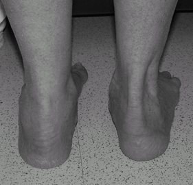 Fig. 1. Clinical photograph showing increased valgus and the too-many-toes sign on the right foot.
P.343
Tests
Lab
None is needed.
Imaging
-
Radiography:
-
Weightbearing foot radiographs to assess the deformity and identify hindfoot arthritis
-
AP view: Lateral navicular subluxation with uncovering of talar head
-
Lateral view: Loss of medial arch height and decreased calcaneal pitch
-
-
Weightbearing ankle radiographs to assess alignment and identify arthritis:
-
Rule out valgus tilt of the talus on the mortise view.
-
-
-
If the diagnosis is in question, use MRI.
-
The PTT may show hypertrophy, thickening, splitting, attenuation, or frank rupture.
-
Pathological Findings
-
Tenosynovitis in an early stage
-
Progressive tendinosis with degeneration and attenuation
-
Continued strain on the tendon can lead to complete rupture.
Differential Diagnosis
-
Benign flatfoot
-
Tarsal tunnel syndrome
-
Inflammatory arthritis of the hindfoot
-
Charcot arthropathy
General Measures
-
Ankle stirrup brace, cast, or boot brace immobilization until acute symptoms subside
-
Rest and assistive devices, such as a cane
-
Patients with tenosynovitis or partial tear may benefit from NSAIDs.
-
Once severe pain improves with immobilization, patients may transition to a semirigid orthotic arch support.
-
More severe deformity may necessitate the use of an ankle-foot orthosis.
Special Therapy
Physical Therapy
Physical therapy is used for muscle strengthening,
motion exercises, ultrasound therapy, proprioception training, and gait
mechanics.
motion exercises, ultrasound therapy, proprioception training, and gait
mechanics.
Medication
NSAIDs
Surgery
-
Surgery is indicated for persistent pain, worsening deformity, or failure of nonsurgical treatments.
-
Stage I: Tenosynovectomy, possible flexor digitorum longus transfer ± medial sliding calcaneal osteotomy
-
Stage II: Multiple components needed, including:
-
± Flexor digitorum longus tendon transfer and medial sliding calcaneal osteotomy or lateral column distraction arthrodesis or subtalar arthrodesis
-
± Plantarflexion cuneiform osteotomy or first tarsometatarsal arthrodesis
-
Achilles lengthening or gastrocnemius recession
-
-
Stage III: Triple arthrodesis ± Achilles lengthening or gastrocnemius recession
-
Stage IV: Pantalar arthrodesis
-
Disposition
Issues for Referral
-
May be treated by general orthopaedist with experience in foot reconstructive surgery
-
Often results in referral to orthopaedic foot and ankle specialist
Prognosis
-
Nonoperative treatment (brace, orthotics)
may be sufficient in sedentary or elderly patients, but if the
condition is left untreated because the patient cannot tolerate the
devices, a high risk of progression of hindfoot and ankle arthritis
occurs (3,4). -
The prognosis is good with various
surgical reconstructions, yielding high rates of patient satisfaction
and functional recovery (5,6).
Complications
-
Weakness
-
Recurrent deformity
-
Arthritis
Patient Monitoring
Patients are followed every 2–3 months until symptoms resolve.
References
1. Johnson KA, Strom DE. Tibialis posterior tendon dysfunction. Clin Orthop Relat Res 1989;239: 196–206.
2. Myerson MS. Adult acquired flatfoot deformity. Treatment of dysfunction of the posterior tibial tendon. J Bone Joint Surg 1996;78A:780–792.
3. Chao W, Wapner KL, Lee TH, et al. Nonoperative management of posterior tibial tendon dysfunction [see comments]. Foot Ankle Int 1996;17:736–741.
4. Friedman
MA, Draganich LF, Toolan B, et al. The effects of adult acquired
flatfoot deformity on tibiotalar joint contact characteristics. Foot Ankle Int 2001;22:241–246.
MA, Draganich LF, Toolan B, et al. The effects of adult acquired
flatfoot deformity on tibiotalar joint contact characteristics. Foot Ankle Int 2001;22:241–246.
5. Myerson
MS, Corrigan J, Thompson F, et al. Tendon transfer combined with
calcaneal osteotomy for treatment of posterior tibial tendon
insufficiency: a radiological investigation. Foot Ankle Int 1995;16:712–718.
MS, Corrigan J, Thompson F, et al. Tendon transfer combined with
calcaneal osteotomy for treatment of posterior tibial tendon
insufficiency: a radiological investigation. Foot Ankle Int 1995;16:712–718.
6. Toolan
BC, Sangeorzan BJ, Hansen ST, Jr. Complex reconstruction for the
treatment of dorsolateral peritalar subluxation of the foot. Early
results after distraction arthrodesis of the calcaneocuboid joint in
conjunction with stabilization of, and transfer of the flexor digitorum
longus tendon to, the midfoot to treat acquired pes planovalgus in
adults. J Bone Joint Surg 1999;81A:1545–1560.
Additional Reading
Anderson RB, Davis WH. Management of the adult flatfoot deformity. In: Myerson MS, ed. Foot and Ankle Disorders. Philadelphia: WB Saunders, 2000:1017–1039.
Basmajian JV, Stecko G. The role of muscles in arch support of the foot. J Bone Joint Surg 1963;45A: 1184–1190.
Frey C, Shereff M, Greenidge N. Vascularity of the PTT. J Bone Joint Surg 1990; 72A:884–888.
Holmes GB, Jr, Mann RA. Possible epidemiological factors associated with rupture of the PTT. Foot Ankle 1992;13:70–79.
Mann RA, Thompson FM. Rupture of the PTT causing flat foot. Surgical treatment. J Bone Joint Surg 1985;67A:556–561.
Pomeroy GC, Pike RH, Beals TC, et al. Acquired flatfoot in adults due to dysfunction of the PTT. J Bone Joint Surg 1999;81A: 1173–1182.
Codes
ICD9-CM
-
726.72 Posterior tibialis tendinitis
-
727.68 Posterior tibial tendon rupture
-
734 Acquired pes planus (flatfoot)
Patient Teaching
Inform the patient that the disorder is degenerative in nature and may progress to worsened deformity, arthritis, and pain.
FAQ
Q: What are the typical foot deformities seen with PTT dysfunction?
A:
Progressive collapse of the medial longitudinal arch occurs with
abduction and supination of the midfoot and valgus of the heel.
Progressive collapse of the medial longitudinal arch occurs with
abduction and supination of the midfoot and valgus of the heel.
Q: What is the most common underlying pathology of PTT dysfunction?
A: The tendon shows chronic degenerative changes, with splitting, attenuation, or frank tearing.
Q: What is the pathognomonic physical finding suggestive of PTT dysfunction?
A:
The too many toes sign, indicating abduction of the midfoot with arch
collapse. The patient is viewed from behind, and the involved side
shows more toes lateral to the fibula than does the uninvolved foot.
The too many toes sign, indicating abduction of the midfoot with arch
collapse. The patient is viewed from behind, and the involved side
shows more toes lateral to the fibula than does the uninvolved foot.