
muscle atrophy or hypertrophy is an important part of the motor
examination. There is normally an appreciable individual variation in
muscular development, but noteworthy changes in the size or shape of
individual muscles or muscle groups, especially when focal or
asymmetric, may be significant.
volume or bulk, and is usually accompanied by changes in shape or
contour. Neurologic conditions likely to cause muscle atrophy are
primarily those that affect the anterior horn cell, the nerve root(s),
the peripheral nerve, or the muscle. Neuromuscular junction disorders
do not cause muscle atrophy. Atrophy may also result from such things
as disuse or inactivity, immobilization, tendonotomy, muscle ischemia,
malnutrition, endocrine disorders, and normal aging.
volume, of muscle tissues. It may result from excessive use of the
muscles (physiologic hypertrophy) or occur on a pathologic basis.
Hypertrophied muscle is not necessarily stronger than normal.
Persistent abnormal muscle contraction may cause hypertrophy. Patients
with myotonia congenita have a diffuse muscularity without significant
increase in strength. Patients with dystonia may develop hypertrophy of
the abnormally active muscle. In cervical dystonia (spasmodic
torticollis), it is common to see hypertrophy of one sternomastoid
muscle. Muscular dystrophies, especially Duchenne dystrophy, often
cause pseudohypertrophy of muscle, with enlargement due to infiltration
of the muscle with fat and connective tissue without an actual increase
in muscle fiber size or number.
muscular development, in part constitutional and in part due to
training, activity, and occupation. Certain individuals have small or
poorly developed muscles, while others show outstanding muscular
development. The sedentary, the elderly, and those with chronic disease
may have small muscles without evidence of wasting or atrophy. Athletes
may develop physiologically hypertrophic muscles. In normal
individuals, the dominant side may exhibit an increase in the size of
the muscles, even of the hand and foot. The appraisal of bulk and
contour should be correlated with the other parts of the motor
examination, especially with the evaluation of strength and tone.
inspection, palpation, and measurement. Inspection generally compares
symmetric parts on the two sides of the body, noting any flattening,
hollowing,
or bulging of the muscle masses. A useful technique for comparing
extremities is to look down the long axis. Hold the patient’s arms
outstretched and close together, comparing “down the barrel” from
fingertips to shoulders for any asymmetry.
consistency. Normal muscles are semi-elastic and regain their shape at
once when compressed. When hypertrophy is present, the muscles are firm
and hard; in pseudohypertrophy they appear enlarged but may feel doughy
or rubbery on palpation. Atrophic muscles are often soft and pulpy in
consistency. When degenerated muscles have undergone fibrotic changes,
they may be hard and firm. Those infiltrated or replaced by fat may
feel pliant and flabby.
hypertrophy. A pronounced difference in muscle size may be recognized
at a glance, especially when confined to one side of the body, one
extremity, or one segment of a limb. Slight differences are more
difficult to detect, and measurements with a tape measure or calipers
may be necessary. Measurements should be made from fixed points or
landmarks, and the sites—such as the distance above or below the
olecranon, anterior superior iliac spine, or patella—recorded. The
extremities should be in the same position and in comparable states of
relaxation. It may also be valuable to measure the length of the limbs.
muscle, to muscles supplied by a specific structure (e.g., a nerve or
root), to those muscles supplied by certain spinal cord segments, or to
one half of the body; or it may be multifocal or generalized. In
atrophy related to arthritis and disuse there may be a pronounced
decrease in volume with little change in strength. In myopathies, on
the other hand, there is often little atrophy in spite of a striking
loss of power.
Neurogenic atrophy follows disease of the anterior horn cell, root, or
peripheral nerve. Atrophy due to other neurologic processes, such as
the hemiatrophy associated with congenital hemiplegia, is not typically
considered neurogenic atrophy even though it is related to nervous
system disease. The term neurogenic atrophy as commonly used implies
disease affecting some part of the lower motor neuron. Myogenic atrophy
is that due to muscle disease, such as muscular dystrophy. As a
generalization, when weakness and wasting are comparable the process is
more likely to be neurogenic; when the weakness is disproportionately
greater than the wasting the process is more likely to be myopathic.
When a muscle appears wasted but is not weak the cause is likely to be
non-neurologic, such as disuse.
or its peripheral processes, the affected muscle lies inert and
flaccid, and no longer contracts voluntarily or reflexively. Muscle
fibers decrease in size, causing wasting or atrophy of the entire
muscle mass. Without timely reinnervation the muscle may become
fibrotic, with an increase in connective tissue and fatty infiltration.
The more abrupt or extreme the interruption of nerve supply, the more
rapid is the wasting. The atrophy may either precede or follow other
signs, such as weakness. In rapidly progressing diseases weakness
precedes atrophy, but in slowly progressive diseases the atrophy may
precede appreciation of weakness. If the pathologic process is confined
to the anterior horn cells or the spinal cord, the neurogenic atrophy
is segmental in distribution. Some conditions cause rapid destruction
of the anterior horn cells and atrophy in the distribution of the
affected spinal cord segments that develops within a short period of
time (e.g., poliomyelitis).
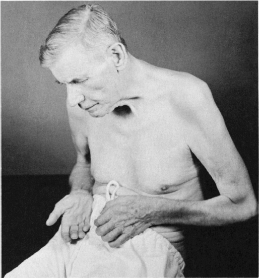
(e.g., amyotrophic lateral sclerosis [ALS]), there is a gradual but
widespread degeneration of the brainstem motor nuclei and anterior horn
cells, causing progressive muscular atrophy that may appear before
weakness is evident
(Figure 20.1).
The distribution of the atrophy is important. To make a diagnosis of
motor neuron disease, it is necessary to demonstrate widespread
denervation in a multiple nerve, multiple root distribution. Eventually
the disease becomes widespread, but it often begins segmentally in one
limb.
 |
|
FIGURE 20.1 • A patient with amyotrophic lateral sclerosis, showing advanced atrophy of the muscles of the hands and shoulders.
|
classical ALS and in progressive spinal muscular atrophy (SMA) of the
Aran-Duchenne type, atrophy is usually first seen in the distal
musculature, then spreads up the limbs to the proximal parts. In
hereditary motor neuron syndromes, the involvement is often proximal.
The proximal distribution and slow progression in SMA type 3 (juvenile
proximal SMA, Kugelberg-Welander disease) may simulate muscular
dystrophy. Segmental atrophy may also follow focal spinal cord lesions
involving the anterior horn cells (e.g., syringomyelia). The rapidity
of the progress depends upon the type of pathologic change.
peripheral nerves leads to atrophy of the muscles supplied by the
diseased or injured component. With severe lesions involving a
peripheral nerve or nerve plexus, atrophy may develop within a short
period of time. Lesions involving single nerve roots usually do not
cause much atrophy, because most muscles are innervated from more than
one level. Marked wasting in a disease that appears consistent with
radiculopathy suggests multiple root involvement. In generalized
peripheral neuropathy, weakness and wasting are usually greatest in the
distal portions of the extremities. The amount of atrophy depends on
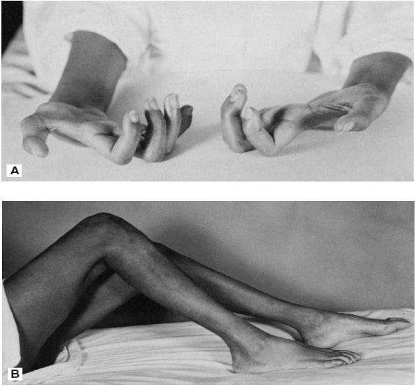
the severity and chronicity of the neuropathy. The hereditary
peripheral neuropathy, Charcot-Marie-Tooth disease (peroneal muscular
atrophy), typically causes marked atrophy in a characteristic
distribution involving the lower legs (inverted champagne bottle
deformity, Figure 20.2). Because of
interruption of autonomic pathways, diseases of the lower motor neuron
may be associated with trophic changes in the skin and subcutaneous
tissues.
followed by atrophy of the paralyzed muscles except for some
generalized loss of muscle volume and secondary wasting because of
disuse, which is seldom severe. With lesions dating from birth or early
childhood, there may be a failure of growth of the contralateral body.
Such congenital hemiatrophy may involve one side of the face or the
face and corresponding half of the body.
 |
|
FIGURE 20.2
• A patient with Charcot-Marie-Tooth disease (peroneal muscular atrophy), showing wasting of distal muscles and contractures of the hands and feet. |
primary muscle disease. In some conditions there may be prominent
wasting without much weakness. In most of these, the primary pathologic
change is type 2 fiber atrophy. Wasting with little weakness occurs in
disuse, aging, cachexia, and some endocrine myopathies. Weakness out of
proportion to wasting occurs in inflammatory myopathy, myasthenia
gravis, and periodic paralysis.
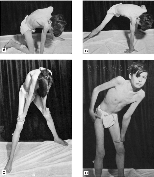
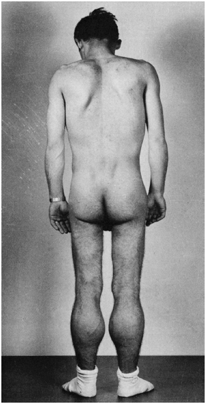
distribution of the wasting parallels the weakness. In
dystrophinopathies, the weakness and atrophy primarily involve the
pelvic and shoulder girdle muscles (Figure 20.3).
As the disease progresses there is increasing wasting of all muscles of
the shoulders, upper arms, pelvis, and thighs. In the face of all of
the atrophy, certain muscles—particularly the calf muscles—are
paradoxically enlarged due to pseudohypertrophy. The limb-girdle
syndromes also primarily involve the pelvic and shoulder girdles. In
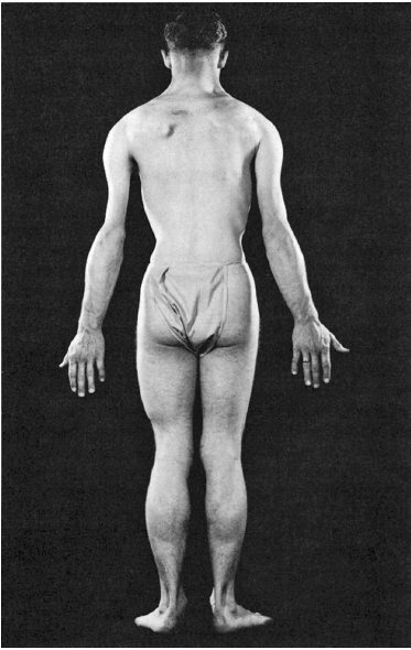
facioscapulohumeral dystrophy, the atrophy predominates in the muscles
of the face, shoulder girdles (especially the trapezius and
periscapular muscles), and upper arms, especially the biceps (Figure 20.4).
Involvement is often asymmetric and occasionally there is
pseudohypertrophy of the deltoid and other shoulder muscles. Some
myopathies cause striking weakness and atrophy involving certain
muscles or muscle groups.
part of the body. It may be rapid in onset and can sometimes simulate
neurogenic atrophy. The degree of muscle wasting is greater than the
degree of weakness, which may be minimal or absent. Muscle atrophy may
accompany malnutrition, weight loss, cachexia, and other wasting
diseases. The loss of muscle mass is typically greater than the degree
of accompanying weakness.
atrophy and other changes in muscle. In thyrotoxic myopathy atrophy is
particularly prone to involve the shoulder girdle and may lead to
scapular
winging. Myopathy due to excess corticosteroids, exogenous or
endogenous, may be associated with muscle wasting. Muscle wasting also
occurs with diabetes. Distal weakness and atrophy are common in
diabetic distal axonopathy. Diabetic amyotrophy is a common syndrome of
bilateral but asymmetric weakness and atrophy that involves the pelvic
and thigh muscles due to radiculoplexopathy. It is usually associated
with severe pain.
 |
|
FIGURE 20.3
• A patient with muscular dystrophy, showing wasting of the musculature in the shoulders and thighs; weakness and atrophy of the glutei cause difficulty in assuming the erect position, and the patient “climbs up on his thighs” (Gowers maneuver) in order to stand erect. |
mistaken for atrophy. Almost any muscle may be congenitally absent, but
some are particularly prone, including the depressor angulii oris,
palmaris longus, trapezius, peroneus tertius, and anterior abdominal
muscles (prune belly syndrome).
atrophy. In true muscle hypertrophy the muscle is enlarged, in
pseudohypertrophy the muscle appears enlarged because it is replaced by
fat and fibrous tissue. Except for physiologic hypertrophy due to
exercise, pseudohypertrophy is
encountered
more commonly than true hypertrophy. Pseudohypertrophy is common in
some forms of muscular dystrophy. Muscle biopsy reveals severe
myopathy, with fatty and connective tissue infiltrations.
Pseudohypertrophy is common in Duchenne and Becker dystrophy; an
alternate term for Duchenne dystrophy is pseudohypertrophic muscular
dystrophy. Certain muscles, particularly the calf muscles and the
infraspinatus, are often strikingly enlarged due to pseudohypertrophy (Figure 20.5).
Comparing the circumference of the calf to the knee is most
informative. In the early stages of the disease, the enlarged muscles
may feel firm and hard and remain strong, and there may actually be an
element of true hypertrophy. With progression, they develop a soft
doughy or rubbery feeling.
 |
|
FIGURE 20.4 • A patient with scapulohumeral muscular dystrophy, showing atrophy of the muscles of the shoulders and upper arms.
|
 |
|
FIGURE 20.5 • A patient with muscular dystrophy, showing pseudohypertrophy of the calf muscles.
|
especially the dominant form (Thomsen disease), because of the
excessive contraction. These patients may have the impressive
muscularity of a bodybuilder; although they may appear strong and
muscular, strength is normal or there is even slight weakness. Muscle
enlargement, either true hypertrophy or pseudohypertrophy, occurs as an
occasional feature in other neuromuscular disorders. Muscle enlargement
may be a manifestation of hypothyroidism. Muscle enlargement may also
occur due to interstitial infiltrates, as in sarcoidosis and
amyloidosis. Loss of body fat may lend the appearance of muscle
enlargement.