
Editors: Frassica, Frank J.; Sponseller, Paul D.; Wilckens, John H.
Title: 5-Minute Orthopaedic Consult, 2nd Edition
Copyright ©2007 Lippincott Williams & Wilkins
> Table of Contents > Accessory Navicular
Accessory Navicular
Kris J. Alden MD, PhD
Description
-
This anatomic variant consists of an accessory ossicle located at the medial edge of the navicular (Fig. 1).
-
Accessory ossicles are derived from unfused ossification centers.
-
Considered an incidental finding on radiographs, but may become symptomatic
-
Classification: 3 major types of accessory navicular adjacent to the posteromedial navicular tuberosity (1)
-
Type I: Small, 2–3-mm sesamoid bone in the PTT; referred to AS “os tibiale externum”
-
Type II:
-
Larger ossicle than type I
-
Secondary ossification center of the navicular bone
-
-
Type III: Enlarged navicular tuberosity, considered a fused variant of a type II, often with pointed shape
-
-
Synonyms: Os tibiale; Os tibiale externum; Naviculare secundum
Pediatric Considerations
Often presents in adolescent patients or young adults, with flatfoot deformity and arch pain
Epidemiology
Incidence
-
4–21% incidence; 89% of cases are bilateral (2).
-
One of the most common accessory ossicles in the foot
-
It is seen over the medial pole of the navicular bone, usually in adolescent patients (3).
-
It is most commonly symptomatic in the 2nd decade of life and causes medial foot pain (4).
-
<1% of patients become symptomatic.
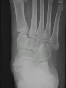 Fig. 1. AP radiograph showing an accessory navicular.
 -
Incidence by classification (1):
-
Type I: ~30% of all accessory navicular bones
-
Type II: ~50%
-
Type III: ~20%
-
Prevalence
-
Usually affects teens and young adults
-
More frequent in females
-
May be seen in older adults as incidental finding or, in rare cases, as symptomatic
Etiology
-
The accessory navicular is a variant of normal anatomy.
-
It may become symptomatic from the bony prominence impinging against shoe wear.
-
The patient may have diffuse medial and plantar arch pain.
-
It may cause problems by destabilizing the insertion and diminishing the pull of the PTT.
-
In patients with associated severe
flatfoot deformity, lateral pain may occur secondary to impingement of
the calcaneus against the fibula. -
A traumatic event can cause injury to the fibrocartilaginous synchondrosis that attaches the ossicle to the main navicular.
Associated Conditions
-
Flatfoot deformity
-
Secondary Achilles tendon contracture
Signs and Symptoms
-
Pain may begin after wearing ill-fitting shoes, with weightbearing activities or athletics, or after trauma to the foot.
-
Characteristics:
-
Pain and tenderness along the medial aspect of the foot in the region of the accessory navicular
-
Pain or weakness when the patient attempts to rise on toes, run, or jump
-
Often increased prominence over the medial end of the navicular
-
History
-
The pain is localized to the medial aspect of the navicular.
-
Symptomatic accessory tarsal navicular may develop in young athletes (5,6,7).
-
Exacerbated by weightbearing, walking, athletic activity, or the wearing of narrow shoes
-
Pain often is relieved by rest.
Physical Exam
-
Tenderness is localized to the medial pole of the navicular.
-
May be exacerbated by abducting and adducting the foot
-
-
Assess the insole of the shoe, which may exacerbate symptoms.
-
Assess the strength of the PTT by manual
resistance testing against plantarflexion-inversion and by determining
the ability to perform multiple single-limb heel rises. -
Assess ankle and subtalar joint motion.
-
Identify contracture of the Achilles tendon.
Tests
Imaging
-
Obtain routine standing AP, external oblique, and lateral radiographs of the foot.
-
Type-II accessory ossicle has smooth borders, is triangular or heart-shaped, and measures 9 × 12 mm in size.
-
The base is situated 1–2 mm from the medial and posterior aspects of the navicular bone.
-
The accessory ossicle may be best visualized on the internal oblique view.
-
Smooth margins with well-formed cortex differentiate this condition from acute fracture.
-
-
Bone scan:
-
May show increased activity over an accessory navicular
-
May be needed if a navicular stress fracture is suspected in the differential diagnosis
-
-
MRI:
-
Useful when plain films are unremarkable
-
Often, a type-II accessory navicular is
attached to the tuberosity by a fibrocartilage or hyaline cartilage
layer, and MRI may show soft-tissue edema consistent with a
synchondrosis sprain or tear. -
Shows altered signal intensity and bone marrow edema, suggestive of chronic stress and/or osteonecrosis (8)
-
Also helpful in showing PTT degeneration
-
Pathological Findings
-
This separate osteocartilaginous fragment is located in place of the normal medial pole of the navicular.
-
The PTT inserts on the accessory navicular, navicular body, and cuneiforms.
Differential Diagnosis
-
Navicular fracture may mimic an acute avulsion fracture of the tuberosity of the navicular.
-
Posterior tibial tendinitis
-
Stress fracture of navicular
P.3
General Measures
-
The patient should rest and avoid athletics or aggravating activities.
-
Anti-inflammatory medication
-
Shoe-wear modification, including use of a softer, wider shoe
-
If flatfoot is present, a medial arch
support may be useful, but often the patient may not tolerate it
because of direct pressure on the ossicle. -
Below-the-knee walking cast or removable fracture boot may be used for 3–6 weeks for persistent symptoms.
-
Physical therapy, including strengthening exercises and cryotherapy, may be helpful.
Medication (Drugs)
No evidence suggests that one NSAID is superior to another.
Surgery
-
If pain is progressive or does not remit with nonoperative treatment, surgical excision may be considered.
-
In the Kidner procedure, the accessory navicular is excised, and the PTT is rerouted into a more plantar position (9).
-
Contemporary surgical treatment:
-
Includes excision of the ossicle and
reattachment of the PTT insertion to the navicular, with suture anchors
or sutures passed through drill holes -
Typically provides satisfactory outcome and good pain relief, particularly in adolescents
-
-
Severe flatfoot deformity with lateral
impingement symptoms may require concomitant osteotomy of the calcaneus
and/or medial column of the foot to improve alignment and decrease
mechanical stress of the PTT insertion.
Complications
Weakness, incomplete pain relief, continued deformity
References
1. Sella EJ, Lawson JP, Ogden JA. The accessory navicular synchondrosis. Clin Orthop Relat Res 1986;209:280–285.
2. Miller TT. Painful accessory bones of the foot. Semin Musculoskelet Radiol 2002;6:153–161.
3. Lawson JP, Ogden JA, Sella E, et al. The painful accessory navicular. Skeletal Radiol 1984;12:250–262.
4. Romanowski CAJ, Barrington NA. The accessory navicular—an important cause of medial foot pain. Clin Radiol 1992;46:261–264.
5. Mygind HB. The accessory tarsal seaphoid [sic]. Clinical features and treatment. Acta Orthop Scand 1953;23:142–151.
6. Ray S, Goldberg VM. Surgical treatment of the accessory navicular. Clin Orthop Relat Res 1983;177:61–66.
7. Veitch JM. Evaluation of the Kidner procedure in treatment of symptomatic accessory tarsal scaphoid. Clin Orthop Relat Res 1978;131:210–213.
8. Demeyere N, De Maeseneer M, Osteaux M. Quiz case. Symptomatic type II accessory navicular. Eur J Radiol 2001;37:60–63.
9. Kidner FC. The prehallux (accessory scaphoid) in its relation to flat-foot. J Bone Joint Surg 1929;11:831–837.
Additional Reading
Coughlin MJ. Sesamoids and accessory bones of the foot. In: Coughlin MJ, Mann RA, eds. Surgery of the Foot and Ankle, 7th ed. St. Louis: Mosby, 1999:437–499.
Kidner FC. The prehallux in relation to flatfoot. JAMA 1933;101:1539–1542.
Kopp FJ, Marcus RE. Clinical outcome of surgical treatment of the symptomatic accessory navicular. Foot Ankle Int 2004;25:27–30.
Codes
ICD9-CM
-
754.61 Pes planus, congenital
-
755.56 Accessory navicular
Patient Teaching
-
Instruct patients on the typically benign nature of the condition.
-
If the condition is secondary to medial pressure from the shoe, suggest a wider, softer shoe.
-
Recommend rest from sports with gradual return when symptoms subside.
FAQ
Q: What is the most common type of accessory navicular seen radiographically?
A: Type-II, with a large accessory ossicle, is the most common.
Q: What are the standard treatment methods for initial management of a symptomatic accessory navicular?
A:
Rest, NSAIDs, restriction from sports, immobilization with a boot brace
or walking cast, physical therapy, and orthotic arch supports.
Rest, NSAIDs, restriction from sports, immobilization with a boot brace
or walking cast, physical therapy, and orthotic arch supports.