
Editors: Frassica, Frank J.; Sponseller, Paul D.; Wilckens, John H.
Title: 5-Minute Orthopaedic Consult, 2nd Edition
Copyright ©2007 Lippincott Williams & Wilkins
> Table of Contents > Lisfranc Fracture-Dislocation
Lisfranc Fracture-Dislocation
Simon C. Mears MD, PhD
Clifford L. Jeng MD
Description
-
Dislocation of the TMT joints of the foot (Fig. 1).
-
Can occur at any age
-
Often accompanied by fractures around the TMT joints
-
-
Classification (1):
-
Type A: Total incongruity of TMT joint
-
Type B: Partial incongruity of TMT joint complex, either medial or lateral
-
Type C: Divergent (1st metatarsal medial, 2nd–5th lateral)
-
Epidemiology
Incidence
Lisfranc injuries account for ~1/3 of midfoot injuries (2).
Risk Factors
-
Car accidents
-
Motorcycle accidents
Etiology
-
The mechanism of injury includes a wide
spectrum of causes from low-energy compression and twisting to
high-energy crush injuries. -
Common cause: Car and motorcycle accidents (3):
-
Pressure on a brake pedal with a plantar flexed foot leads to the Lisfranc pattern of injury (4).
-
-
Sporting events (5)
Associated Conditions
-
Comminuted fractures of the metatarsal bases or cuneiforms
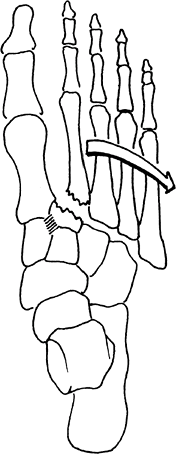 Fig. 1. Lisfranc dislocation occurs at the midfoot joints, usually with substantial trauma.
 -
Severe soft-tissue injury
-
Compartment syndrome
-
Open fractures
Signs and Symptoms
-
Pain
-
Swelling
-
Deformity
-
Ecchymosis
-
Difficulty bearing weight
-
Tenderness over midfoot
-
Possible spontaneous reduction
Physical Exam
-
Rotational stress on the forefoot causes pain at Lisfranc joint.
-
Palpation over the 2nd metatarsal base also can cause pain.
Tests
Imaging
-
Radiography:
-
Plain films usually are diagnostic.
-
AP, lateral, and oblique projections are mandatory.
-
On the AP view, the medial margin of the 2nd metatarsal base should be aligned with the middle cuneiform.
-
On the oblique view, the medial base of the 4th metatarsal should be aligned with the medial margin of the cuboid.
-
On the lateral view, an unbroken line should run from the dorsum of the 1st and 2nd metatarsals to the corresponding cuneiform.
-
-
Avulsion fracture of the 2nd metatarsal
base (“fleck” fracture) and compression fracture of the cuboid are
pathognomonic of this condition (6). -
If the diagnosis is uncertain on plain
film, especially if the Lisfranc joint has reduced spontaneously,
stress radiography with fluoroscopy may be helpful in further defining
the instability pattern.
-
-
CT is an important adjunct to plain radiographs.
-
Shows small fractures and displacements that are not visible on plain films
-
In 1 study, radiographs missed the Lisfranc injury in 24% of cases and CT scan revealed the injury (7).
-
Differential Diagnosis
-
Soft-tissue contusion
-
Ligament sprain
-
Isolated metatarsal or midfoot fractures
General Measures
-
Before the patient is taken to the operating room:
-
Compartment syndrome and neurovascular injury should be assessed.
-
The foot is splinted and kept elevated until surgery.
-
-
The goal of treatment is to achieve and maintain anatomic reduction of the joints while the ligaments heal.
-
Usually requires surgical intervention
-
-
Before and after surgery: Ice, elevation, and a compression dressing
-
Foot pumps may help reduce foot swelling.
Special Therapy
Physical Therapy
-
Patients should be referred for gait training on a nonweightbearing basis postoperatively.
-
Edema control and ROM of the toes and ankle are important to decrease late stiffness.
Surgery
-
Open reduction and internal fixation of joints:
-
Through 2–3 dorsal longitudinal incisions
-
Fixation may consist of Kirschner wires or 3.5-mm cortical screws.
-
If Kirschner wires are used as fixation,
they can be removed in the office at 6 weeks, and the patient may begin
protected weightbearing. -
If screws are used as fixation,
unprotected weightbearing is not permitted until the screws have been
removed, at 10–12 weeks after surgery.
-
-
-
Fusion of the joints with 3.5-mm cortical screws has been advocated by some as primary treatment (8) or as a salvage procedure for later arthritis of the midfoot (9).
P.231
Prognosis
-
Patients with anatomic reduction generally have good results.
-
In 1 study, 11 of 24 patients had a good to excellent result (10).
-
-
Outcomes are worse with nonanatomic reduction and extensive joint injury (11).
-
Patients with worker compensation claims have poorer outcomes (12).
-
The role of joint fusion is controversial.
-
Recently, a randomized study showed that joint fusion gave better results than did reduction and fixation (8).
-
-
Patients with posttraumatic arthritis can undergo salvage procedures with arthrodesis.
Complications
-
Traumatic arthritis
-
Fixed deformity
-
For injuries diagnosed late (7–8 weeks):
-
Poor prognosis
-
Patients may be candidates for primary arthrodesis (13).
-
Patient Monitoring
Follow-up radiographs (at 1-month intervals) should be taken to check for maintained alignment of the Lisfranc complex.
References
1. Hardcastle
PH, Reschauer R, Kutscha-Lissberg E, et al. Injuries to the
tarsometatarsal joint. Incidence, classification and treatment. J Bone Joint Surg 1982;64B:349–356.
PH, Reschauer R, Kutscha-Lissberg E, et al. Injuries to the
tarsometatarsal joint. Incidence, classification and treatment. J Bone Joint Surg 1982;64B:349–356.
2. Richter
M, Wippermann B, Krettek C, et al. Fractures and fracture dislocations
of the midfoot: occurrence, causes and long-term results. Foot Ankle Int 2001;22:392–398.
M, Wippermann B, Krettek C, et al. Fractures and fracture dislocations
of the midfoot: occurrence, causes and long-term results. Foot Ankle Int 2001;22:392–398.
3. Jeffers RF, Tan HB, Nicolopoulos C, et al. Prevalence and patterns of foot injuries following motorcycle trauma. J Orthop Trauma 2004;18: 87–91.
4. Smith BR, Begeman PC, Leland R, et al. A mechanism of injury to the forefoot in car crashes. Traffic Inj Prev 2005;6:156–169.
5. Nunley JA, Vertullo CJ. Classification, investigation, and management of midfoot sprains: Lisfranc injuries in the athlete. Am J Sports Med 2002;30:871–878.
6. Pearse EO, Klass B, Bendall SP. The “ABC” of examining foot radiographs. Ann R Coll Surg Engl 2005;87:449–451.
7. Haapamaki VV, Kiuru MJ, Koskinen SK. Ankle and foot injuries: analysis of MDCT findings. AJR Am J Roentgenol 2004;183:615–622.
8. Ly
TV, Coetzee JC. Treatment of primarily ligamentous Lisfranc joint
injuries: primary arthrodesis compared with open reduction and internal
fixation. A prospective, randomized study. J Bone Joint Surg 2006;88A:514–520.
TV, Coetzee JC. Treatment of primarily ligamentous Lisfranc joint
injuries: primary arthrodesis compared with open reduction and internal
fixation. A prospective, randomized study. J Bone Joint Surg 2006;88A:514–520.
9. Komenda GA, Myerson MS, Biddinger KR. Results of arthrodesis of the tarsometatarsal joints after traumatic injury. J Bone Joint Surg 1996;78A: 1665–1676.
10. O’Connor PA, Yeap S, Noel J, et al. Lisfranc injuries: patient- and physician-based functional outcomes. Int Orthop 2003;27:98–102.
11. Kuo RS, Tejwani NC, Digiovanni CW, et al. Outcome after open reduction and internal fixation of Lisfranc joint injuries. J Bone Joint Surg 2000;82A:1609–1618.
12. Calder JDF, Whitehouse SL, Saxby TS. Results of isolated Lisfranc injuries and the effect of compensation claims. J Bone Joint Surg 2004; 86B:527–530.
13. Aronow MS. Treatment of the missed Lisfranc injury. Foot Ankle Clin 2006;11:127–142.
Additional Reading
Sands AK, Grose A. Lisfranc injuries. Injury 2004;35:B71–B76.
Codes
ICD9-CM
825.25 Metatarsal fracture
Patient Teaching
Patients must be warned about the risks of traumatic arthritis and fixed deformity, which may require later arthrodesis.
FAQ
Q: How long will it take to recover from a Lisfranc injury?
A:
Lisfranc injuries are severe injuries to the midfoot and commonly
require surgery. Recovery takes at least a year. Patients may require
hardware removal. ~25% develop arthritis and may require later fusion.
Lisfranc injuries are severe injuries to the midfoot and commonly
require surgery. Recovery takes at least a year. Patients may require
hardware removal. ~25% develop arthritis and may require later fusion.