
Editors: Frassica, Frank J.; Sponseller, Paul D.; Wilckens, John H.
Title: 5-Minute Orthopaedic Consult, 2nd Edition
Copyright ©2007 Lippincott Williams & Wilkins
> Table of Contents > Clubfoot
Clubfoot
Paul D. Sponseller MD
Description
-
Clubfoot, or talipes equinovarus, is a complex deformity seen at birth.
-
It can be divided into 3 elements (1–4):
-
Equinus of the heel
-
Varus and internal rotation of the hindfoot
-
Adductus of the forefoot
-
-
Equinus (“horse”) is used to describe a plantarflexed position because horses walk with their heels in complete plantarflexion, and varus (“turned in”) refers to the adducted component.
-
The affected child thus bears weight along the lateral part of the foot, rather than on the sole.
-
Clubfoot can be divided into 2 categories:
-
An isolated, idiopathic type
-
Associated with other congenital
deformities (amniotic band syndrome, arthrogryposis, myelodysplasia,
diastrophic dwarfism, Larson syndrome, Freeman-Sheldon syndrome):-
These clubfeet have a tendency to be more
severe and refractory to nonoperative therapy and more often require
surgical correction. -
The talar neck is deviated medially and in a plantar direction.
-
The foot is smaller than normal, and shortening, usually <1 cm compared with the contralateral, normal side, may occur.
-
-
Epidemiology
Incidence
-
~1 of every 1,000 births (1)
-
Male:Female ratio: ~2:1
Risk Factors
-
A parent with the disorder
-
A sibling with the disorder
-
Presence of other disorders associated with clubfoot, including:
-
Amniotic band syndrome
-
Arthrogryposis
-
Myelodysplasia
-
Mobius syndrome
-
Freeman-Sheldon syndrome
-
Larsen syndrome
-
Diastrophic dwarfism
-
-
Drugs, such as aminopterin, taken during pregnancy
Genetics
-
The disorder is widely believed to be polygenic with variable penetrance.
-
If a child has a clubfoot, a 2–6% chance exists that the next sibling will have the disorder (1).
-
If a parent has clubfoot, a 10% chance exists that each child will inherit the disorder (1).
Etiology
The more common isolated, idiopathic form is thought to
be of a genetic origin, although the genetic basis has not been
identified.
be of a genetic origin, although the genetic basis has not been
identified.
Associated Conditions
Other congenital deformities (see conditions discussed earlier)
Signs and Symptoms
-
Signs and symptoms of clubfoot are related to the findings on physical examination.
-
In general, the newborn’s foot looks excessively turned inward.
-
In older children, uncorrected clubfeet
may cause gait disturbances, problems with shoe fit, and painful
callosities along the lateral border of the foot.
-
-
The patient may be forced to walk on the dorsum (top) of the foot.
-
Pain within the foot may develop in the older child or adult.
Physical Exam
-
The physical examination reveals equinus
of the heel, supination or varus of the hindfoot, and adductus of the
midfoot and forefoot (2), which creates a foot described as “kidney shaped,” with a prominent medial crease along the plantar aspect of the foot. -
The foot projects medially from the leg and resembles a club (Figs. 1 and 2).
-
Check the flexibility of the foot and the correctability of its position.
-
Verify the function of the ankle and toe muscles.
-
Check for calf hypoplasia, which is a typical feature.
Tests
Imaging
-
In routine care, radiographs may be
unnecessary unless a suspicion exists for underlying bony fusion or if
surgery is contemplated. -
Simulated standing AP and lateral views are most important.
-
Because of the deformity caused by clubfoot, these views may be difficult to take and may require multiple attempts.
-
The foot should be corrected to a position as close to neutral as possible and held in this position with a Plexiglas plate.
-
-
The AP view:
-
The forefoot is noted to be in adduction.
-
In a normal foot:
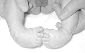 Fig. 1. Bilateral clubfeet in a newborn.
-
The talus aligns roughly with the first metatarsal, and the calcaneus aligns roughly with the 5th metatarsal.
-
The angle thus formed between the axes of these 2 bones is the anterior talocalcaneal angle, or Kite angle.
-
Normal range for this angle is 20–40°.
-
-
In clubfoot:
-
The axes of the calcaneus and talus are nearly parallel, and thus the angle is much smaller.
-
-
-
Lateral view:
-
The position of the foot is in equinus.
-
In a normal foot, the talocalcaneal angle is 35–50°.
-
In a clubfoot, that angle is considerably smaller because the 2 bones are nearly parallel in all planes.
-
-
The assessment of these radiographic
abnormalities in a clubfoot is used to determine whether a correction
is acceptable after any method of correction.
Pathological Findings
-
The essential bony change is medial deviation of the talar neck with subluxation of the talonavicular joint.
-
Histologic study also has revealed smaller muscle fibers than normal on cross-section.
-
Increased thickness of fascia and joint capsules is present on the medial side of the foot.
Differential Diagnosis
-
The differential diagnosis of clubfoot is limited because of the dramatic and characteristic appearance of this deformity.
-
Some cases of metatarsus adductus varus
are so severe that they are confused with clubfoot, but the equinus
component of clubfoot is absent in metatarsus adductus, and the
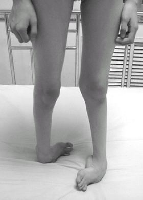
distinction should be easily made.![]() Fig. 2. Untreated clubfeet in a 9-year-old.
Fig. 2. Untreated clubfeet in a 9-year-old.
P.81
General Measures
-
The 1st step in management of clubfoot is taping or casting.
-
Should be started as soon as possible after birth
-
Can correct a clubfoot deformity and (for the most part) help restore normal function; it is most successful in mild cases.
-
Procedure:
-
The heel is firmly stabilized, the
forefoot is brought laterally out of adductus, the foot is dorsiflexed,
and the heel is externally rotated into valgus. -
This position allows the tight tendons to stretch so that additional correction may be obtained the next time.
-
Adhesive tape or a cast from above the knee to the toes (Fig. 3) (sometimes known as the “Ponseti method”) (1,2,3) is applied to hold the foot in this position, which essentially corrects all 3 components of clubfoot.
-
During the immediate postnatal term, the
cast or taping is changed every few days; after the 1st week, the
tapings are changed once to twice weekly in the office, until the
position of the foot is acceptable or improvement has ceased.
-
-
By ~8 weeks of age, the child’s foot and
leg are large enough that an above-the-knee cast may be applied with
the foot in the corrected position. -
Occasionally, an Achilles tenotomy is needed.
-
After the foot is corrected (which may
take 2–3 months), it should be held with reverse shoes and a Denis
Browne bar between the legs full-time for 2 months and at night and nap
times for 2–4 years.
-
Special Therapy
Physical Therapy
-
Some clinicians believe that certain
stretching exercises, particularly of the heel cord and medial ankle
structures, may be of some benefit, but exercises alone usually are not
sufficient. -
These exercises are most beneficial in maintaining the correction obtained by cast treatment.
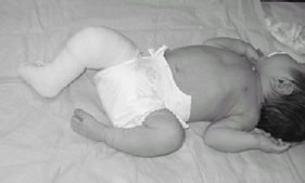 Fig. 3. Above-the-knee corrective cast for clubfoot.
Medication
First Line
The drug botulinum toxin shows promise in combination with casting and splinting.
Surgery
-
If cast treatment fails, surgery is needed.
-
Surgery is best performed when the child is 4–8 months old.
-
The general principle involves:
-
Releasing the tight medial structures by lengthening the flexor tendons and the PTT
-
Releasing the posterior structures by transecting the posterior joint capsule and lengthening the Achilles tendon
-
-
A more extensive release may be required.
-
The calcaneus also can be repositioned laterally, and a more complete correction can be achieved.
-
Pins are placed (and remain for 6 weeks) to help stabilize the correction.
-
-
About 10–20% of children require repeat surgery in later years (4).
Disposition
Issues for Referral
The patient should be referred to an orthopaedic surgeon who has expertise in treating clubfoot.
Prognosis
-
Adequate treatment, whether nonoperative
or surgical, likely will eliminate the positional deformity and enable
to patient to ambulate adequately on the affected foot. -
Certain elements of the deformity, such
as the small size of the foot, the calf hypoplasia, and the minimal
shortening, cannot be corrected, but these rarely have a large effect
on the patient’s functional ability.
Complications
-
Residual deformity
-
Rocker-bottom foot
-
Overcorrection into valgus
-
Stiffness
-
Pain in later childhood or adulthood
Patient Monitoring
-
Children need regular follow-up care for several years to monitor for recurrence.
-
Idiopathic clubfoot may recur up to age 6–7 years.
-
Most recurrences occur in the 1st few years, and may be treated with repeat casting.
References
1. Ponseti IV. The Ponseti technique for correction of congenital clubfoot [letter]. J Bone Joint Surg 2002;84A:1889–1890.
2. Morcuende JA, Abbasi D, Dolan LA, et al. Results of an accelerated Ponseti protocol for clubfoot. J Pediatr Orthop 2005;25:623–626.
3. Morcuende
JA. Congenital idiopathic clubfoot: prevention of late deformity and
disability by conservative treatment with the Ponseti technique. Pediatr Ann 2006;35:128–136.
JA. Congenital idiopathic clubfoot: prevention of late deformity and
disability by conservative treatment with the Ponseti technique. Pediatr Ann 2006;35:128–136.
4. Vitale MG, Choe JC, Vitale MA, et al. Patient-based outcomes following clubfoot surgery: a 16-year follow-up study. J Pediatr Orthop 2005;25:533–538.
Codes
ICD9-CM
754.51 Clubfoot
Patient Teaching
-
Clubfeet tend to be slightly shorter and less supple than normal feet.
-
Parents should be reassured that adequately corrected clubfeet have good long-term function.
-
Parents also must monitor for signs of recurrence.
FAQ
Q: Will a child with a clubfoot be able to participate in sports?
A: With appropriate treatment, most children should be able to participate.