
Editors: Frassica, Frank J.; Sponseller, Paul D.; Wilckens, John H.
Title: 5-Minute Orthopaedic Consult, 2nd Edition
Copyright ©2007 Lippincott Williams & Wilkins
> Table of Contents > Blount Disease
Blount Disease
Paul D. Sponseller MD
Description
-
Bowing of the legs can be a normal stage of growth for infants and toddlers.
-
This phase is called “physiologic bowing” and resolves spontaneously by approximately 2 years of age.
-
The vast majority of children presenting with bowed legs have this benign physiologic state.
-
-
More rarely, bowed legs may be pathologic.
-
The most common cause of this condition is Blount disease.
-
It is an abnormality of the proximal tibial growth plate secondary to overload.
-
It causes progressive varus alignment of the knees (bowed legs) in children or adolescents (1,2) (Fig. 1).
-
-
Classification:
-
Infantile form: Presents in children 0–4 years old
-
Juvenile form: Presents at >4–9 years of age in obese children
-
Adolescent form: Presents in children >10 years old; has excellent prognosis with surgery
-
-
Synonyms: Infantile tibia vara (0–4
years); Juvenile tibia vara (>4–9 years); Adolescent tibia vara
(10–16 years); Pathologic bowlegs
General Prevention
-
Weight control
-
Extremely early standing or walking should not be encouraged.
-
Early bracing sometimes is effective.
Epidemiology
Incidence
-
Infantile tibia vara is the most common cause of pathologic bowing in young children and accounts for <1% of all bowed legs (2).
-
The juvenile form is much less common: Only 60 reports in the literature in the United States.
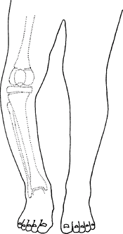 Fig. 1. In Blount disease, the varus is focal at the upper tibia.
 -
The adolescent form is becoming more commonly recognized.
Prevalence (1,2)
-
The infantile form is more common in girls.
-
The juvenile or adolescent form is more common in boys.
-
The disorder is more common in African American children than those of other races.
Risk Factors
-
African American ethnicity
-
Obesity
-
Early age of walking
-
Varus greater on the tibial than the femoral side is a risk factor for infantile and juvenile Blount disease (1).
Genetics
-
No genetic pattern has been proved.
-
More likely, patients inherit a body habitus that predisposes them to the disorder.
Pathophysiology
-
The growth plate shows islands of densely
packed cartilage cells with more hypertrophy than normal, islands of
almost acellular fibrous cartilage, and abnormal groups of capillaries (2). -
Biopsy is not indicated.
Etiology
Decreased growth of the proximal medial tibial growth
plate (physis) causes varus angulation (bowing) and internal rotation
of the proximal tibia secondary to weight-related overload of this
portion of the growth plate.
plate (physis) causes varus angulation (bowing) and internal rotation
of the proximal tibia secondary to weight-related overload of this
portion of the growth plate.
Associated Conditions
Obesity
Signs and Symptoms
-
Patients with infantile tibia vara
usually present between 14 and 40 months of age with increasingly bowed
legs (usually bilateral involvement). -
Adolescent presentation also involves
progressive varus deformity (bowing), but many of these patients also
have medial knee pain and often only 1 leg is affected. -
If untreated, the infantile form may progress to become severe.
-
The juvenile and adolescent forms rarely become as severe.
-
Some internal tibial torsion usually is present along with the bowing.
Physical Exam
-
Record the patient’s height, weight, and percentiles.
-
The finding of short stature suggests rickets or a skeletal dysplasia.
-
Note the location of any pain.
-
Record the gap between the medial sides of the knees and check knee ROM and ligamentous laxity.
-
Assess tibial torsion by the thigh–foot angle.
-
Perform a routine knee examination,
observe gait, and measure the foot progression angle (angle of the feet
with the direction of walking).
Tests
Lab
-
Testing is indicated if rickets is suspected.
-
In Blount disease, calcium, phosphorus, alkaline phosphatase, and renal function tests are all normal (2).
Imaging
-
Radiography:
-
Appropriate radiographs: A long leg AP view of the tibia and femur to evaluate the tibiofemoral angle and mechanical axis (1–5).
-
The radiograph should show the whole limb from the hip to the ankle, and it should be a true AP view of the knee (4).
-
The metaphyseal–diaphyseal angle differentiates Blount disease and physiologic varus:
-
<11° is physiologic varus.
-
≥16° indicates Blount disease.
-
Values between 11° and 16° signify a risk of potential Blount disease.
-
-
Reveals a medial physeal bar (disappearance of the growth plate with metaphysis–epiphysis fusion) in more advanced disease
-
-
CT or MRI:
-
Can be useful in delineating the physeal damage that later may form a bar.
-
Patients with adolescent Blount disorder (Fig. 2)
show less deformation of the epiphysis and rarely form a bar, but they
usually have some deformity on the femoral as well as on the tibial side
-
Differential Diagnosis
-
Physiologic bowed legs
-
Hypophosphatemic rickets
-
Trauma to metaphysis or growth plate
-
Osteochondroma
-
Metaphyseal chondrodysplasia
-
Focal fibrocartilaginous dysplasia
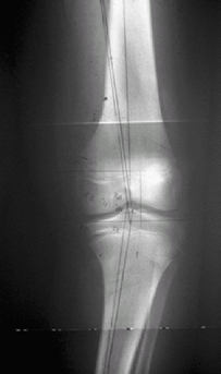 Fig. 2. Adolescent Blount disease. Note the physeal widening and relative preservation of the epiphysis of the tibia.
P.41
General Measures
-
Children <3 years old who present with
Blount disease may be braced, preferably with a long brace from the hip
to the ankle that is locked at the knee (2). -
Full-time bracing (22 hours a day) puts a
corrective valgus stress on the knee (more knock-kneed) and decreases
the stress on the medial physis.-
If bracing is begun when the deformity is mild, this treatment allows the growth plate to “catch up” the growth medially.
-
-
If the patient is being braced, full weightbearing is encouraged.
-
If bracing fails to correct the
deformity, or if a patient >3 years old presents with moderate to
severe deformity, an osteotomy is needed (6,7). -
If osteotomy is performed, the patient is
kept nonweightbearing until healing (8–12 weeks) of the osteotomy; then
full weightbearing can be resumed. -
Hemiepiphysiodesis is also an alternative in juveniles if the deformity is moderate (6).
Activity
-
No activity restriction on unbraced patients
-
Bracing restricts participation in sports.
Special Therapy
Physical Therapy
-
Does not help improve knee varus
-
Crutch training after osteotomy
-
Regaining knee ROM after osteotomy
Surgery
-
Osteotomy (cutting and realigning the proximal tibia) will decrease the stress on the medial physis and can allow healing.
-
If a physeal bar has formed:
-
An osteotomy often is combined with completion of the closure of the proximal tibial physis.
-
The whole growth plate should be fused to prevent the lateral portion of the growth plate from causing recurrent varus (5); patients with advanced cases may have a secondary deformity of the distal femur and/or the distal tibia.
-
-
If substantial leg-length inequality
develops, it may be treated by lengthening the short limb or by growth
plate closure of the longer limb. -
Adolescent Blount disease may be treated
with tethering (“stapling”) of the lateral sides of the growth plate to
allow the bone to correct itself. -
In more advanced cases, osteotomy of the tibia and/or the femur is needed.
Prognosis
-
Because the recurrence rate is higher in
patients treated after 4 years of age (70–75%) than in patients treated
before age 4 (20–30%) (2), early osteotomy (before age 4) should be performed if bracing is not successful. -
Patients with late treatment or incomplete treatment have an increased risk of arthritis of the knee.
Complications
-
Recurrence of deformity leads to abnormal limb alignment and degenerative arthritis.
-
Limb-length inequality may result.
-
Postosteotomy complications include neurovascular complications and compartment syndrome.
Patient Monitoring
-
Patients should be followed until skeletal maturity.
-
The interval between visits is determined by the severity of the disease.
References
1. Bowen
RE, Dorey FJ, Moseley CF. Relative tibial and femoral varus AS a
predictor of progression of varus deformities of the lower limbs in
young children. J Pediatr Orthop 2002;22:105–111.
RE, Dorey FJ, Moseley CF. Relative tibial and femoral varus AS a
predictor of progression of varus deformities of the lower limbs in
young children. J Pediatr Orthop 2002;22:105–111.
2. Schoenecker PL, Rich MM. The lower extremity. In: Morrissy RT, Weinstein SL, eds. Lovell and Winter’s Pediatric Orthopaedics, 6th ed. Philadelphia: Lippincott Williams & Wilkins, 2006:1157–1211.
3. Doyle BS, Volk AG, Smith CF. Infantile Blount disease: long-term follow-up of surgically treated patients at skeletal maturity. J Pediatr Orthop 1996;16:469–476.
4. Gordon JE, Heidenreich FP, Carpenter CJ, et al. Comprehensive treatment of late-onset tibia vara. J Bone Joint Surg 2005;87A:1561–1570.
5. Stanitski DF, Stanitski CL, Trumble S. Depression of the medial tibial plateau in early-onset Blount disease: myth or reality? J Pediatr Orthop 1999;19:265–269.
6. Gordon JE, King DJ, Luhmann SJ, et al. Femoral deformity in tibia vara. J Bone Joint Surg 2006;88A:380–386.
7. Henderson RC, Kemp GJ, Jr, Greene WB. Adolescent tibia vara: alternatives for operative treatment. J Bone Joint Surg 1992;74A:342–350.
Codes
ICD9-CM
732.4 Blount’s disease
Patient Teaching
-
The patient’s family must understand the benefit of regular monitoring and weight reduction.
-
If they elect to use bracing in patients
with infantile Blount disease, the braces must be worn 22 hours a day
to exert their corrective effect on growth.
FAQ
Q: How can Blount disease be distinguished from physiologic bowing of the tibia?
A:
They can be distinguished by the metaphyseal–diaphyseal angle in young
children (<3 years old) being >11° and by the shape of the tibial
deformation in older children.
They can be distinguished by the metaphyseal–diaphyseal angle in young
children (<3 years old) being >11° and by the shape of the tibial
deformation in older children.
Q: What is the cause of Blount disease?
A: The cause is overload of the medial growth plate of the upper tibia.
Q: Is surgery always necessary?
A: Surgery is recommended for all patients who are symptomatic or who have varus of >10°.
Q: Does adolescent Blount disease develop from infantile Blount disease?
A: No, adolescent Blount disease develops spontaneously in older-aged children than does infantile Blount disease.