
Editors: Morrey, Bernard F.; Morrey, Matthew C.
Title: Master Techniques in Orthopaedic Surgery: Relevant Surgical Exposures, 1st Edition
Copyright ©2008 Lippincott Williams & Wilkins
> Table of Contents > Section I – Upper Extremity > 3 – Elbow
3
Elbow
Bernard F. Morrey
Facility with exposures to the elbow characterized by
flexibility and extensibility is an essential prerequisite to the
execution of the full spectrum of elbow surgery which is discussed in
detail in Master Techniques in Orthopedic Surgery: The Elbow (1).
In this chapter we emphasize how limited exposures to the elbow can be
expanded to address broadened pathology and perform more complex
procedures. Specific details of exposure and technique are found in the
above cited volume.
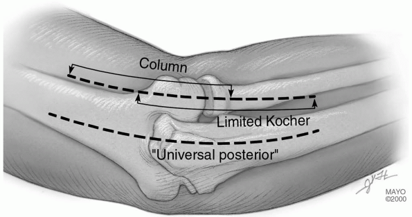
There are two conceptual incision types: an extensile
posterior or posterior/lateral or a limited, specific for-purpose
exposure (Fig. 3-1).
For extensile exposures, a straight posterior or posterior lateral
incision is used. We term the posterior exposure the “universal”
incision since both medial and lateral elbow pathology can be addressed
through a posterior skin incision by elevating skin flaps (Fig. 3-2).
For fear of injuring the ulnar nerve, a posterior incision of variable
length (12 to 18 cm) is placed just medial or lateral to the tip of the
olecranon and not directly over the cubital tunnel.
 |
|
FIGURE 3-1
|
 |
|
FIGURE 3-2
|
P.62
OLECRANON OSTEOTOMY
Indications
Reduction and fixation, distal humeral, and comminuted fractures (C3).
Landmarks
Tip of olecranon, medial epicondyle, ulnar nerve in cubital tunnel, nonarticular portion of olecranon.
Position
The patient is supine with arm across the chest.
Technique
-
Skin incision: direct posterior from 6 to
8 cm proximal to the tip of the olecranon, over subcutaneous border of
ulna; distal as required (Fig. 3-3A). -
Elevate skin flaps medially and laterally to the epicondyles (Fig. 3-3B).
-
Identify ulnar nerve and incise medial ulnohumeral capsule.
-
The anconeus is released from the
triceps, the ulnohumeral capsule is incised, and the nonarticular
portion of the greater sigmoid notch is identified (Fig. 3-3C).-
Alternate:
Laterally, we prefer to protect and elevate the anconeus distal to
proximal thereby protecting its origin from the triceps fascia (see
later).
-
-
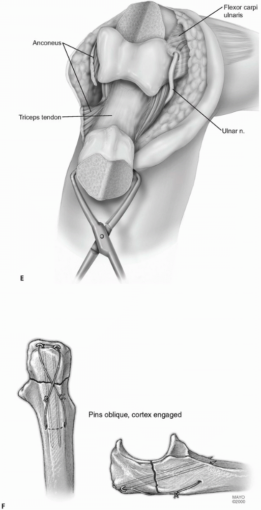
A Chevron osteotomy is performed, apex distal with a depth of 5 to 10 mm (Fig. 3-3D). Protect the ulnar nerve medially. Use osteotome to crack last few millimeters to assure accurate subsequent reduction.
-
The olecranon fragment is reflected proximally exposing the distal humerus (Fig. 3-3E).
-
Repair uses the AO-K-wire, tension band technique with the wires in the anterior cortex, not down the canal (Fig. 3-3F).
P.63
 |
|
FIGURE 3-3
|
 |
|
FIGURE 3-3 (Continued)
|
P.64
 |
|
FIGURE 3-3 (Continued)
|
P.65
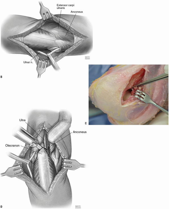
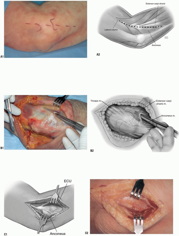
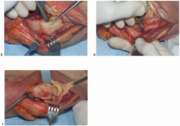
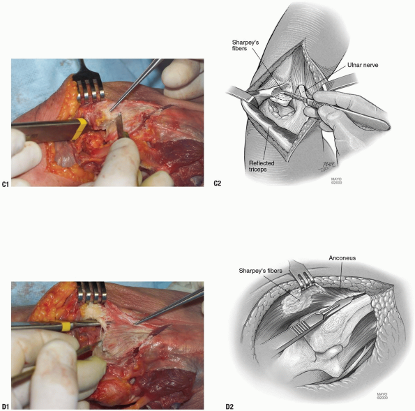
Mayo Olecranon Osteotomy of the Elbow—Anconeus Preserving
Concern with regard to transecting the anconeus
attachment to the triceps has prompted the development of an olecranon
osteotomy that preserves the anconeus origin and viability.
attachment to the triceps has prompted the development of an olecranon
osteotomy that preserves the anconeus origin and viability.
Position
The patient is supine with the arm across the chest.
Technique
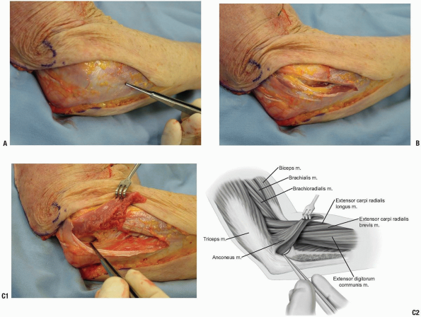
-
The exposure is as required by the
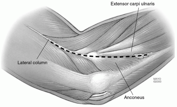
pathology. Deep exposure is at the Kocher’s interval between the
extensor carpi ulnaris and anconeus (Fig. 3-4A). -
The interval is entered and the anconeus is identified and isolated (Fig. 3-4B).
-
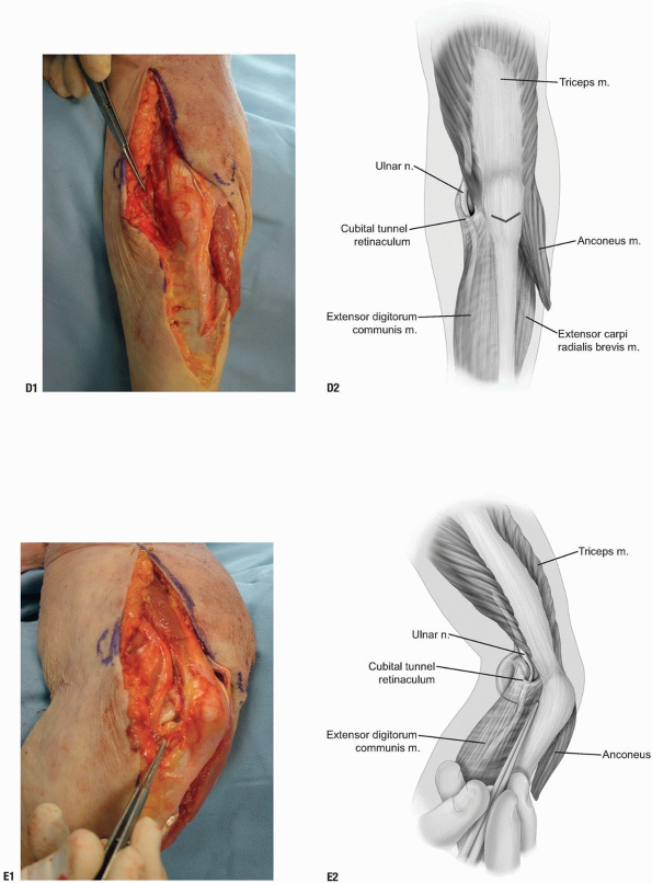
The anconeus is elevated from its bed by
sharp dissection leaving the attachment of its origin at the fascial
expansion of the triceps and the mid-portion of the sigmoid notch is
identified laterally (Fig. 3-4C). -
Medially the ulnar nerve is identified (Fig. 3-4D) and the mid-portion of the articulation is exposed (Fig. 3-4E).
-
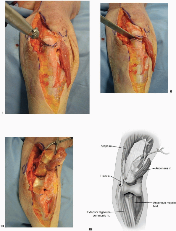
A V-shaped osteotomy is carried out as above with an oscillating saw (Fig. 3-4F). The osteotomy is completed with an osteotome (Fig. 3-4G).
-
The osteotomized olecranon along with the attached anconeus is elevated proximally (Fig. 3-4H).
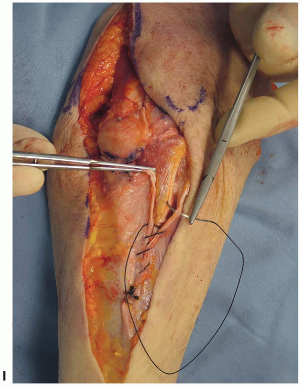
-
Closure consists of the standard AO
reattachment of the olecranon. The anconeus is brought back to its
insertion on the ulna. The fascia over the anconeus is closed with a
running 2-0 absorbable suture (Fig. 3-4I).
 |
|
FIGURE 3-4
|
P.66
 |
|
FIGURE 3-4 (Continued)
|
P.67
 |
|
FIGURE 3-4 (Continued)
|
P.68
 |
|
FIGURE 3-4 (Continued)
|
Pearls/Pitfalls/Comments
-
Pearl: The ulnar nerve does not need to be mobilized unless dictated by the pathology.
-
Comments:
The attractiveness of this exposure is that the anconeus dissection can
be done very safely and quickly. This does preserve the anconeus
triceps continuity in the event that a later reconstructive procedure
may be necessary that uses the anconeus. -
Pitfalls: Avoid osteotomy in rheumatoid arthritis as the thin olecranon compromises healing if an osteotomy is carried out (2). The transverse osteotomy of McAusland is associated with an approximately 5% nonunion rate (2).
Although for fractures the Chevron osteotomy may improve these results
and decrease the nonunion rate, I personally have not had the clinical
need to osteotomize the olecranon in the last 14 years, and osteotomy
should be avoided if the olecranon is resorbed.
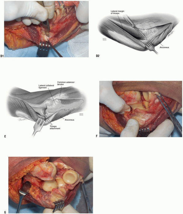
LATERAL EXPOSURES
The central concept is predicated on raising
subcutaneous flaps both medially and laterally. This in turn is
strongly dependent on recognizing the flexibility of extending limited
incisions as needed.
subcutaneous flaps both medially and laterally. This in turn is
strongly dependent on recognizing the flexibility of extending limited
incisions as needed.
A limited proximal lateral approach exposes the
supracondylar column. A limited distal approach enters Kocher’s
interval and exposes the radial head and the lateral collateral
ligament. Connecting the two defines the extensile Kocher exposure (Fig. 3-5).
supracondylar column. A limited distal approach enters Kocher’s
interval and exposes the radial head and the lateral collateral
ligament. Connecting the two defines the extensile Kocher exposure (Fig. 3-5).
 |
|
FIGURE 3-5
|
P.69
The Column Exposure (3)
Indications
Anterior-posterior capsular release for stiff elbow.
Landmarks
Lateral epicondyle, the common extensor tendon, the extensor carpi radialis longus, and the anterior capsule.
Position
The patient is supine with arm across the chest.
Technique
-
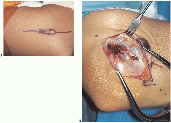
Incision: the skin incision starts over
the lateral column 5 cm proximal to the lateral epicondyle and extends
distally 2 cm past the epicondyle (Fig. 3-6A). -
The extensor carpi radialis longus is
identified and elevated from the lateral column and epicondyle and the
anterior capsule is visualized (Fig. 3-6B). -
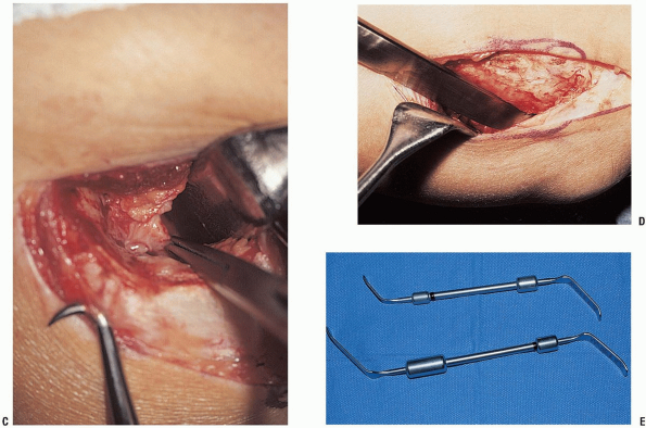
An incision is made in the capsule just superior to the collateral ligament (Fig. 3-6C).
-
If the posterior joint needs to be exposed the triceps is easily elevated (Fig. 3-6D).
 |
|
FIGURE 3-6
|
P.70
 |
|
FIGURE 3-6 (Continued)
|
Pearls/Pitfalls/Comments
-
A periosteal elevator is used to elevate
the brachialis muscle off the anterior capsule which can be safely
performed since the arthrotomy provides accurate spatial orientation
from lateral to medial across the joint. -
Special contoured retractors have been designed making the soft tissue retractor easier (Fig. 3-6E).
-
If an extensile exposure is anticipated,
a posterior incision is made. The same deep exposure can be
accomplished by extending the posterior lateral skin incision and
elevating the lateral skin cutaneous flap.
Limited Kocher Exposure of the Elbow
Indications
Simple excision of the radial head and repair of lateral ulnar collateral ligament.
Landmarks
Lateral epicondyle, radial head, and interval between anconeus and extensor carpi ulnaris.
Position
The patient is supine with arm across the chest.
Technique
-
Incision: from the subcutaneous border of
the ulna obliquely across the posterolateral aspect of the elbow ending
just proximal to the lateral epicondyle (Fig. 3-7A).-
Note: This follows Kocher’s interval.
-
-
The interval between the anconeus and extensor carpi ulnaris is identified and entered (Fig. 3-7B).
-
For excision of the radial head, the
extensor carpi ulnaris and a small portion of the supinator muscle are
dissected free of the capsule and retracted anteriorly (Fig. 3-7C).
P.71
 |
|
FIGURE 3-7
|
P.72
 |
|
FIGURE 3-7 (Continued)
|
Distal Extension
Landmarks
-
The lateral epicondyle, posterior border
of the extensor carpi ulnaris, anterior edge of the anconeus, and the
crista supinatoris. -
The anconeus is elevated from the ulna and the tubercle of the supinator is palpated (Fig. 3-7D).
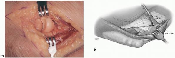
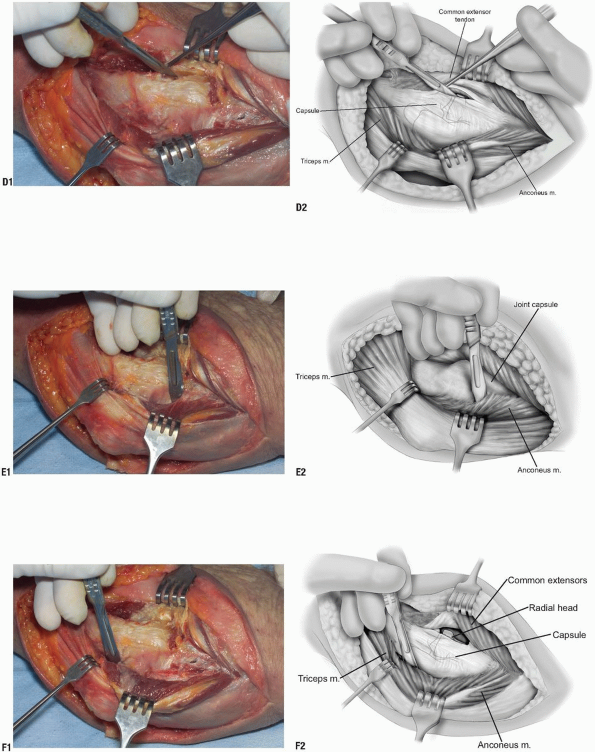
Extensile Posterior Lateral Exposure (Kocher)
We have found that the described surgical exposures to
the elbow sufficient for virtually all reconstructive procedures and
all may be executed with a posterior skin incision. The classic
extensile approach described by Kocher implies that the anterior
capsule has been incised and the lateral collateral ligament has been
released (4).
the elbow sufficient for virtually all reconstructive procedures and
all may be executed with a posterior skin incision. The classic
extensile approach described by Kocher implies that the anterior
capsule has been incised and the lateral collateral ligament has been
released (4).
Indications
Reconstructive procedures, including open reduction
internal fixation, total elbow arthroplasty (unlinked), and
interposition arthroplasty.
internal fixation, total elbow arthroplasty (unlinked), and
interposition arthroplasty.
Landmarks
The proximal lateral column and the distal Kocher interval.
Position
The patient is supine with arm across the chest.
Technique
The basic interval is the connection of the above two
exposures, the column, and the distal limited Kocher. The lateral
collateral ligament is released and the triceps may be elevated from
the posterior aspect of the humerus by extending the skin incision 6 to
7 cm proximal to the lateral epicondyle (see Fig. 3-1).
exposures, the column, and the distal limited Kocher. The lateral
collateral ligament is released and the triceps may be elevated from
the posterior aspect of the humerus by extending the skin incision 6 to
7 cm proximal to the lateral epicondyle (see Fig. 3-1).
-
Enter Kocher’s interval and elevate the extensor carpi ulnaris.
-
The common extensor tendon is identified and reflected anteriorly exposing the capsule (Fig. 3-8A,B).
-
The insertion of the extensor carpi
radialis longus and the distal fibers of the brachioradialis muscle are
released from the lateral column of the distal humerus (Fig. 3-8C). -
The anterior capsule is entered (Fig. 3-8D) and released to the extent necessary to expose the anterior joint.
-
Proceed as shown in Figure 3-8B completely elevating the anconeus from the ulna and from its humeral attachment (Fig. 3-8E).
-
The triceps is easily elevated from the
posterior humerus in the normal situation and even in posttraumatic
contractures it can be elevated with a periosteal elevator without much
additional difficulty (Fig. 3-8F). -
The lateral collateral ligament is
released from the humeral origin as a separate structure or, if prior
surgery has caused scarring, it is released with the common extensor
tendon complex (Fig. 3-8G). -
The anterior and posterior capsules are
then completely incised. We have found it necessary to release
approximately 25% of lateral triceps attachment to the ulna to allow
the triceps to invert with the maneuver (Fig. 3-8H). -
A varus supinatory stress is applied to
the elbow, which then opens like a book hinging on the medial ulnar
collateral ligament and common flexor muscles (Fig. 3-8I).
The triceps remains attached to the ulna. Inspect the ulnar nerve to be
sure it is not being compressed. If it is, release it from the cubital
tunnel.
P.73
 |
|
FIGURE 3-8
|
P.74
 |
|
FIGURE 3-8 (Continued)
|
P.75
 |
|
FIGURE 3-8 (Continued)
|
Mayo Modified Extensile Kocher Posterior-Lateral Exposure
The Mayo (R.S. Bryan) modification of the Kocher
approach consists of reflection and release of the extensor mechanism
from the tip of the olecranon in a fashion similar to that described
for the Mayo approach (5). If reflected, the
triceps must be securely reattached to bone. Further, when the Mayo
modified Kocher release has been performed, the ulnar nerve must be
exposed and released as necessary to avoid compression with varus
angular forearm manipulation.
Indictions
More extensile exposure is required than has been obtained with the previous steps.
P.76
 |
|
FIGURE 3-9
|
Technique
-
Incision: medial and lateral skin flaps
are elevated using the knife in a flattened disposition to avoid
cutting through the skin (Fig. 3-9A,B) and is protected or translocated according to the merits of the case. -
The triceps muscle is elevated laterally and the humeral attachment of the triceps muscle is released (Fig. 3-9C).
-
The triceps-anconeus muscle sleeve is sharply reflected from the tip of the olecranon (Fig. 3-9D).
-
The entire extensor mechanism, including anconeus, is thus reflected from lateral to medial (Fig. 3-9E).
-
Note: Lateral collateral ligament detachment from the humerus is optional depending on the pathology (Fig. 3-9F).
-
-
Flexing the elbow rotating the ulna and
removing the tip of the olecranon exposes the articular surface and the
posterior humerus (Fig. 3-9G).
P.77
 |
|
FIGURE 3-9 (Continued)
|
P.78
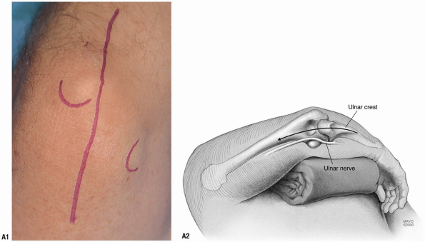
POSTERIOR-MEDIAL EXPOSURE
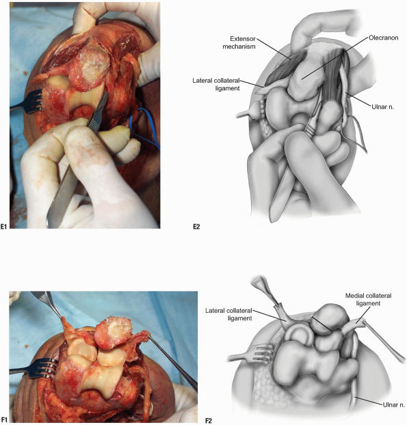
The Mayo Approach (Bryan-Morrey).
Indications
Ankylosis release, total elbow arthroplasty, open
reduction internal fixation (ORIF) medial column, and distal humeral
fractures.
reduction internal fixation (ORIF) medial column, and distal humeral
fractures.
Position
The patient is supine with arm across the chest.
Landmarks
Medial epicondyle, olecranon, and subcutaneous borer of ulna.
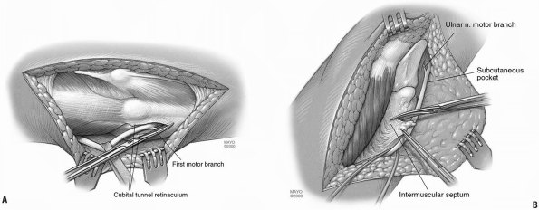
Technique
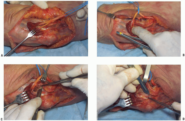
-
Incision: 7 cm proximal and 7 cm distal and just medial to the tip of the olecranon.
-
The ulnar nerve is released from the margin of the triceps and elevated from its bed (Fig. 3-10A).
The cubital tunnel retinaculum is split and the nerve is released to
the first motor branch. A subcutaneous pocket is developed, the
intermuscular septum is removed (Fig. 3-10B), and the nerve is translated anteriorly. -
A sleeve of tissue consisting of the forearm fascia and ulnar periosteum is elevated from the medial margin of the ulna.
-
The attachment of the triceps to the olecranon is released by sharp dissection (Fig. 3-10C).
-
The distal forearm fascia and ulnar periosteum are elevated from the ulna.
-
The extensor mechanism and capsule
continue to be reflected from the lateral epicondyle and the anconeus
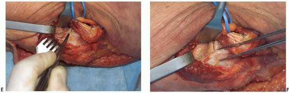
is released from the ulna (Fig. 3-10D).P.79-
Pearls/Pitfalls:
For linked total elbow arthroplasty, the lateral and medial collateral
ligaments are released and the extensor mechanism is reflected lateral
to the epicondyle (Fig. 3-10E). The elbow is flexed and the tip of the olecranon is removed to expose the joint (Fig. 3-10F).
-
 |
|
FIGURE 3-10
|
 |
|
FIGURE 3-10 (Continued)
|
P.80
 |
|
FIGURE 3-10 (Continued)
|
P.81
 |
|
FIGURE 3-11 (Continued)
|
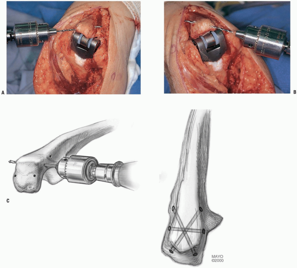
Comment
In every instance in which the triceps has been
completely reflected, it is necessary to securely reattach the
insertion site to the olecranon with a crisscross type of suture.
completely reflected, it is necessary to securely reattach the
insertion site to the olecranon with a crisscross type of suture.
-
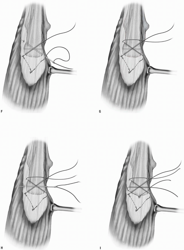
Drill holes about 3 cm in length are placed in a cruciate fashion in the olecranon from proximal to distal (Fig. 3-11A,B).
-
A third transverse hole is drilled through the olecranon to secure a second stabilizing suture (Fig. 3-11C).
-
The margin of the triceps is first grasped with an Alis clamp and brought over the olecranon.
-
A no. 5 nonabsorbable suture is introduced with a straight needle from distal lateral to proximal medial.
-
The suture is first brought through the
tip of the olecranon and passes through the triceps tissue at its
anatomic attachment site with the elbow in 90 degrees (Fig. 3-11D).P.82-
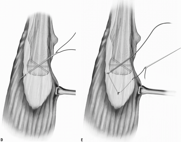
Note: We
prefer to displace the tendon somewhat medially at the time of
reattachment after the Bryan-Morrey reflection and somewhat laterally
after the modified Kocher release.
-
-
A locked suture is first placed in the
tendon followed by a second locked suture placed more proximally in the
contoured portion of the tendon. The suture passes through the triceps
tendon opposite its locked attachment site (Fig. 3-11E). -
The suture then enters the opposite hole in the olecranon now being passed from proximal to distal (Fig. 3-11F).
After the suture has emerged from the second hole in the olecranon, it
is brought back over the top of the ulna through the soft tissue distal
expansion of the extensor sleeve (Fig. 3-11G).-
Note: Care is taken to tie this stitch off to the side of the subcutaneous border of the ulna to avoid irritation or skin erosion.
-
-
To snugly stabilize the triceps insertion
against the olecranon, a second suture is placed transversely across
the ulna; again, beginning on the side from which the triceps
reflection began (Fig. 3-11H).
It is brought back across the triceps tendon in a transverse fashion
with a locked stitch in the mid/lateral portion of the tendon (Fig. 3-11I). The suture then passes through the lateral margin of the triceps of flexion, again with the knots off the subcutaneous border.All sutures are tied with the elbow in 90 degrees of flexion, again with the knots off the subcutaneous border.
 |
|
FIGURE 3-11 (Continued)
|
P.83
 |
|
FIGURE 3-11 (Continued)
|
P.84
MEDIAL EXPOSURES
There are two relevant exposures to the medial aspect of
the elbow. The first is a focused exposure to allow identity and
management of coronoid fractures. The second is a more extensile medial
approach which affords an opportunity to release the anterior and
posterior elbow capsules as well as manage fractures and a broader
spectrum of pathology.
the elbow. The first is a focused exposure to allow identity and
management of coronoid fractures. The second is a more extensile medial
approach which affords an opportunity to release the anterior and
posterior elbow capsules as well as manage fractures and a broader
spectrum of pathology.
Focused Medial Exposure of the Coronoid
While this is a limited exposure, it can be modified to a more extensile exposure as described below.
Indications
Coronoid fracture, specifically application of a buttress plate.
Landmark
Medial epicondyle, ulnar nerve, and flexor carpi ulnaris.
Position
The patient is supine with arm on an elbow board.
Technique
-
Skin incision proceeds from 5 cm proximal
to 7 cm distal to the medial epicondyle passing posterior to the
epicondyle near the midline. -
The medial epicondyle is identified along
with the ulnar nerve. The ulnar nerve is mobilized from its bed, the
cubital tunnel retinaculum is released (Fig. 3-12A). -
The flexor carpi ulnaris is split
allowing the ulnar nerve to be further mobilized. The sublime tubercle
is palpated in the depths of the wound (Fig. 3-12B). -
Sharp dissection frees the muscle mass from the anterior (Fig. 3-12C) and posterior (Fig. 3-12D) aspects of the capsule.
-
The capsule is further identified with a periosteal elevator. The sublime tubercle is identified and the capsule is entered.
-
Releasing the capsule allows clear identity of the coronoid just anterior to the medial collateral ligament (Fig. 3-12E).
-
The dissection may be extended distally
as necessary to apply the buttress plate or otherwise provide internal
fixation for the coronoid. -
Extension proximally allows adequate exposure to reconstruct the collateral ligament (Fig. 3-12F).
P.85
 |
|
FIGURE 3-12
|
 |
|
FIGURE 3-12 (Continued)
|
Medial Column (“Over the Top,” “Hotchkiss”) (6)
Indications
Access to the coronoid with an intact radial head,
anterior capsule release if ulnar nerve pathology is also to be
addressed, anterior and posterior medial ectopic bone excision, and
anterior, posterior capsule excision.
anterior capsule release if ulnar nerve pathology is also to be
addressed, anterior and posterior medial ectopic bone excision, and
anterior, posterior capsule excision.
-
Note
-
It is not a good approach if there is
need of excision of heterotopic bone from the lateral elbow joint or if
access to the radial head is needed. -
Conversion or extension between the Bryan-Morrey, Mayo, and the Hotchkiss approach is readily accomplished but rarely indicated.
-
Landmarks
The medial supracondylar ridge of the humerus, the
medial intermuscular septum, the origin of the flexor pronator muscle
mass, and the ulnar nerve.
medial intermuscular septum, the origin of the flexor pronator muscle
mass, and the ulnar nerve.
Position
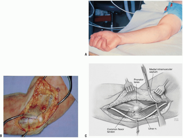
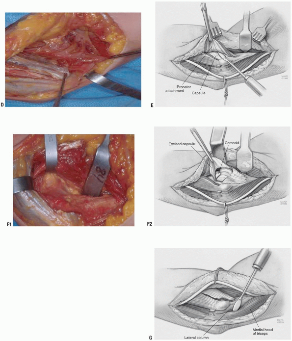
The patient is supine with extremity supported by a hand or elbow table (Fig. 3-13A).
P.86
Technique
-
Skin incision 5 cm distal and proximal to medial epicondyle.
-
The medial intermuscular septum is
identified. Anterior to the septum superficial to the fascia (and not
in the subdermal tissue), the medial antebrachial cutaneous nerve is
identified and protected (Fig. 3-13B).
The line of reflection is identified distally at the raphe between the
flexor carpi ulnaris and the pronator teres. The intermuscular septum
is identified proximally.-
Note: If
the patient has had previous surgery, the ulnar nerve is usually most
easily identified proximally before proceeding distally.
-
-
The ulnar nerve is mobilized. The medial
intermuscular septum is exposed anteriorly and posterior and then
released for a distance of about 5 cm proximally (Fig. 3-13C). -
Locate the medial supracondylar ridge and begin elevating the anterior brachialis muscle with a periosteal elevator.
-
Subperiosteally elevate enough of the
anterior structures of the distal humeral region to allow the placement
of a wide retractor. The median nerve, brachial artery, and vein are
superficial to the brachialis muscle and need not be identified. -
The flexor pronator muscle mass is
divided in line with its fibers along the raphe which separates the
pronator and flexor carpi ulnaris muscles, leaving a portion of the
flexor carpi ulnaris tendon attached to the epicondyle (Fig. 3-13D).-
Note: A
small cuff of fibrous origin can be left on the supracondylar ridge as
the muscle is elevated to facilitate reattachment when closing.
-
-
The pronator muscle is elevated from the
capsule encountering the brachialis muscle which has been mobilized and
retracted laterally (Fig. 3-13E).P.87-
Note: A proximal, transverse incision in the lacertus fibrosis may also be needed to adequately mobilize the brachialis muscle.
-
-
As the pronator muscle is elevated from the capsule, the entire anterior capsule is exposed (Fig. 3-13F).
-
If necessary, the posterior capsule may be exposed by elevating the triceps from its lateral distal humeral attachment (Fig. 3-13G).
 |
|
FIGURE 3-13
|
 |
|
FIGURE 3-13 (Continued)
|
P.88
RESULTS
There have been limited attempts to document the
efficacy of one or the other of the various types of triceps-sparing
approaches. In the original description we compared the clinical result
of the Mayo approach to that of the triceps splitting or transverse
release of the triceps attachment (5). There
were no triceps disruptions after approximately 75 procedures done with
the triceps being released in continuity (Mayo approach) compared with
an approximately 20% complication rate when the triceps was released
transversely. Wolfe and Ranawat (7) have also
observed no instances of triceps insufficiency with their modification
of this approach. The use of the Mayo medial exposure was also shown to
have improved triceps strength after total elbow arthroplasty (8).
This manner of exposing the elbow was found to be associated with
approximately 20% greater extension strength than with the Campbell
fascial turn-down (Van Gorder) type of exposure.
COMPLICATIONS
One beauty of the previously described exposures is that
they are relatively free of complication. Today most problems are
related to the pathology rather than to the surgical approach.
they are relatively free of complication. Today most problems are
related to the pathology rather than to the surgical approach.
Difficult ankylosis release procedures are associated
with a significant amount of swelling as often occurs in patients
undergoing total elbow arthroplasty. Wound healing is generally not a
problem, however, and is related to the presence of prior incisions and
the magnitude of the dissection, as is typical for release of the stiff
elbow. The elevation of the large medial and lateral flaps does not
retard healing but occasionally can give rise to subcutaneous seroma.
Rarely does this need to be addressed or drained.
The infection rate after total elbow arthroplasty has
been reduced at our institution from a high of 11% in 1970 to
approximately 3% over the last 10 years (9).
This reduction is coincident with adopting the Mayo approach to the
elbow, but other technique changes have occurred in this period,
including using antibiotic-impregnated cement and splinting the elbow
in extension.
Injury to the ulnar nerve appears to be more common in
those instances in which the ulnar nerve is not exposed and the elbow
is flexed on the medial collateral ligament, as with the classical
extensile Kocher approach (9,10).
Simply exposing the ulnar nerve, although it decreases the
complication, does not completely obviate it. The theoretical
disadvantage of the Mayo approach, which allows translocation of the
ulnar nerve, is that this maneuver devascularizes the nerve and the
dissection itself may cause ulnar nerve irritation. Having used this
particular exposure in more than 500 cases, the incidence of permanent
ulnar nerve injury with motor dysfunction is less than 1%. I am,
therefore, comfortable exposing and moving the ulnar nerve in a
subcutaneous pocket as an essential and integral part of the Mayo
triceps-sparing approach.
Although posterior interosseous nerve palsy is known to occur with some approaches to the radial head (11, 12, 13), the complication is virtually unheard of when the joint is exposed through Kocher’s interval.
Triceps disruption is very uncommon with either the Mayo
modified extensile Kocher exposure or the Mayo medial-to-lateral type
of approach. The incidence of triceps disruption after total elbow
replacement, therefore, is less than 1% in our experience (14).
If, however, the triceps should become disrupted after either of the
procedures described earlier, if adequate tissue is present, it may be
reattached as described for the primary procedure (14).
If the remaining tissue is inadequate, triceps power is restored by
either an anconeus slide or an Achilles tendon allograft reconstruction
(15).
REFERENCES
1. Morrey BF. Surgical exposures. In: Morrey BF, ed. Master Techniques in Orthopedic Surgery: The Elbow, 2nd ed. Philadelphia: Lippincott Williams & Wilkins, 2002.
2. Morrey BF. Surgical exposures of the elbow. In: Morrey BF, ed. The Elbow and Its Disorders, 3rd ed. Philadelphia: WB Saunders, 2000:109-134.
3. Mansat P, Morrey BF. The column procedure: A limited lateral approach for extrinsic contracture of the elbow. J Bone Joint Surg 1998;80A(11):1603-1615.
4. Kocher T. Text-book of Operative Surgery, 3rd ed. London: A and C Black, 1911.
5. Bryan RS, Morrey BF. Extensive posterior exposure of the elbow: a triceps-sparing approach. Clin Orthop 1982;166:188.
P.89
6. Kasparyan NG, Hotchkiss RN. Dynamic skeletal fixation in the upper extremity. Hand Clin 1997;13:643-663.
7. Wolfe SW, Ranawat CS. The osteo-anconeus flap: an approach for total elbow arthroplasty. J Bone Joint Surg 1990;72A:684.
8. Morrey BF, Askew LJ, An KN. Strength function after elbow arthroplasty. Clin Orthop 1988;234:43-50.
9. Morrey BF, Bryan RS. Complications of total elbow arthroplasty. Clin Orthop 1982;170:204-212.
10. Ewald FC, Jacobs MA. Total elbow arthroplasty. Clin Orthop 1984;182:137.
11. Hoppenfield S, deBoer P. Surgical Exposures in Orthopaedics: The Anatomic Approach. Philadelphia: J.B. Lippincott Co., 1984.
12. Kaplan EB. Surgical approaches to the proximal end of the radius and its use in fractures of the head and neck of the radius. J Bone Joint Surg 1941;23:86.
13. Strachan JH, Ellis BW. Vulnerability of the posterior interosseous nerve during radial head resection. J Bone Joint Surg 1971;53B:320.
14. Celli A, Arash A, Adams RA, et al. Triceps insufficiency following total elbow arthroplasty. J Bone Joint Surg Am 2005;87(9):1957-1964.
15. Sanchez-Sotelo
J, Morrey BF. Surgical techniques for reconstruction of chronic
insufficiency of the triceps—Rotation flap using anconeus and tendo
Achillis allograft. J Bone Joint Surg 2002;84B(8):1116-1120.
J, Morrey BF. Surgical techniques for reconstruction of chronic
insufficiency of the triceps—Rotation flap using anconeus and tendo
Achillis allograft. J Bone Joint Surg 2002;84B(8):1116-1120.