Systems Anatomy
Authors: Doyle, James R.
Title: Hand and Wrist, 1st Edition
Copyright ©2006 Lippincott Williams & Wilkins
> Table of Contents > Section I – Basic Anatomy > 1 – Anatomy > 1.2 – Systems Anatomy
1.2
Systems Anatomy
The following figures and discussion represent and are
designed to provide an overall perspective on the deeper anatomy—the
skeletal, muscular, vascular, and neural anatomy—of the upper
extremity. The perspective spans from the shoulder and neck for
discussions of the skeletal, neural, and vascular anatomy, and from the
elbow for the muscular system.
designed to provide an overall perspective on the deeper anatomy—the
skeletal, muscular, vascular, and neural anatomy—of the upper
extremity. The perspective spans from the shoulder and neck for
discussions of the skeletal, neural, and vascular anatomy, and from the
elbow for the muscular system.
Skeletal Anatomy
The osseous structures of the upper limb include the
humerus, the radius and ulna, eight carpal bones, five metacarpals, and
14 phalanges. The upper extremity skeleton is depicted in Figure 1.2-1.
humerus, the radius and ulna, eight carpal bones, five metacarpals, and
14 phalanges. The upper extremity skeleton is depicted in Figure 1.2-1.
P.8
P.9
P.10
 |
|
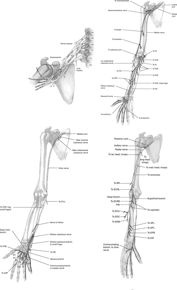
Figure 1.2-5
Schematic illustration of the brachial plexus, associated major branches, peripheral branches and muscles they innervate. A, nerve to subclavius; B, lateral pectoral nerve; C, subscapular nerve; D, thoracodorsal nerve; E, medial antebrachial cutaneous nerve; F, medial brachial cutaneous nerve; G, medial pectoral nerve. |
P.11
 |
|
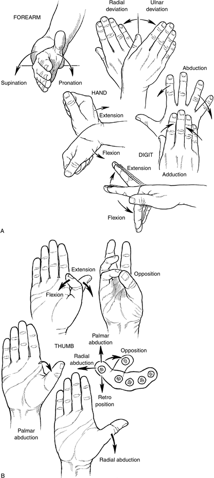
Figure 1.3-1 Currently accepted terminology and depiction of movement in the forearm, wrist, fingers: wrist, forearm (A), thumb (B).
|
P.12
|
Table 1.3-1 Anatomic Basis for Movement in the Upper Extremity
|
||||||||||||||||||||||||||||||||||||||||||||||||||||||||||||||||||||||||||||||||||||||||||||||||||||||||||||||||||||||||||||||||||||||||||||||||||||||||||||||||||||
|---|---|---|---|---|---|---|---|---|---|---|---|---|---|---|---|---|---|---|---|---|---|---|---|---|---|---|---|---|---|---|---|---|---|---|---|---|---|---|---|---|---|---|---|---|---|---|---|---|---|---|---|---|---|---|---|---|---|---|---|---|---|---|---|---|---|---|---|---|---|---|---|---|---|---|---|---|---|---|---|---|---|---|---|---|---|---|---|---|---|---|---|---|---|---|---|---|---|---|---|---|---|---|---|---|---|---|---|---|---|---|---|---|---|---|---|---|---|---|---|---|---|---|---|---|---|---|---|---|---|---|---|---|---|---|---|---|---|---|---|---|---|---|---|---|---|---|---|---|---|---|---|---|---|---|---|---|---|---|---|---|---|---|---|---|
|
||||||||||||||||||||||||||||||||||||||||||||||||||||||||||||||||||||||||||||||||||||||||||||||||||||||||||||||||||||||||||||||||||||||||||||||||||||||||||||||||||||
P.13
Muscular Anatomy
The muscles of the forearm are depicted in Figure 1.2-2 and the hand in Figure 1.2-3.
Vascular Anatomy
The vascular anatomy from the axillary artery to the hand is depicted in Figure 1.2-4.
Neural Anatomy
The brachial plexus, median, ulnar, and radial nerves and the muscles they innervate are depicted in Figure 1.2-5.
