
Editors: Chelly, Jacques E.
Title: Peripheral Nerve Blocks: A Color Atlas, 3rd Edition
Copyright ©2009 Lippincott Williams & Wilkins
> Table of Contents > Section V
– Pediatric Peripheral Blocks > 46 – Technical Considerations for
Pediatric Regional Anesthesia
– Pediatric Peripheral Blocks > 46 – Technical Considerations for
Pediatric Regional Anesthesia
46
Technical Considerations for Pediatric Regional Anesthesia
Giorgio Ivani
Valeria Mossetti
A. Nerve Mapping in Children
Equipment
For many years there has been a lack of specific
material to perform regional anesthesia in infants and children. Radial
artery catheterization sets, epidural kits, and peripheral and central
venous catheter sets have been used for continuous peripheral nerve
blocks. Nowadays for a safe performance it is mandatory to use
dedicated pediatric tools. Any peripheral nerve can be blocked either
by infiltrating a local anesthetic within a compartment space through
which the nerve runs or by precisely locating the nerve. Compartment
blocks such as intercostal block, intrapleural block, fascia iliaca
compartment block, penile block, etc., depend on the localization of
the fascial plane; when the relevant fascia is unique with no
underlying vital structure, different needles such as an IM needle, can
be safely used. When there are several fascial planes or a danger of
damaging important anatomical structures, such as with an ilioinguinal
block, only a short-beveled or pinpoint needle should be selected.
Precise localization of a plexus or a nerve trunk must not be performed
by seeking paresthesias with standard IM needles because of the danger
of direct nerve damage; only short-beveled needles, insulated and
connected to a nerve stimulator are suitable. For most peripheral nerve
blocks in children, 21- to 23-gauge, 35- to 50-mm long needles are
used, depending on the type of block and on the age of the child.
Eliciting a motor response using a nerve stimulator is the most useful
and safe technique for performing a pediatric nerve block. Since the
plexuses in children are quite superficial, especially the brachial
plexus at the axilla, before introducing the needle, try to detect the
position of the plexus with the transcutaneous technique. After having
introduced the tip of the needle, connect it to the neurostimulator and
set it to stimulate at a frequency of 2 Hz. Starting with a current of
1 to 1.5 mA, the needle is advanced until distinct contractions of the
nerves to be blocked are noticed. The optimum nerve location is
achieved by adjusting the needle so that these contractions are still
visible with currents of 0.4 mA. It is now possible
P.322
to
inject the bolus dose of local anesthetic. Remember that, as nerves are
thin and very closely linked to each other without sheaths dividing
them, one twitch of a single nerve is enough to administer the drug
without looking for a multiple-twitch technique.
Mapping of Nerves in Pediatric Regional Anesthesia
In the new century, regional anesthesia in the pediatric
field has reached worldwide acceptance. Safety and efficacy have been
evidenced in a major survey showing that pediatric regional anesthesia
has a low rate of complications and no major sequelae or deaths. Light
sedation or anesthesia plus a block offers optimal pain control
throughout surgery, allowing also good postoperative analgesia.
Peripheral blocks are employed more and more, but thus far they are
still less common than central blocks even though in the same survey
their safety was superior—no complications in more than 9,000 blocks.
One of the problems connected with the performance of a
peripheral block, even with the mandatory use of a nerve stimulator
(NS), is a thorough knowledge of the anatomy of children; specifically,
the closeness of different structures—nerves, veins, arteries. The
small distance between the skin and the nerves can cause, in
inexperienced hands, severe injuries while detecting a plexus with the
needle.
Adrian Bosenberg published in 2002 a simple but very
effective method to improve experience and reduce mistakes during the
performance of a peripheral block. The technique, called nerve mapping,
requires the use of the unblunted tip of the negative electrode of the
NS. It involves increasing the mA of the NS up to 3 mA or more; the
skin is touched close to the nerve plexus, causing stimulation until
motor responses are elicited, and then the voltage is reduced to detect
the best point to perform the block.
In the same year (2002) Urmey and Grossi described the
same technique in adults using a device with a needle-through passage
obtaining an even more successful performance; in this case they
employed a higher voltage, 4 to 5 mA, due to the thickness of the skin
in adults; in 2003 they described a modified tool for the same
technique.
same technique in adults using a device with a needle-through passage
obtaining an even more successful performance; in this case they
employed a higher voltage, 4 to 5 mA, due to the thickness of the skin
in adults; in 2003 they described a modified tool for the same
technique.
 |
|
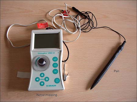
Figure 46-1. Pen-like nerve stimulator.
|
P.323
More recently new devices have been produced by
industrial companies using a pen-like stimulator instead of the
negative electrode allowing an easier mapping. The NS must be set at 3
mA, 2 Hz, 1 ms (while 0.1 ms is usually used for performing the block
with the needle); changing slightly the position of the tip of the pen,
a motor response is elicited. The best position can then be chosen (and
the best motor response according to the needs of surgery) and marked
with a pen (Fig. 46-1).
Nerve mapping is a painless method as the mAs used
produce a motor but not a sensory response, and can also be performed
in an awake patient. Moreover, children are very often under sedation
or anesthesia so that any stress is avoided; in the meantime we can
teach the anatomy of a plexus allowing young doctors to detect the
nerves without any skin damage.
In this way we can find the different nerve responses of
a plexus; radial, medial, ulnar, and musculocutaneous nerves can be
elicited for the upper arm, and femoral and sciatic with its
components—peroneal and tibial—for the lower limb.
a plexus; radial, medial, ulnar, and musculocutaneous nerves can be
elicited for the upper arm, and femoral and sciatic with its
components—peroneal and tibial—for the lower limb.
The success rate of blocks in children can be increased,
keeping in mind that malformations may make it difficult to place the
needle (e.g., arthrogryposis).
keeping in mind that malformations may make it difficult to place the
needle (e.g., arthrogryposis).
We use this technique for the axillary and the
parascalene approach of the brachial plexus, the femoral approach, and
all the more distal detection of nerves (i.e., popliteal level)
including the “small blocks.”
parascalene approach of the brachial plexus, the femoral approach, and
all the more distal detection of nerves (i.e., popliteal level)
including the “small blocks.”
It takes only a few minutes, is not time consuming, and
can be considered one of the tools for daily clinical practice—a new
and extremely useful technique that can increase the efficacy and
safety of peripheral blocks in children.
can be considered one of the tools for daily clinical practice—a new
and extremely useful technique that can increase the efficacy and
safety of peripheral blocks in children.
Suggested Readings
Bosenberg A, Raw R, Boezaart AP. Surface mapping of peripheral nerves in children with a nerve stimulator. Paed Aneasth 2002;12(5):398–403.
Giaufre
E., Dalens B, Gombert A. Epidemiology and morbidity of regional
anesthesia in children. A one year prospective survey of the French
Language Society of Pediatric Anesthesiologists. Anesth Analg 1996;83:904–912.
E., Dalens B, Gombert A. Epidemiology and morbidity of regional
anesthesia in children. A one year prospective survey of the French
Language Society of Pediatric Anesthesiologists. Anesth Analg 1996;83:904–912.
Urmey
WF, Grossi P. Percutaneous electrode guidance: a noninvasive technique
for prelocation of peripheral nerves to facilitate peripheral plexus or
nerve block. Reg Anesth Pain Med 2002; 27:261–267.
WF, Grossi P. Percutaneous electrode guidance: a noninvasive technique
for prelocation of peripheral nerves to facilitate peripheral plexus or
nerve block. Reg Anesth Pain Med 2002; 27:261–267.
Urmey
WF, Grossi P. Percutaneous electrode guidance and subcutaneous
stimulating electrode guidance: modifications of the original
technique. Reg Anesth Pain Med 2003;28:253–255.
WF, Grossi P. Percutaneous electrode guidance and subcutaneous
stimulating electrode guidance: modifications of the original
technique. Reg Anesth Pain Med 2003;28:253–255.
P.324
B. Local Anesthetics
Local anesthetics are tertiary amines and are divided
into esters, metabolized by plasma cholinesterases (neonates and
infants up to 6 months have half of the adult levels of this enzyme),
and amides, metabolized by the liver and bound by plasma proteins
(neonates and infants up to 3 months have a reduced hepatic blood flow
and immature degradation pathways). In children, a large amount of
anesthetic remains unmetabolized and active in comparison with adults;
moreover, neonates and infants are at greater risk of toxic effects due
to lower levels of albumin and α-1 acid glycoprotein. As the nerve
fibers in children are small and myelination is not complete, the
minimum concentration necessary to obtain nerve block may be reduced
and we can expect to use lower concentrations of local anesthetic. The
toxic effects of local anesthetics are dependent on the total dose of
drug administered and on the rapidity of absorption into the
bloodstream. Few local anesthetics have been studied systematically or
approved for use in pediatric age groups. Local anesthetics like
mepivacaine, lidocaine, and bupivacaine are still largely used.
Although adequate dose guidelines are available, case reports on toxic
plasma concentrations (mainly concerning bupivacaine) have been
described. Recently two new aminoamide local anesthetics, ropivacaine
and levobupivacaine, have been introduced and are showing interesting
characteristics in the pediatric area, too (Table 46-1).
Ropivacaine and levobupivacaine have similar characteristics: both of
them are isomers, S-(-) enantiomers whose main pharmacological aspects,
in comparison with the racemic mixture, are the minor cardio and
nervous affinity and toxicity, and a differential neural blockade with
less motor than sensation block. Currently, for these two new local
anesthetics specific dosage limits have been reported in the pediatric
population. Although ropivacaine and levobupivacaine have similar
characteristics, there are differences between them as evidenced in
several investigations in adults. Levobupivacaine, as S-enantiomer of
bupivacaine, maintains similar properties of the racemic formula in
terms of liposolubility, protein binding, and MLAC
(levobupivacaine/bupivacaine potency ratio of 0.98) but with less motor
block. Ropivacaine, on the contrary, showed in adults 40% less potency
in comparison with levobupivacaine and bupivacaine, thus partially
reducing the advantage of a lower toxicity. On the other hand, looking
at the studies in children, some differences appear between
ropivacaine, levobupivacaine, and bupivacaine in comparison with
adults’ results.
Ropivacaine
Studies confirm an equianalgesic effect of 0.2% solution
vs. 0.25% bupivacaine; this effect is probably linked to the biphasic
vascular action of ropivacaine—vasoconstriction at lower concentrations
is no more detectable at higher concentrations. Moreover this action
adds safety delaying the uptake from the action sites. There is only
one case report of inadvertent I.V. injection of ropivacaine—a
continuous infusion through the I.V. line instead of
P.325
through
the epidural line. Authors claim no clinical signs of toxicity because
of the very low dose but probably also thanks to the increased safety
of this isomer.
|
Table 46-1. Local Anesthetics Commonly Used and Usual Doses for the Peripheral Nerve Block
|
||||||||||||||||||||||||
|---|---|---|---|---|---|---|---|---|---|---|---|---|---|---|---|---|---|---|---|---|---|---|---|---|
|
Levobupivacaine
So far there are very few studies in children except
some with data concerning both single-shot and continuous infusion,
pharmacokinetics, and dose response. One of the main characteristics of
these isomers that is significant in children is the reduced motor
block. At the end of surgery the motor impairment, even for a short
time, is stressful both for children and parents; the use of
levoenantiomers reduces this motor block, concentration dependent: it
is not evident at 0.2%–0.25% while it increases with higher
concentrations. Levoisomers have a vasoconstrictive activity at low
concentration while they give vasodilation at higher concentration; in
children normally a low concentration is used (0.1–0.2%) so that
probably we have a longer duration of analgesia. There are, so far,
very few studies comparing ropivacaine, levobupivacaine, and
bupivacaine; some results show that onset time and analgesic duration
were similar while the motor block impairment is statistically longer
with bupivacaine in comparison with the two isomers.
In conclusion, levobupivacaine and ropivacaine appear to
have similar characteristics in children: same analgesic duration, same
reduced motor blockade, same dose required. Probably many more studies
are needed to verify differences, if any, but even if there is a
minimal difference in MLAC, in daily clinical practice adequate doses
are used to obtain 100% success (that is, a higher concentration and
dose), thus minimizing the hypothetic difference between the drugs.
-
It is mandatory to have a good
theoretical knowledge and a knowledge of various techniques before
approaching a peripheral block in children. -
All peripheral blocks should be performed
in a sedated child, therefore in a room with resuscitation equipment
and after the placement of an intravenous line and standard monitoring
(electrocardiogram, saturated oxygen, heart and respiratory rates,
blood pressure). -
Use only pediatric equipment that should be stocked in a cart specifically designed for peripheral nerve blocks.
-
All peripheral nerve blocks should be
performed under aseptic conditions. After defining the anatomic
landmarks and, when possible, detecting the plexus position with the
transcutaneous stimulation, the gloved anesthesiologist prepares the
material in an aseptic manner. The site of puncture is cleaned with an
antiseptic solution. -
Using the nerve stimulator to elicit the
motor response and setting it to stimulate at a frequency of 2 Hz with
a current of 1.5 mA, the needle is advanced until distinct contractions
of the region to be blocked are noticed. The optimum nerve location is
achieved by adjusting the needle so that these contractions are still
visible with currents of 0.4 mA. -
Make the aspiration test before any
injection and after the injection of the test dose (0.5 to 1 mL). Check
that the motor response disappears and that no anomalies appear on the
ECG monitor for 30 to 40 seconds after this injection. -
Inject slowly (no more than 10 mL/min)
since toxicity depends mainly on the plasma peak concentration rather
than the total amount of local anesthetic injected. -
Inject with low pressure to avoid, in case of intraneural injection, irreversible damage to the nerve.
-
Follow accurate drug dose guidelines.
-
If doubt arises about any part of the
procedure (abnormal resistance, pain, ECG, neuro-anomalies) the
injection should be immediately stopped. -
Be aware of any possible complications and know how to treat them.
-
Do not attempt to perform the same procedure more than three times in the same patient.
P.326
Suggested Readings
Dalens B. Regional anaesthesia in infants, children and adoloscents. Baltimore: Williams & Wilkins, 1995:550.
De Negri P, Ivani G, Tirri T. New local anesthetics for pediatric anesthesia. Curr Opin Anaesthesiol 2005;18(3):289–292.
Ecoffey C. Local anesthetics in pediatric anesthesia: an update. Minerva Anestesiol 2005;71(6): 357–360. Review.
Giaufre
E, Dalens B, Gombert A. Epidemiology and morbidity of regional
anesthesia in children. A one year prospective survey of the French
Language Society of Pediatric Anesthesiologists. Anesth Analg 1996;83:904–912.
E, Dalens B, Gombert A. Epidemiology and morbidity of regional
anesthesia in children. A one year prospective survey of the French
Language Society of Pediatric Anesthesiologists. Anesth Analg 1996;83:904–912.
Hadzic
A, Dilberovic F, Shah S. Combination of intraneural injection and high
injection pressure leads to severe fascicular injury and neurologic
deficits in dogs. Reg Anesth Pain Med 2004; 5:417–423.
A, Dilberovic F, Shah S. Combination of intraneural injection and high
injection pressure leads to severe fascicular injury and neurologic
deficits in dogs. Reg Anesth Pain Med 2004; 5:417–423.
Ivani G. Paediatric regional anaesthesia, a practical approach. Firenze: SEE, 2001.
Ivani
G, De Negri P, Lonnquist PA. Caudal anesthesia for minor pediatric
surgery: a prospective randomized comparison of ropivacaine 0.2% vs.
levobupivacaine 0.2%. Paediatr Anaesth 2005; 15(6):491–494.
G, De Negri P, Lonnquist PA. Caudal anesthesia for minor pediatric
surgery: a prospective randomized comparison of ropivacaine 0.2% vs.
levobupivacaine 0.2%. Paediatr Anaesth 2005; 15(6):491–494.