
Editors: Berquist, Thomas H.
Title: Musculoskeletal Imaging Companion, 2nd Edition
Copyright ©2007 Lippincott Williams & Wilkins
> Table of Contents > Chapter 12 – Marrow Disorders
Chapter 12
Marrow Disorders
Thomas H. Berquist
Protocols
Key Facts
-
Routine radiographs
-
Anteroposterior (AP) and lateral views of involved structures
-
-
Radionuclide scans
-
Technetium-99m whole-body images
-
-
Magnetic resonance imaging (MRI)
-
Field of view: smallest practical for area of interest
-
Coil: phased array
-
Matrix: 256 × 256 or 192 × 256
-
Acquisitions: 1 to 2
-
Sequences:
-
T1-weighted spin-echo 500/10
-
Fast spin-echo T2-weighted with fat suppression (2800/105, ETL 17)
-
Short T1 inversion recovery (STIR) 5600/109/165
-
-
Suggested Reading
Mirowitz
SA, Apicella P, Reinus WR, et al. MR imaging of bone marrow lesions:
Relative conspicuousness on T1-weighted, fat suppressed T2-weighted,
and STIR images. AJR Am J Roentgenol 1994;162:215–221.
SA, Apicella P, Reinus WR, et al. MR imaging of bone marrow lesions:
Relative conspicuousness on T1-weighted, fat suppressed T2-weighted,
and STIR images. AJR Am J Roentgenol 1994;162:215–221.
Schellinger
D, Lin CS, Fertikh D, et al. Normal lumbar vertebrae: Anatomic, age,
sex variance in subjects at proton MR spectroscopy-initial experience. Radiology 2000;215:910–916.
D, Lin CS, Fertikh D, et al. Normal lumbar vertebrae: Anatomic, age,
sex variance in subjects at proton MR spectroscopy-initial experience. Radiology 2000;215:910–916.
P.779
Normal Marrow: Basic Concepts
Key Facts
-
Bone marrow consists of trabeculae (15%)
and cellular constituents (85%) (erythrocytic, leukocytic, fat cells,
and reticulum cells). -
Hemopoietic cells (blood cells) comprise red marrow. The remainder, or inactive marrow, is yellow marrow.
-
Red marrow is 40% water, 40% fat, and 20% protein. Yellow marrow is 80% fat, 15% water, and 5% protein.
-
At birth, nearly the entire skeleton is
composed of red marrow. Red marrow converts to yellow marrow in a
predictable pattern from peripheral to central. The adult marrow
pattern is usually attained by 25 years of age. -
Adult marrow pattern is primarily red in
the axial skeleton (skull, spine, pelvis, ribs, and sternum) and
proximal humeri and femora. -
MRI features of normal marrow are
predictable on T1- and T2-weighted sequences. Yellow marrow has signal
intensity similar to subcutaneous fat on T1-weighted sequences and low
signal intensity on T2-weighted sequences. Red marrow has lower
intensity than fat on T1-weighted and signal intensity near that of fat
(high) on T2-weighted sequences. STIR sequences show red marrow as even
higher intensity compared with T2-weighted sequences.
P.780
 |
|
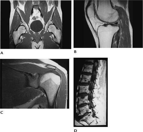
FIGURE 12-1 Marrow patterns. T1-weighted images of the pelvis (A), knee (B), shoulder (C), and spine (D). The marrow in the pelvis (A) is predominantly fatty in this adult except for small areas of red marrow in the acetabular regions (arrows). There is also fatty high signal intensity marrow in the knee (B) in an older adult and epiphysis of the shoulder (C)
in this 20-year-old patient. There is red marrow in the humeral shaft and glenoid. The sagittal spine image shows predominantly yellow marrow with areas of low signal intensity because of multiple metastases. |
Suggested Reading
Vogler JB, Murphy WA. Bone marrow imaging. Radiology 1988;168:679–693.
P.781
Marrow Reconversion
Key Facts
-
Hematopoietic demand may result in conversion of yellow to red marrow.
-
The sequence of red marrow conversion
occurs in reverse of the red to yellow marrow conversion, occurring as
one develops adult marrow pattern. -
Reconversion begins in the axial skeleton and moves peripherally.
-
Reconversion is symmetric, but not necessarily uniform.
-
Patients without bone marrow disorders may develop expanded red marrow termed “hyperplasia.”
-
Marrow reconversion, or hyperplasia, may be the result of multiple conditions:
-
Chronic anemia
-
Chronic infection
-
Marrow replacement diseases
-
Cyanotic heart disease
-
Marathon running
-
Smoking
-
-
Imaging of red marrow reconversion is
best accomplished with MRI. Signal intensity changes may be difficult
to differentiate from infiltrative diseases. T1-weighted images show
areas of decreased signal intensity. T2-weighted and STIR sequences
show signal intensity higher than fat.
P.782
 |
|
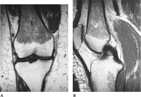
FIGURE 12-2 Marrow hyperplasia in a long-distance runner. Coronal (A) and sagittal (B)
T1-weighted images showing low intensity marrow in the femoral diaphysis and metaphysis with a focal area of hyperplasia in the tibia. Cortical bone is normal, and there are no soft tissue abnormalities. |
Suggested Reading
Shellock
FG, Morris E, Deutsch AL, et al. Hemopoietic marrow hyperplasia: High
prevalence on MR images of the knee in asymptomatic marathon runners. AJR Am J Roentgenol 1992;158:335–338.
FG, Morris E, Deutsch AL, et al. Hemopoietic marrow hyperplasia: High
prevalence on MR images of the knee in asymptomatic marathon runners. AJR Am J Roentgenol 1992;158:335–338.
Vande
Berg BC, Levouvet FE, Moyson P, et al. MR assessment of red marrow
distribution and composition in the proximal femur: Correlation with
clinical and laboratory parameters. Skel Radiol 1997;26:589–596.
Berg BC, Levouvet FE, Moyson P, et al. MR assessment of red marrow
distribution and composition in the proximal femur: Correlation with
clinical and laboratory parameters. Skel Radiol 1997;26:589–596.
P.783
Myeloid Depletion
Key Facts
-
In patients with myeloid depletion, the hemopoietic (red) marrow is replaced by fatty (yellow) marrow.
-
Fat replacement may be diffuse or focal, depending on the extent and duration of the process.
-
Conditions leading to myeloid depletion include
-
Aplastic anemia
-
Chemotherapy
-
Radiation
-
Marrow toxins
-
Viral infections
-
-
Radiation changes are focal, so fat
replacement occurs in the treated region. Changes occur as early as 3
to 7 weeks. With low doses, recovery may occur. Patients receiving more
than 50 Gy usually are irreversible. -
Fatty marrow has fat signal intensity on T1- and T2-weighted sequences.
 |
|
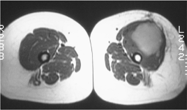
FIGURE 12-3
Radiation therapy. Axial T1-weighted image showing a malignant soft tissue mass with fatty marrow in the femur. Compare with normal lower signal intensity red marrow in the opposite femur. |
P.784
 |
|
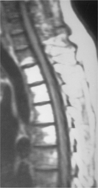
FIGURE 12-4 Radiation changes with fatty marrow in the upper thoracic spine seen on T1-weighted magnetic resonance (MR) image.
|
Suggested Reading
Cavenagh
EC, Weinberger E, Shal DW, et al. Hemopoietic marrow reconversion in
pediatric patients undergoing spinal irradiation. MR depiction. AJNR Am J Neuroradiol 1995;16:461–467.
EC, Weinberger E, Shal DW, et al. Hemopoietic marrow reconversion in
pediatric patients undergoing spinal irradiation. MR depiction. AJNR Am J Neuroradiol 1995;16:461–467.
P.785
Marrow Ischemia
Key Facts
-
Bone marrow ischemic changes usually are focal (see Chapters 4, 5, and 7).
-
Causes of bone marrow ischemia and osteonecrosis are numerous.
-
Trauma
-
Exogenous steroids
-
Systemic diseases
-
Gaucher disease
-
Sickle cell anemia
-
Idiopathic
-
-
Focal changes are most common in the hip, knee, and shoulder.
-
Avascular necrosis occurs in the subchondral epiphysis, and bone infarction occurs in the metaphysis and diaphysis.
-
Image changes may be evident on
radiographs in advanced cases. MRI is the technique of choice for
imaging early ischemic changes. Contrast-enhanced images are useful to
evaluate subtle changes in flow.
 |
|
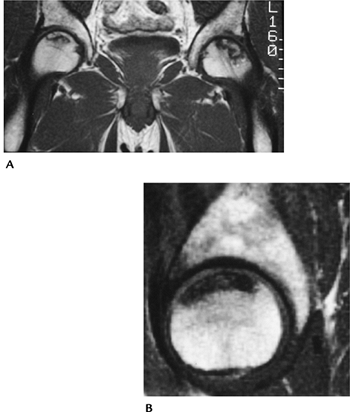
FIGURE 12-5 Avascular necrosis of the hip. Coronal (A) and sagittal (B) T1-weighted MR images showing avascular necrosis, which is more extensive on the left.
|
P.786
 |
|
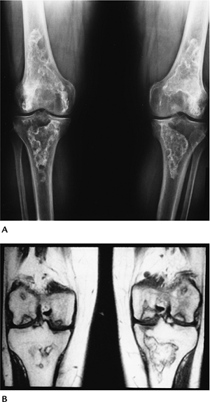
FIGURE 12-6 Bone infarction. (A) Standing radiographs of the knees show serpiginous marginal calcifications characteristic of bone infarction in both knees. (B) Coronal T1-weighted images of the knees demonstrate the areas of infarction and subchondral avascular necrosis.
|
Suggested Reading
Deely DM, Schweitzer ME. MR imaging of bone marrow disorders. Radiol Clin North Am 1997;35:193–212.
P.787
Marrow Infiltration: Basic Concepts
Key Facts
-
There are numerous marrow infiltrative
processes, including inflammatory conditions, neoplastic processes
(metastasis, myeloma), lipidosis, histiocytosis, and
hyperlipoproteinemias. -
Table 12-1 summarizes infiltrative marrow conditions.
-
Routine radiographs may demonstrate marrow expansion, lytic lesions, sclerotic lesions, or mixed lytic and sclerotic changes.
-
MR images demonstrate low signal
intensity on T1-weighted sequences and increased signal on T2-weighted
or STIR sequences in most cases. Contrast enhancement is variable and
nonspecific.
|
TABLE 12-1 INFILTRATIVE MARROW DISORDERS
|
|
|---|---|
|
Suggested Reading
Resnick D. Lipidosis, histiocytosis, and hyperlipoproteinemias. In: Resnick D, ed. Diagnosis of bone and joint disorders, 4th ed. Philadelphia: WB Saunders; 2002:2233–2290.
P.788
Marrow Infiltration: Lipidosis
Key Facts
-
There are multiple lipidoses. The more common disorders are listed in Table 12-1.
-
Gaucher disease is a rare familial
disorder of cerebroside metabolism resulting in lipid accumulation in
reticuloendothelial cells. The condition affects males and females in
childhood or early adulthood. Patients present with hepatosplenomegaly.
Infiltration occurs in other regions, including marrow. Radiographs
demonstrate marrow expansion, endosteal scalloping, osteonecrosis, and
pathologic fractures. Changes are most common in the axial skeleton and
proximal long bones. MR images demonstrate decreased signal intensity
on T1- and T2-weighted sequences because of fibrosis and
glucocerebroside deposition. -
Niemann-Pick disease is a rare genetic
disorder with deposition of sphingomyelin in the reticuloendothelial
system. Males and females are equally affected. Many are Jewish. -
There is an acute fatal type and chronic
forms with and without central nervous system involvement. Most
patients present in infancy with jaundice and hepatosplenomegaly. -
Radiographic features are similar to those for Gaucher disease.
-
Fabry disease is a hereditary sex-linked disorder with deposition of ceramide trihexoside.
-
Patients present with skin lesions,
fever, pain, and paresthesias. Patients may die in later adult life
because of renal or cardiac involvement. Radiographic features include
periarticular swelling and avascular necrosis. -
Gangliosidosis is an inherited liposomal
storage disorder caused by lack of ganglioside β-galactosidase.
Ganglioside accumulates in the nervous system and other tissues.
Clinical features include mental retardation, a cherry spot in the
retina, hepatomegaly, and bony abnormalities. Radiographic features
include osteopenia, delayed bone maturation, thickened calvarium,
horizontal ribs, flattening of vertebral bodies, flared iliac, and thin
long bones.
P.789
 |
|
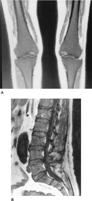
FIGURE 12-7 Gaucher disease. Coronal T1-weighted images of the knees (A) and sagittal T1-weighted images of the spine (B) showing low signal intensity throughout the marrow.
|
Suggested Reading
Resnick D. Lipidosis, histiocytosis, and hyperlipoproteinemias. In: Resnick D, ed. Diagnosis of bone and joint disorders, 4th ed. Philadelphia: WB Saunders; 2002:2233–2290.
P.790
Langerhans Cell Histiocytosis
Key Facts
-
Three conditions constitute Langerhans cell histiocytosis (histiocytosis X): (i) eosinophilic granuloma (see Chapter 10), (ii) Hand-Schüller-Christian disease, and (iii) Letterer-Siwe disease.
-
Common to all is the presence of Langerhans cells containing cytoplasmic inclusion bodies.
-
Eosinophilic granuloma is the mildest form presenting with single or multiple bone lesions.
-
This condition constitutes 70% of the
disorders. Patients present with pain, swelling, and, on occasion,
fever and elevated white counts. Radiographic features include lytic
lesions with endosteal scalloping and periosteal reaction. Most common
sites of involvement are the skull, mandible, ribs, spine, and long
bones. Vertebral involvement leads to marked compression. -
Differential diagnosis includes osteomyelitis, Ewing sarcoma, and lymphoma.
-
-
Hand-Schüller-Christian disease presents
with diabetes insipidus, exophthalmus, and single or multiple bone
lesions. This condition is most common in children. Multisystem
involvement includes otitis media, skin lesions, hepatosplenomegaly,
and anemia. Neural, lung, and renal involvement are common. Skeletal
lesions are similar to eosinophilic granuloma, but more disseminated.
The disease is fatal in up to 30% of cases. -
Letterer-Siwe disease is an acute
disorder common in children younger than 3 years. Multisystem
involvement (liver, spleen, skin, anemia) is typical. Most patients
survive only 1 to 2 years. Radiographically, there are multiple lytic
lesions, most commonly involving the skull. -
Erdheim-Chester disease is a disorder of adults (both male and female).
-
Cardiac and pulmonary involvement is common because of release of cholesterol from cells.
-
Radiographic features include long bone
sclerosis and prominent trabeculae with sparing of the axial skeleton.
Bilateral symmetric involvement is common. Histologic changes resemble
Hand-Schüller-Christian disease.
-
P.791
 |
|
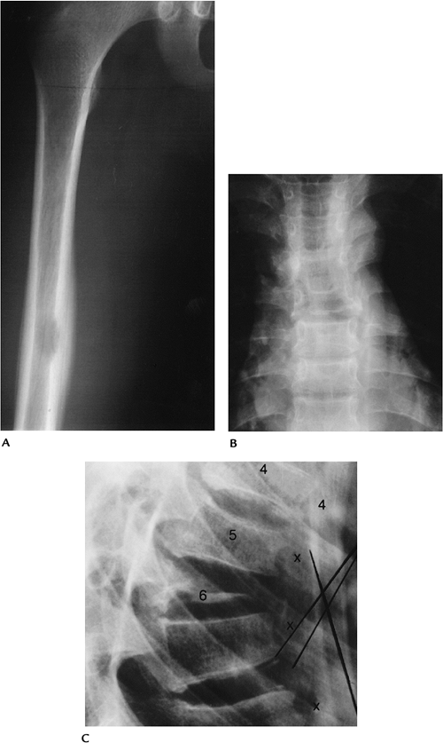
FIGURE 12-8 Eosinophilic granuloma. (A) AP radiograph of the femur showing a lytic lesion with endosteal scalloping and periosteal reaction. AP (B) and lateral (C) radiographs of the spine with marked compression (vertebra plana) of T6.
|
P.792
 |
|
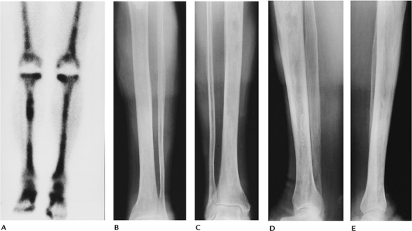
FIGURE 12-9 Erdheim-Chester disease. (A) Radionuclide bone scan showing increased tracer in both femora and tibiae. AP (B,C) and lateral (D,E) radiographs of the tibiae showing sclerotic areas with prominent trabeculae.
|
P.793
Suggested Reading
Resnick D. Lipidosis, histiocytosis, and hyperlipoproteinemias. In: Resnick D, ed. Diagnosis of bone and joint disorders, 4th ed. Philadelphia: WB Saunders; 2002:2233–2290.
P.794
Hyperlipoproteinemias
Key Facts
-
Hyperlipoproteinemias present with increased plasma cholesterol and triglycerides. Five types are described.
-
Type I: deficiency in lipoprotein lipase.
Patients are 10 to 30 years old, with lipemia retinalis,
hepatosplenomegaly, abdominal pain, and pancreatitis. -
Type II: The most common form, it
presents with increased low-density lipoproteins and β-lipoproteins.
Patients present in early childhood with xanthomas and premature
vascular disease. -
Type III: β-lipoprotein or pre–β-lipoprotein disorders. Patients present with xanthomas and premature vascular disease.
-
Type IV: increased low-density
lipoproteins and pre–β-lipoproteins. May be secondary to diabetes,
pancreatitis, alcoholism, Gaucher disease, and so forth. Patients
present after age 20 years with xanthomas, gout, and coronary artery
disease. -
Type V: similar to Type IV but with hepatosplenomegaly and paresthesias.
-
-
Radiographic features include
-
Osseous xanthomas
-
Soft tissue xanthomas
-
Gout
-
Soft tissue swelling about joints
-
P.795
 |
|
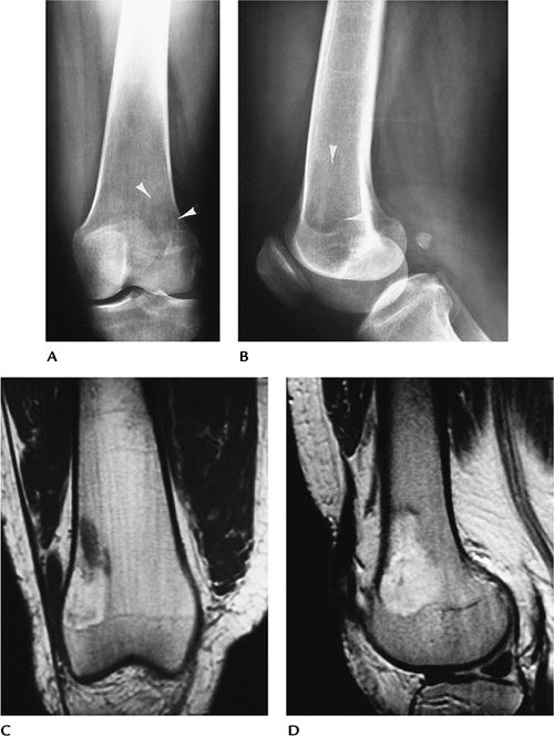
FIGURE 12-10 Osseous xanthomas (Type IV hyperlipoproteinemia). (A,B) Routine radiographs showing a poorly defined lytic lesion in the distal femur (arrowheads). Coronal T1-weighted (C) and sagittal T2-weighted (D) MR images showing a poorly defined lesion that has fatty intensity on T1-weighted image (C) and areas of increased intensity on T2-weighted image (D).
|
Suggested Reading
Resnick D. Lipidosis, histiocytosis, and hyperlipoproteinemias. In: Resnick D, ed. Diagnosis of bone and joint disorders, 4th ed. Philadelphia: WB Saunders; 2002:2233–2290.
P.796
Myelofibrosis
Key Facts
-
Myelofibrosis is an uncommon disorder resulting in fibrous replacement of marrow and extramedullary hematopoiesis.
-
Patients typically are middle-aged or elderly and present with fatigue, hepatosplenomegaly, and purpura.
-
Secondary gout is evident in 5% to 20%.
-
Radiographic features include bone sclerosis and splenomegaly.
-
MR images demonstrate low signal intensity on T1- and T2-weighted sequences.
-
Differential diagnosis includes Paget disease and metastasis.
P.797
 |
|
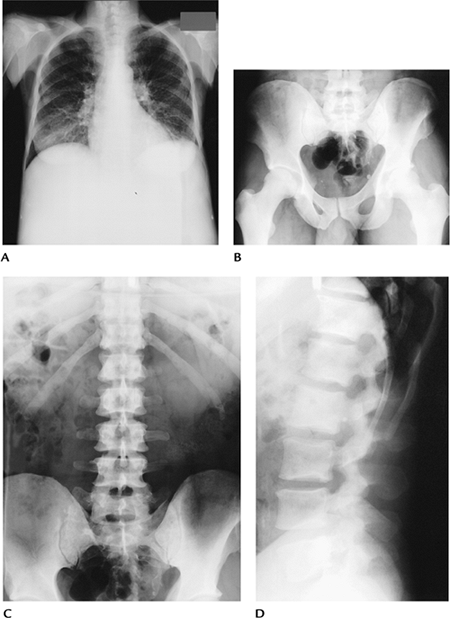
FIGURE 12-11 Myelofibrosis. (A) Chest radiograph showing increased bone density and marked splenomegaly. (B) AP radiograph of the pelvis showing scattered bone sclerosis. AP (C) and lateral (D) radiographs showing increased bone density.
|
P.798
 |
|
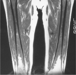
FIGURE 12-12 Myelofibrosis. T1-weighted MR image of the femora showing low signal intensity, except distally.
|
Suggested Reading
Lanir A, Aghai E, Simon JS, et al. MR imaging in myelofibrosis. J Comput Assist Tomogr 1986;10:634–636.
P.799
Compression Fractures: Benign versus Malignant
Key Facts
-
Vertebral compression fractures are common, especially in elderly females.
-
Differentiation of traumatic or
osteopenic compression fractures from metastasis or myeloma may be
difficult with conventional imaging studies (radiographs, radionuclide
scans). -
MRI is more useful for differentiating
compression fractures caused by osteopenia or trauma from malignancy.
MR features suggesting malignancy:-
Focal signal abnormality in multiple vertebrae
-
Diffuse signal abnormality involving the vertebral body and posterior elements
-
Epidural mass
-
Convex posterior vertebral margin on T1-weighted images
-
-
Benign MR features:
-
Preservation of normal marrow signal
-
Isointense marrow on T2-weighted sequences
-
Isolated vertebral involvement
-
Diffusion-weighted MR images may be most
specific. Signal attenuation is evident with osteoporotic compression,
and increased signal is evident with malignant lesions or diffusion
images (spin-echo or fat-suppressed spin-echo sequences). Dynamic
gadolinium-enhanced sequences may also detect pathologic marrow more
readily than conventional MR sequences.
-
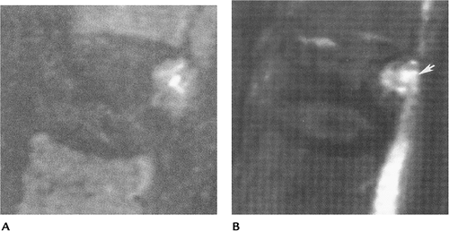 |
|
FIGURE 12-13 Steroid-induced osteoporosis in a 75-year-old man. (A) Sagittal T2-weighted nondiffusion image showing vertebral compression with mixed decreased and increased signal intensity. (B) Diffusion-weighted spin-echo image showing an area of increased intensity (arrow) caused by lung metastasis. (From
Spuentriep
E, Buecher A, Adam G, et al. Diffusion-weighted MR imaging for differentiation of benign fracture edema and tumor infiltration of the vertebral body. AJR Am J Roentgenol 2001;176:351–358, with permission. ) |
P.800
Suggested Reading
Rahmouni
A, Montazel JL, Divine M, et al. Bone marrow with diffuse tumor
infiltration in patients with lymphoproliferative diseases: Dynamic
gadolinium-enhanced MR imaging. Radiology 2003;229:710–717.
A, Montazel JL, Divine M, et al. Bone marrow with diffuse tumor
infiltration in patients with lymphoproliferative diseases: Dynamic
gadolinium-enhanced MR imaging. Radiology 2003;229:710–717.
Spuentriep
E, Buecher A, Adam G, et al. Diffusion-weighted MR imaging for
differentiation of benign fracture edema and tumor infiltration of the
vertebral body. AJR Am J Roentgenol 2001;176:351–358.
E, Buecher A, Adam G, et al. Diffusion-weighted MR imaging for
differentiation of benign fracture edema and tumor infiltration of the
vertebral body. AJR Am J Roentgenol 2001;176:351–358.