
Editors: Chapman, Michael W.
Title: Chapman’s Orthopaedic Surgery, 3rd Edition
Copyright ©2001 Lippincott Williams & Wilkins
> Table of Contents > SECTION
IV – SPORTS MEDICINE > Ankle > CHAPTER 94 – TENDON LACERATIONS
AND OTHER TENDON DISORDERS ABOUT THE ANKLE
IV – SPORTS MEDICINE > Ankle > CHAPTER 94 – TENDON LACERATIONS
AND OTHER TENDON DISORDERS ABOUT THE ANKLE
CHAPTER 94
TENDON LACERATIONS AND OTHER TENDON DISORDERS ABOUT THE ANKLE
George Lian
G. Lian: Department of Orthopaedic Surgery, University of California, Davis, School of Medicine, Sacramento, California 95816.
TENDON LACERATIONS
Lacerations to tendons about the foot and ankle can be
caused by stepping on or dropping a sharp object such as glass onto the
foot, which usually results in a clean sharp isolated tendon
laceration. More violent crushing-type lacerations occur with motor
vehicle accidents, power saw and lawnmower injuries, or dropping
objects on the dorsum of the foot, all of which cause more extensive
associated injuries (4,6).
Floyd et al. (4) showed that all
tendon lacerations, with the possible exception of the flexor digitorum
tendons, benefit from primary repair. They found in a number of cases
that surgical exploration revealed additional significant injuries that
were not appreciated in the clinical examination.
tendon lacerations, with the possible exception of the flexor digitorum
tendons, benefit from primary repair. They found in a number of cases
that surgical exploration revealed additional significant injuries that
were not appreciated in the clinical examination.
PATHOPHYSIOLOGY
The tendon lacerations discussed here involve the following tendons: flexor hallucis longus (FHL), flexor digitorum
P.2468
longus (FDL), extensor digitorum longus (EDL), extensor hallucis longus
(EHL), anterior tibial (AT), and posterior tibial (PT) tendons.
The FHL muscle originates from the deep posterior
compartment of the leg. The FHL tendon passes beneath the sustenaculum
tali and continues distally, crossing deep to the FDL tendon at the
flexor knot of Henry. At this point the tendon produces a tendinous
slip that inserts into the FDL tendon (16). The
FHL tendon is covered by two synovial sheaths. The first provides
coverage from the ankle joint to a point just distal to the flexor knot
of Henry; the second provides coverage from the first metatarsal base
to the tendon insertion.
The EDL, EHL, and AT tendons originate from the anterior
compartment in the leg. Each muscle produces a tendon that is enclosed
in a synovial sheath and is retained in position by a thickened section
of crural fascia known as the extensor retinaculum. There are
individual sheaths for the anterior tibial tendon and extensor hallucis
longus tendon and a common sheath for the extensor digitorum longus
tendons (17).
The anatomic features of the posterior tibial tendon are described in the POSTERIOR TIBIAL TENDON DYSFUNCTION section of Chapter 118.
The subcutaneous positions of the tendons about the foot
and ankle predispose them to injury by direct trauma. Laceration of the
plantar or dorsal aspect of the foot may produce disruption of
tendinous, vascular, or neurologic structures.
and ankle predispose them to injury by direct trauma. Laceration of the
plantar or dorsal aspect of the foot may produce disruption of
tendinous, vascular, or neurologic structures.
PRINCIPLES OF TREATMENT
Injuries associated with foot lacerations are commonly
missed. Establish the full extent of the injury by careful physical
examination, appropriate radiographic imaging, and, when necessary,
surgical exploration. When primary repair is indicated, techniques used
are similar to those for flexor tendon injuries in the hand.
missed. Establish the full extent of the injury by careful physical
examination, appropriate radiographic imaging, and, when necessary,
surgical exploration. When primary repair is indicated, techniques used
are similar to those for flexor tendon injuries in the hand.
ASSESSMENT
Document the mechanism of injury. Determine the exact
anatomic area of injury. Check the vascular and neurologic status and
follow this by an analysis of tendon function. Remember that the FHL
tendon provides a tendinous slip to the FDL tendon. If the FHL tendon
is disrupted proximal to this slip, active flexion at the hallux
interphalangeal joint may be produced by the FDL tendon. Therefore, the
presence of active hallux interphalangeal joint plantarflexion does not
rule out FHL tendon injury.
All lacerations require plain radiographic imaging to rule out concomitant bony injury and the presence of foreign material.
CLASSIFICATION
Tendon lacerations can be classified on the basis of
mechanism of injury, degree of injury (incomplete versus complete),
location of injury (insertional versus noninsertional, relation to
synovial sheath, relation to retaining structures), and by the presence
of associated neurovascular or tendinous injury. With regard to
laceration of the FHL tendon, the integrity of the flexor hallucis
brevis is an important consideration.
PREOPERATIVE MANAGEMENT
In the emergency department, provide routine wound care
and give appropriate tetanus prophylaxis. Generally speaking, isolated
tendon laceration about the foot may be treated by nonsurgical or
surgical means. Repair primarily complete laceration of the Achilles,
PT, AT, peroneal, EHL, and FHL tendons when local conditions are
favorable. Functional (partial) lacerations as well as laceration of
the brevis tendons are typically not repaired. Do a meticulous
debridement and evaluation of associated structures and repair if
needed.
OPERATIVE TECHNIQUES
Surgical Exploration and Repair of FHL Tendon Laceration
-
Use general or regional anesthesia and a
proximal thigh tourniquet. Enlarge the plantar wound with proximal and
distal extensions as required. If possible, avoid the weight-bearing
surface of the foot. -
Inspect the FHL tendon, the FHB muscle
and tendon, and surrounding neurovascular structures. If the FHL tendon
laceration is distal to the tendinous connection with the FDL tendon,
retraction of the FHL tendon is limited. Identify both ends of the
tendon and meticulously debride the wound; then perform a primary
tendon repair. Use a single-core 2-0 nonabsorbable suture. Repair the
peripheral edges of the tendon with a smaller polydioxanone (PDS,
Ethicon, Inc., Wayne, NJ) or nylon suture. After completion of the
repair, verify excursion of the tendon. -
If primary repair cannot be accomplished,
tenodese the adjacent FDL tendon to the proximal and distal ends of the
FHL tendon. In the unusual event of a nonrepairable FHL tendon
laceration accompanied by disruption of both heads of the FHB muscle,
anastomose the proximal FHB muscle and the distal end of the FHL tendon. -
Because of the risk of formation of a
painful neuroma, we do not repair digital nerve lacerations primarily
but rather transplant them to a protected position, such as into a
muscle belly. Repair the surgical extensions to the foot wound and
immobilize the foot in slight plantarflexion. -
Postoperatively, do routine wound care to
assure a viable wound with eventual successful closure. Over the course
of 2 to 3 weeks, bring the foot gradually up to a neutral position.
Begin passive range of motion at the hallux interphalangeal and the
metatarsophalangeal joints. At 6 weeks postoperatively, start weight
bearing in a removable cast boot. At 8 weeks postoperatively, begin a
strengthening program followed by gradual resumption of activities
without the cast boot.
P.2469
Surgical Exploration and Repair of the EDL, EHL, and AT Tendons
-
Use general or regional anesthesia and a
proximal thigh tourniquet. Extend the dorsal wound as necessary in
proximal and distal directions. If possible, keep the incision in line
with, but not over, the injured tendon to minimize adhesions with
overlying skin. -
Identify the proximal and distal ends of
the lacerated tendons and systematically inspect surrounding
structures. After meticulous debridement and irrigation of the wound,
perform a primary tendon repair. Use a core suture of nonreactive,
nonabsorbable material within the tendon substance to provide primary
apposition, supplemented with a whipstitch of the tendon ends with a
smaller running nylon or PDS suture (Ethicon, Inc. Wayne, NJ). If
end-to-end or side-to-side repair cannot be performed, use a proximal
or distal Z-lengthening to provide additional length. -
Close the surgical wound extensions
primarily and immobilize the foot in a neutral to slightly dorsiflexed
position. The addition of toe dorsiflexion may also be required. -
Postoperatively, provide routine wound
care as discussed for FHL tendon lacerations. At 6 weeks
postoperatively, begin range-of-motion exercises and weight bearing in
a removable cast boot. At 8 weeks postoperatively, begin a
strengthening program. Finally, discontinue the cast boot once local
warmth and swelling have resolved. Maintain range of motion. Motion is
often limited as a result of formation of local adhesions.
COMPLICATIONS
Placing sutures in a traumatic wound increases the risk
of infection. Therefore, perform primary tendon repair only if local
wound conditions are favorable. Digital nerve injury may be complicated
by neuroma formation or the onset of reflex sympathetic dystrophy.
Minimize the risk of posttraumatic neuroma formation by transplanting
the proximal end of the lacerated digital nerve into a local muscle
belly. If a painful neuroma occurs, treatment with desensitization
modalities is indicated. Occasionally, referral to a pain management
specialist is required.
CONCLUSIONS
When local conditions permit, we prefer primary tendon
repair, especially in the pediatric and athletic population. Because of
their propensity to form neuromas, we do not repair digital nerve
lacerations but transplant the proximal nerve to a well-protected
location.
repair, especially in the pediatric and athletic population. Because of
their propensity to form neuromas, we do not repair digital nerve
lacerations but transplant the proximal nerve to a well-protected
location.
FLEXOR HALLUCIS LONGUS TENDON
Injury to the FHL tendon at the ankle has been recognized mostly in runners and dancers (2,3,7,8,50,12,15,19).
The FHL tendon enters the foot through a fibro-osseous tunnel along the
posteromedial ankle. This is defined superiorly by the posterolateral
process of the talus, and it extends inferiorly to the sustentaculum
tali on the medial side of the hindfoot. The tendon undergoes a change
in direction within this tunnel from vertical proximally to horizontal
distally (11). As a result, it can become irritated and develop intrasubstance tears that cause it to swell and become tethered.
CLINICAL PRESENTATION
Pain is present in the posterior ankle deep to the
Achilles tendon. Deep palpation in this area may cause tenderness. The
pain may be aggravated by passive motion of the great toe. With
significant swelling, the tendon may become entrapped and cause
triggering, leading to clawing of the great toe (2,7,8,10,12,15,19).
Entrapment can be verified by comparing great toe dorsiflexion with the
ankle in the plantarflexed position and when dorsiflexed. With
entrapment, dorsiflexion of the toe is limited with the ankle also
dorsiflexed but possible when the ankle is plantarflexed (9).
RADIOGRAPHS
Routine radiographs will be unremarkable. The presence
of a large posterior process or os trigonum may cloud the diagnosis.
Magnetic resonance imaging (MRI) is a useful test for evaluating
questionable cases (11,19)
where fluid can be identified about the tendon in the fibro-osseous
tunnel and intrasubstance tears in the tendon may also be present.
of a large posterior process or os trigonum may cloud the diagnosis.
Magnetic resonance imaging (MRI) is a useful test for evaluating
questionable cases (11,19)
where fluid can be identified about the tendon in the fibro-osseous
tunnel and intrasubstance tears in the tendon may also be present.
NONOPERATIVE TREATMENT
Nonoperative treatment consists of activity limitation
and nonsteroidal antiinflammatory medications for 10 to 14 days.
Immobilization in a short-leg walking cast with extended toe plate to
prevent great toe motion may be helpful as well.
and nonsteroidal antiinflammatory medications for 10 to 14 days.
Immobilization in a short-leg walking cast with extended toe plate to
prevent great toe motion may be helpful as well.
P.2470
OPERATIVE TREATMENT
Treat athletes who do not obtain relief with
conservative methods or those who have persistent triggering of the
great toe with release of the fibro-osseous tunnel of the FHL. The
medial approach is favored because it allows access to the entire
fibro-osseous canal. Full return of function can be expected with this
procedure (2,8,10,12,14,19).
Surgical Release of the FHL Sheath
-
Use a general or regional anesthetic and
a thigh tourniquet. Place a bump under the opposite thigh to aid in
exposure of the medial side of the posterior ankle. -
Make a curving longitudinal incision in
line with the flexor tendons posterior to the medial malleolus. Begin 5
cm above the malleolus and extend distally to the level of the
navicular bone. -
Divide the flexor retinaculum in line
with the incision and identify the FDL by passively moving the toes.
Enter the interval behind the FDL tendon and in front of the posterior
tibial artery. -
Identify the FHL tendon and protect the
neurovascular bundle, which is posterior. Retract the FDL tendon
anteriorly. Passive motion of the great toe will aid in identifying the
FHL tendon. -
Split the sheath from proximal to distal
to the level of the sustentaculum tali. Identify the neurovascular
bundle over the course of this dissection to avoid injuring it. Inspect
the tendon for any tears and repair them as indicated. -
Close the wound by repairing the flexor
retinaculum with #0 absorbable sutures. Close the subcutaneous layer
and skin and apply a posterior splint.
Postoperatively, use crutches for 1 week and then remove
skin sutures. Then start weight bearing as tolerated. Start
strengthening therapy but avoid training activities for 6 weeks.
skin sutures. Then start weight bearing as tolerated. Start
strengthening therapy but avoid training activities for 6 weeks.
ANTERIOR TIBIALIS TENDON
ANATOMY
The anterior tibialis (AT) muscle originates from the
proximal tibia and interosseous membrane. It becomes tendinous at the
level of the distal tibial metaphysis. The anterior tibialis tendon
passes over the front of the ankle and runs with a straight course
under the superior extensor retinaculum to attach to the dorsum of the
medial cuneiform and base of the first metatarsal. It is the primary
dorsiflexor of the ankle and also helps invert the foot (3).
Despite its subcutaneous course, the tendon is rarely injured, probably
because it does not change direction against a bony fulcrum and because
of its excellent blood supply (5,20).
CLINICAL PRESENTATION
Tendinitis can occur from overuse, particularly from
hiking downhill, and has been described from direct pressure against
the tendon by ski boots (20). Rupture of the
anterior tibialis tendon is an unusual injury that occurs in two
distinct populations. The most common presentation is in the elderly,
who usually have an atraumatic rupture and typically note the insidious
onset of a foot drop (9,13). The second presentation is an acute traumatic rupture in older athletes, but it can also occur in young athletes (3,13,18).
Patients with tendinitis have tenderness over the course
of the tendon and pain on resisted dorsiflexion of the ankle. A rupture
can be diagnosed by a visual or palpable defect in the tendon, foot
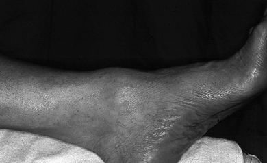
drop, and weakness of ankle dorsiflexion (Fig. 94.1).
of the tendon and pain on resisted dorsiflexion of the ankle. A rupture
can be diagnosed by a visual or palpable defect in the tendon, foot
drop, and weakness of ankle dorsiflexion (Fig. 94.1).
 |
|
Figure 94.1.
Rupture of anterior tibialis tendon. The ruptured anterior tibialis tendon has retracted, leaving a soft-tissue prominence on the dorsum of the foot. |
RADIOGRAPHS
Plain radiographs are unremarkeable. The rupture can be easily identified with an MRI scan (11,13). This is essentially a clinical diagnosis, however, and an MRI is usually not necessary.
TREATMENT
Treat tendinitis with activity limitation and
nonsteroidal medications. If pain is caused by direct pressure against
the tendon, remove the irritant.
nonsteroidal medications. If pain is caused by direct pressure against
the tendon, remove the irritant.
Chronic rupture in the older patient may be satisfactorily treated with a brace or may even be left untreated (1).
The acute rupture is probably best treated with surgical
reconstruction. In cases in which it is recognized early, a direct
repair of the tendon is possible (9,13,18,20). If it has been unrecognized for several months, reconstruction
The acute rupture is probably best treated with surgical
reconstruction. In cases in which it is recognized early, a direct
repair of the tendon is possible (9,13,18,20). If it has been unrecognized for several months, reconstruction
P.2471
with either an EHL tendon transfer or interposition of an EDL tendon
graft may be used, allowing return to recreational activities (13).
REFERENCES
Each reference is categorized according to the following
scheme: *, classic article; #, review article; !, basic research
article; and +, clinical results/outcome study.
scheme: *, classic article; #, review article; !, basic research
article; and +, clinical results/outcome study.
+ 1. Bernstein RM. Spontaneous Rupture of the Tibialis Anterior Tendon. Am J Orthop 1995;24:354.
+ 2. Cowell HR, Elener V. Bilateral Tendonitis of the Flexor Hallucis Longus in a Ballet Dancer. J Pediatr Orthop 1982;2:582.
+ 3. Frey C, Shereff M. Tendon Injuries About the Ankle in Athletes. Clin Sports Med 1988;7:103.
+ 4. Floyd DW, Heckman JD, Rockwood CA. Tendon Lacerations in the Foot. Foot Ankle 1983;4:8.
! 5. Geppert MJ, Sobel M, Hannafin JA. Microvasculature of the Tibialis Anterior Tendon. Foot Ankle 1993;14:261.
+ 6. Griffiths JC. Tendon Injuries About the Ankle. J Bone Joint Surg 1965;47B:686.
# 7. Hamilton WG. Foot and Ankle Injuries in Dancers. In: Mann RA, Coughlin MJ, eds. Surgery of the Foot and Ankle. St. Louis: CV Mosby, 1993;1241.
+ 8. Hamilton
WG. Stenosing Tenosynovitis of the Flexor Hallucis Longus Tendon and
Posterior Impingement upon the Os Trigonum in Ballet Dancers. Foot Ankle 1982;3:74.
WG. Stenosing Tenosynovitis of the Flexor Hallucis Longus Tendon and
Posterior Impingement upon the Os Trigonum in Ballet Dancers. Foot Ankle 1982;3:74.
# 9. Jones DC. Tendon Disorders of the Foot and Ankle. J Am Acad Orthop Surg 1993;1:87.
+ 10. McCarroll JR, Ritter MA, Becker TE. Triggering of the Great Toe: A Case Report. Clin Orthop 1983;175:184.
+ 11. Mink JH, Deutsch AL, Kerr R. Tendon Injuries of the Lower Extremity: Magnetic Resonance Assessment. Top Magn Reson Imag 1991;3:23.
+ 12. Newman NM, Fowles JV. A Case of “Trigger Toe.” Can J Surg 1984;27:378.
+ 13. Ouzounian TJ, Anderson R. Anterior Tibial Tendon Rupture. Foot Ankle 1995;16:406.
+ 14. Sammarco GJ, DiRaimondo CV. Chronic Peroneus Brevis Tendon Lesions. Foot Ankle 1989;9:163.
+ 15. Sammarco GJ, Miller EH. Partial Rupture of the Flexor Hallucis Longus Tendon in Classical Ballet Dancers. J Bone Joint Surg 1979;61:149.
# 16. Sarrafian SK. Myology. In: Sarrafian SK, ed. Anatomy of the Foot and Ankle, 2nd ed. Philadelphia: JB Lippincott, 1993;240.
# 17. Sarrafian SK. Tendon sheaths and bursae. In: Sarrafian SK, ed. Anatomy of the Foot and Ankle, 2nd ed. Philadelphia: JB Lippincott, 1993;283.
+ 18. Stuart MJ. Traumatic Disruption of the Anterior Tibial Tendon while Cross-Country Skiing: A Case Report. Clin Orthop 1992;281:193.
+ 19. Trepman E, Mizel MS, Newberg AH. Partial Rupture of the Flexor Hallucis Longus Tendon in a Tennis Player: A Case Report. Foot Ankle 1995;16:227.
+ 20. Trevino S, Baumhauer JF. Tendon Injuries of the Foot and Ankle. Clin Sports Med 1992;11:727.